Review
. 2022 Nov 11;10(4):47.
doi: 10.3390/jdb10040047.

Coordination of Cilia Movements in Multi-Ciliated Cells

Affiliations
Review

Coordination of Cilia Movements in Multi-Ciliated Cells

Masaki Arata et al. J Dev Biol. .

Abstract

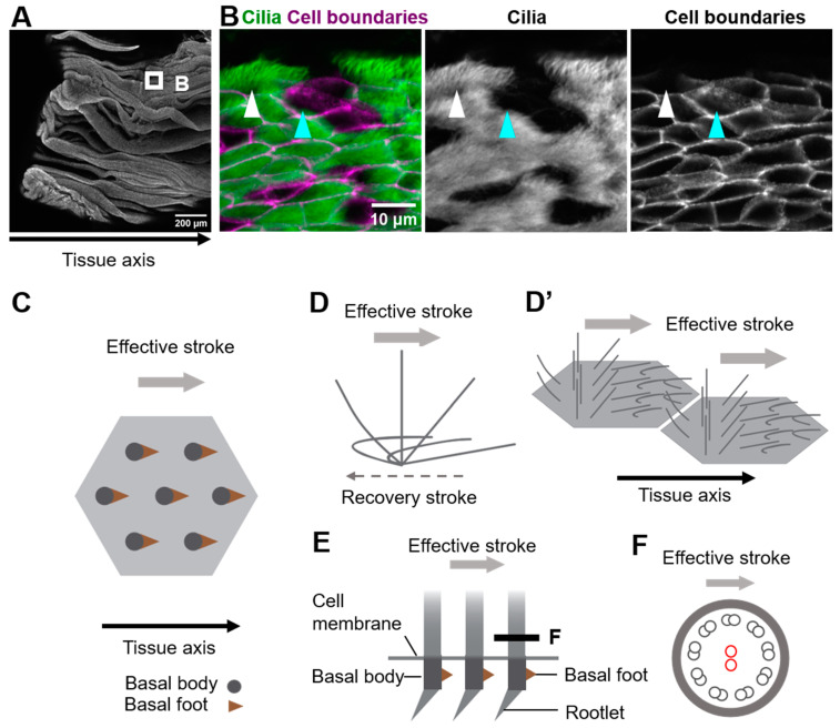
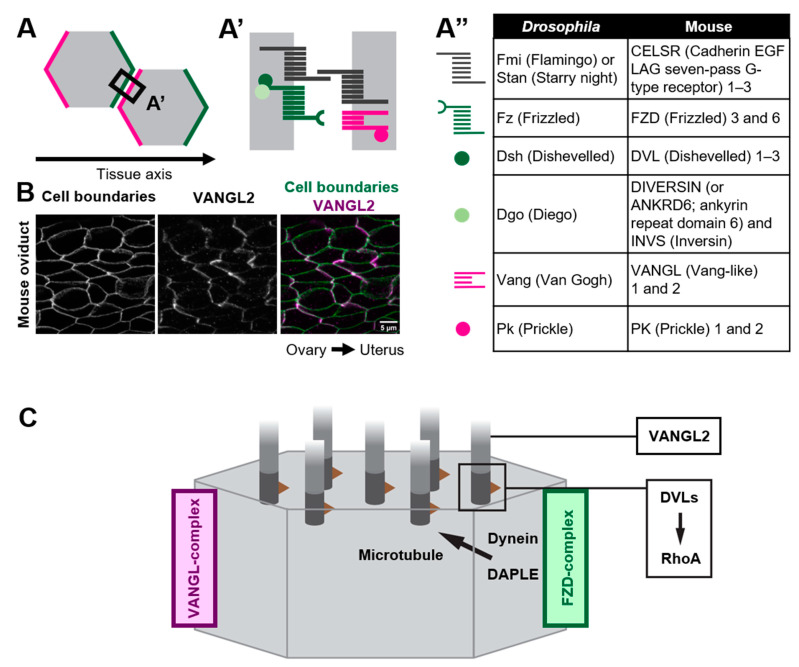
Multiple motile cilia are formed at the apical surface of multi-ciliated cells in the epithelium of the oviduct or the fallopian tube, the trachea, and the ventricle of the brain. Those cilia beat unidirectionally along the tissue axis, and this provides a driving force for directed movements of ovulated oocytes, mucus, and cerebrospinal fluid in each of these organs. Furthermore, cilia movements show temporal coordination between neighboring cilia. To establish such coordination of cilia movements, cilia need to sense and respond to various cues, including the organ's orientation and movements of neighboring cilia. In this review, we discuss the mechanisms by which cilia movements of multi-ciliated cells are coordinated, focusing on planar cell polarity and the cytoskeleton, and highlight open questions for future research.

Keywords: cytoskeleton; motile cilia; multi-ciliated cells; planar cell polarity.


Conflict of interest statement

The authors declare no conflict of interest.

Figures

Figure 1
Figure 1
Coordination of cilia movements in multi-ciliated cells. (A) Ovary end of the oviduct was opened longitudinally and stained with E-cadherin. (B) Cilia (green; acetylated tubulin) and cell boundaries (magenta; E-cadherin) of the oviduct epithelium are visualized. The oviduct epithelium is composed of multi-ciliated cells (white arrowhead) and secretory cells (cyan arrowhead). At the apical surface of those multi-ciliated cells, about 150 cilia are formed on average. (C) Representative apical views of multi-ciliated cells. Gray circles and brown triangles indicate basal bodies and basal feet, respectively (also see Figure 1(E)). Note that the basal foot points in the same direction as the effective stroke (gray arrow). (D) Schematic representation of the movement of an individual cilium. Cilia repeat cyclic movements comprised of a fast effective stroke (gray arrow), and a slow recovery stroke (a backward motion; dotted arrow). (D’) A schematic of cilia movements in multi-ciliated cells. Apical surfaces of two multi-ciliated cells are shown. Note that the orientation of effective stroke (or recovery stroke) is consistent with that of the tissue axis. The phase of the beating cycles of cilia shifts between neighboring cilia and generates a metachronal wave, which is a wave-like propagation of cilia movements. (E) Lateral view of the basal region of cilia. The basal body has two appendages, the basal foot and the rootlet. (F) Cross-sectional view of the cilium along the gray line in (E). 9 + 2 microtubules are shown in small circles. The central pair of 9 + 2 microtubules (red circles) run perpendicular to the direction of the effective stroke (gray arrow).
Figure 2
Figure 2
Polarized distribution of core PCP proteins along the tissue axis. (AA”) Core PCP proteins form an asymmetric complex at cell boundaries. (A,A’) FZD-containing complex (FZD-complex; green) and VANGL-containing complex (VANGL-complex; magenta) are segregated to opposite cell boundaries (note that their distribution is polarized along the tissue axis). Extracellular domain of CELSRs provides intercellular bridges between the FZD-complex and the VANGL-complex, thus enabling the coupling of cell polarity at a multicellular level. (A”) A list of members of core PCP proteins in Drosophila and their counterparts in mice. (B) Mouse oviduct epithelium was stained for E-cadherin and a core PCP protein, VANGL2. E-cadherin labels cell boundaries. Note the zigzag pattern of VANGL2 signals which highlights polarity in the cell-boundary distribution of VANGL2 along the ovary–uterus axis. (C) Mechanisms by which core PCP proteins control the coordinated movements of cilia.

References

    1. Brooks E.R., Wallingford J.B. Multiciliated Cells. Curr. Biol. 2014;24:R973–R982. doi: 10.1016/j.cub.2014.08.047. - DOI - PMC - PubMed
    1. Boutin C., Kodjabachian L. Biology of Multiciliated Cells. Curr. Opin. Genet. Dev. 2019;56:1–7. doi: 10.1016/j.gde.2019.04.006. - DOI - PubMed
    1. Reiter J.F., Leroux M.R. Genes and Molecular Pathways Underpinning Ciliopathies. Nat. Rev. Mol. Cell Biol. 2017;18:533–547. doi: 10.1038/nrm.2017.60. - DOI - PMC - PubMed
    1. Meunier A., Azimzadeh J. Multiciliated Cells in Animals. Cold Spring Harb. Perspect. Biol. 2016;8:a028233. doi: 10.1101/cshperspect.a028233. - DOI - PMC - PubMed
    1. Spassky N., Meunier A. The Development and Functions of Multiciliated Epithelia. Nat. Rev. Mol. Cell Biol. 2017;18:423–436. doi: 10.1038/nrm.2017.21. - DOI - PubMed

LinkOut - more resources