

Crystallographic and Physicochemical Analysis of Bovine and Human Teeth Using X-ray Diffraction and Solid-State Nuclear Magnetic Resonance
- PMID: 36412897
- PMCID: PMC9680385
- DOI: 10.3390/jfb13040254
Crystallographic and Physicochemical Analysis of Bovine and Human Teeth Using X-ray Diffraction and Solid-State Nuclear Magnetic Resonance
Abstract
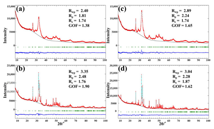
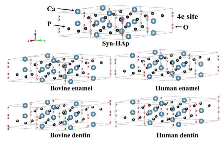
Dental research often uses bovine teeth as a substitute for human teeth. The aim of this study was to evaluate differences in the crystalline nanostructures of enamel and dentin between bovine and human teeth, using X-ray diffraction (XRD) and solid-state nuclear magnetic resonance (NMR). The crystallite size (crystallinity) and microstrains were analyzed using XRD with the Rietveld refinement technique and the Halder-Wagner method. The 31P and 1H NMR chemical environments were analyzed by two-dimensional (2D) 1H-31P heteronuclear-correlation (HETCOR) magic-angle spinning (MAS) NMR spectroscopy. Enamel had a greater crystallite size and fewer microstrains than dentin for both bovine and human teeth. When compared between the species, the bovine apatite had a smaller crystallite size with more microstrains than the human apatite for both dentin and enamel. The 2D HETCOR spectra demonstrated that a water-rich layer and inorganic HPO4- ions were abundant in dentin; meanwhile, the hydroxyl group in the lattice site was more dominant in enamel. A greater intensity of the hydroxyl group was detected in human than in bovine for both dentin and enamel. For 31P projections, bovine dentin and bovine enamel have wider linewidths than human dentin and human enamel, respectively. There are differences in the crystallite profile between human and bovine. The results of dental research should be interpreted with caution when bovine teeth are substituted for human teeth.
Keywords: X-ray diffraction; apatites; dentin; enamel; heteronuclear; solid-state nuclear magnetic resonance.
Conflict of interest statement
The authors declare no conflict of interest.
Figures

Similar articles
-
Structural differences in enamel and dentin in human, bovine, porcine, and ovine teeth.Ann Anat. 2018 Jul;218:7-17. doi: 10.1016/j.aanat.2017.12.012. Epub 2018 Mar 28. Ann Anat. 2018. PMID: 29604387
-
Solid-state NMR spectroscopy measurement of fluoride reaction by bovine enamel and dentin treated with silver diammine fluoride.Dent Mater. 2022 May;38(5):769-777. doi: 10.1016/j.dental.2022.04.017. Epub 2022 Apr 18. Dent Mater. 2022. PMID: 35450704
-
The crystallographic properties of the mineral phases of enamel and dentin in normal deciduous and permanent teeth.Zhonghua Kou Qiang Yi Xue Za Zhi. 2002 May;37(3):219-21. Zhonghua Kou Qiang Yi Xue Za Zhi. 2002. PMID: 12419150
-
Viability of Bovine Teeth as a Substrate in Bond Strength Tests: A Systematic Review and Meta-analysis.J Adhes Dent. 2018;20(6):471-479. doi: 10.3290/j.jad.a41636. J Adhes Dent. 2018. PMID: 30564794
-
Functional role of inorganic trace elements in dentin apatite tissue-Part 1: Mg, Sr, Zn, and Fe.J Trace Elem Med Biol. 2022 May;71:126932. doi: 10.1016/j.jtemb.2022.126932. Epub 2022 Jan 15. J Trace Elem Med Biol. 2022. PMID: 35101699 Review.
Cited by
-
Validation of Age Estimation Using the Compositional Variation of Dental Hard Tissue: An X-ray Diffraction Analysis.Cureus. 2024 Jul 29;16(7):e65696. doi: 10.7759/cureus.65696. eCollection 2024 Jul. Cureus. 2024. PMID: 39211662 Free PMC article.
-
Spectroscopic and dosimetric comparison of tooth enamel separation methods for EPR retrospective dosimetry.Heliyon. 2024 Apr 30;10(9):e30571. doi: 10.1016/j.heliyon.2024.e30571. eCollection 2024 May 15. Heliyon. 2024. PMID: 38742072 Free PMC article.
-
The Effects of Khat Chewing among Djiboutians: Dental Chemical Studies, Gingival Histopathological Analyses and Bioinformatics Approaches.Bioengineering (Basel). 2024 Jul 15;11(7):716. doi: 10.3390/bioengineering11070716. Bioengineering (Basel). 2024. PMID: 39061798 Free PMC article.
-
A Comparison between Porous to Fully Dense Electrodeposited CuNi Films: Insights on Electrochemical Performance.Nanomaterials (Basel). 2023 Jan 25;13(3):491. doi: 10.3390/nano13030491. Nanomaterials (Basel). 2023. PMID: 36770452 Free PMC article.
References
-
- Ortiz-Ruiz A.J., de Dios Teruel-Fernández J., Alcolea-Rubio L.A., Hernández-Fernández A., Martínez-Beneyto Y., Gispert-Guirado F. Structural differences in enamel and dentin in human, bovine, porcine, and ovine teeth. Ann. Anat. Anat. Anz. Off. Organ Anat. Ges. 2018;218:7–17. doi: 10.1016/j.aanat.2017.12.012. - DOI - PubMed
Grants and funding
LinkOut - more resources
Full Text Sources
