. 2023 Jan 10;8(1):e163855.
doi: 10.1172/jci.insight.163855.

Skeletal muscle mitochondrial inertia is associated with carnitine acetyltransferase activity and physical function in humans

Affiliations

Skeletal muscle mitochondrial inertia is associated with carnitine acetyltransferase activity and physical function in humans

Rodrigo F Mancilla et al. JCI Insight. .

Abstract

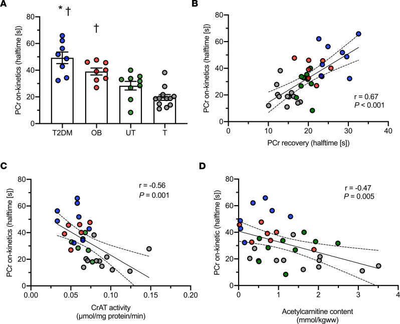
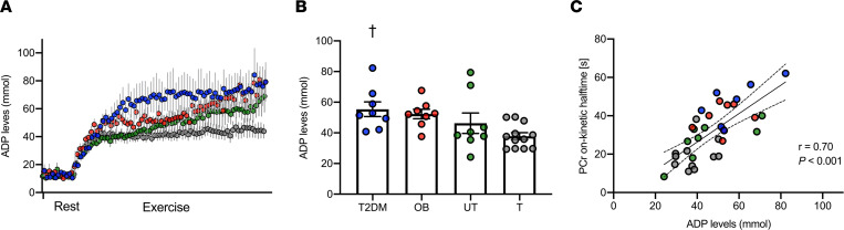
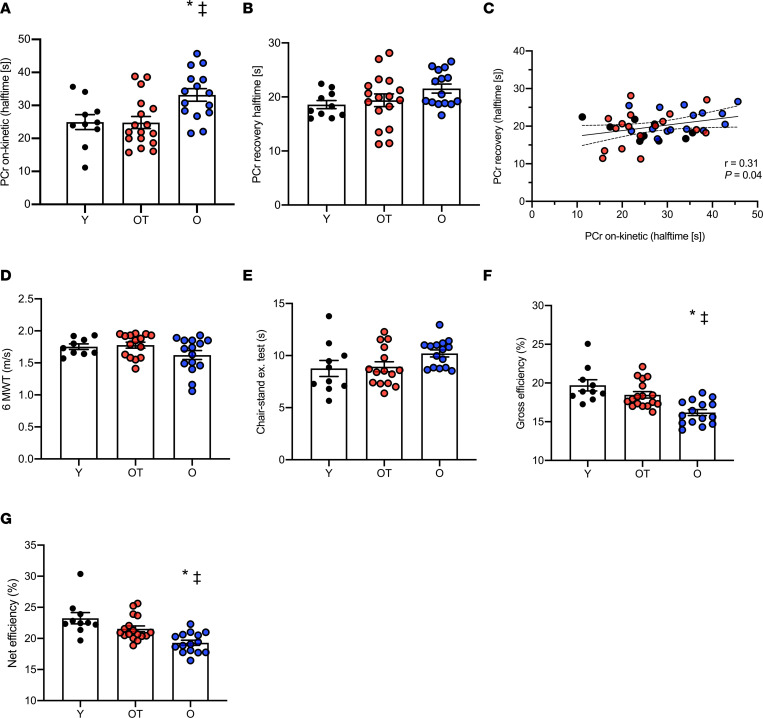
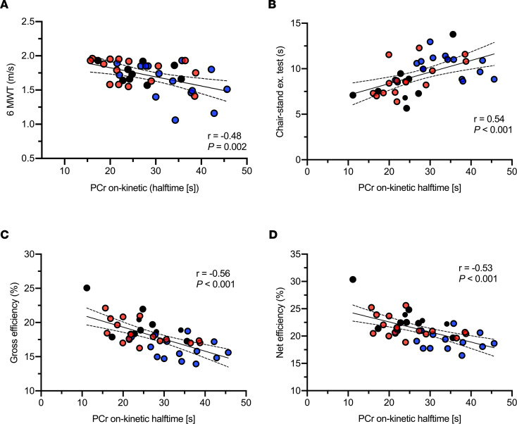
BACKGROUNDAt the onset of exercise, the speed at which phosphocreatine (PCr) decreases toward a new steady state (PCr on-kinetics) reflects the readiness to activate mitochondrial ATP synthesis, which is secondary to Acetyl-CoA availability in skeletal muscle. We hypothesized that PCr on-kinetics are slower in metabolically compromised and older individuals and are associated with low carnitine acetyltransferase (CrAT) protein activity and compromised physical function.METHODSWe applied 31P-magnetic resonance spectroscopy (31P-MRS) to assess PCr on-kinetics in 2 cohorts of volunteers. Cohort 1 included patients who had type 2 diabetes, were obese, were lean trained (VO2max > 55 mL/kg/min), and were lean untrained (VO2max < 45 mL/kg/min). Cohort 2 included young (20-30 years) and older (65-80 years) individuals with normal physical activity and older, trained individuals. Previous results of CrAT protein activity and acetylcarnitine content in muscle tissue were used to explore the underlying mechanisms of PCr on-kinetics, along with various markers of physical function.RESULTSPCr on-kinetics were significantly slower in metabolically compromised and older individuals (indicating mitochondrial inertia) as compared with young and older trained volunteers, regardless of in vivo skeletal muscle oxidative capacity (P < 0.001). Mitochondrial inertia correlated with reduced CrAT protein activity, low acetylcarnitine content, and functional outcomes (P < 0.001).CONCLUSIONPCr on-kinetics are significantly slower in metabolically compromised and older individuals with normal physical activity compared with young and older trained individuals, regardless of in vivo skeletal muscle oxidative capacity, indicating greater mitochondrial inertia. Thus, PCr on-kinetics are a currently unexplored signature of skeletal muscle mitochondrial metabolism, tightly linked to functional outcomes. Skeletal muscle mitochondrial inertia might emerge as a target of intervention to improve physical function.TRIAL REGISTRATIONNCT01298375 and NCT03666013 (clinicaltrials.gov).FUNDINGRM and MH received an EFSD/Lilly grant from the European Foundation for the Study of Diabetes (EFSD). VS was supported by an ERC starting grant (grant 759161) "MRS in Diabetes."

Keywords: Aging; Diabetes; Metabolism; Mitochondria; Skeletal muscle.


Conflict of interest statement

Conflict of interest: The authors have declared that no conflict of interest exists.

Figures

Figure 1
Figure 1. Pronounced skeletal muscle mitochondrial inertia in metabolically compromised individuals.
(A) Halftime of PCr on-kinetics. (B) Linear association between PCr on-kinetics and PCr recovery after exercise. (C) Linear association between PCr on-kinetics and CrAT protein activity. (D) Linear association between PCr on-kinetics and in vivo skeletal muscle acetylcarnitine content. [s], seconds; T2DM, patients with type 2 diabetes; OB, individuals with obesity; UT, lean untrained; T, trained. Circles in blue, T2DM (n = 8); circles in red, OB (n = 8); circles in green, UT (n = 11); circles in gray, T (n = 12). Data are shown as individual points and mean ± SEM. * P < 0.05 versus untrained individuals, †P < 0.05 versus trained individuals. PCr on-kinetics could not be measured in one T2DM patient due to unreliable steady state and, therefore, are excluded for the analysis. Statistical test were 1-way ANOVA with Bonferroni post hoc correction and Pearson’s correlation.
Figure 2
Figure 2. Skeletal muscle mitochondrial inertia coexists with elevated ADP accumulation in muscle tissue during exercise.
(A) ADP levels in muscle tissue increases upon exercise onset. (B) ADP levels when PCr utilization reaches a new steady state at the onset of exercise. (C) Linear association between PCr on-kinetics and ADP levels when PCr reached a new steady state during exercise. [s], seconds; T2DM, patients with type 2 diabetes; OB, individuals with obesity; UT, lean untrained; T, trained. Circles in blue, T2DM (n = 8); circles in red, OB (n = 7); circles in green, UT (n = 8); circles in gray, T (n = 12). Data are shown as individual points and mean ± SEM. †P < 0.05 versus trained volunteers. Statistical test were 1-way ANOVA with Bonferroni post hoc correction and Pearson’s correlation.
Figure 3
Figure 3. Skeletal muscle mitochondrial inertia and reduced functional outcomes in older sedentary individuals.
(A) Halftime of PCr on-kinetics. (B) Halftime of PCr recovery after exercise. (C) Linear association between PCr on-kinetics and PCr recovery after exercise. (D) Walking speed upon performing the 6-minute walking test (6MWT). (E) Seating and standing upon performing the chair-stand test. (F) Gross exercise efficiency. (G) Net exercise efficiency upon performing a submaximal cycling test. [s], seconds; Y, young; OT, older trained; O, older with normal physical activity. Circles in black, Y (n = 10); circles in red, OT (n = 17); circles in blue, O (n = 15). Data are shown as individual points and mean ± SD. P < 0.05 versus Y individuals, *P < 0.05 versus OT individuals. One participant from Y and 2 participants from OT did not perform the 6MWT due to scheduling issues. Two other participants from OT did not perform the chair-stand test due to scheduling issues. Statistical test were 1-way ANOVA with Bonferroni post hoc correction and Pearson’s correlation.
Figure 4
Figure 4. Skeletal muscle mitochondrial inertia is associated with physical function and exercise efficiency in humans.
(AD) Linear association between PCr on-kinetics and walking speed (A), chair seating-standing exercise performance (B), gross (C), and net exercise efficiency (D). [s], seconds; Y, young; OT, older trained; O, older with normal physical activity. Circles in black, Y (n = 10); circles in red, OT (n = 17); circles in blue, O (n = 15). Statistical test was Pearson’s correlation.

References

    1. Vestergaard S, et al. Fatigue in a representative population of older persons and its association with functional impairment, functional limitation, and disability. J Gerontol A Biol Sci Med Sci. 2009;64(1):76–82. - PMC - PubMed
    1. Lewsey SC, et al. Exercise intolerance and rapid skeletal muscle energetic decline in human age-associated frailty. JCI Insight. 2020;5(20):e141246. doi: 10.1172/jci.insight.141246. - DOI - PMC - PubMed
    1. Calderón-Larrañaga A, et al. Multimorbidity and functional impairment-bidirectional interplay, synergistic effects and common pathways. J Intern Med. 2019;285(3):255–271. doi: 10.1111/joim.12843. - DOI - PMC - PubMed
    1. Hargreaves M, Spriet LL. Skeletal muscle energy metabolism during exercise. Nat Metab. 2020;2(9):817–828. doi: 10.1038/s42255-020-0251-4. - DOI - PubMed
    1. Jones AM, et al. Influence of endurance training on muscle [PCr] kinetics during high-intensity exercise. Am J Physiol Regul Integr Comp Physiol. 2007;293(1):R392–R401. doi: 10.1152/ajpregu.00056.2007. - DOI - PubMed

Publication types

MeSH terms

Associated data