. 2022 Nov 29;119(48):e2209149119.
doi: 10.1073/pnas.2209149119. Epub 2022 Nov 21.

VpdC is a ubiquitin-activated phospholipase effector that regulates Legionella vacuole expansion during infection

Affiliations

VpdC is a ubiquitin-activated phospholipase effector that regulates Legionella vacuole expansion during infection

Xiao Li et al. Proc Natl Acad Sci U S A. .

Abstract

Intravacuolar pathogens need to gradually expand their surrounding vacuole to accommodate the growing number of bacterial offspring during intracellular replication. Here we found that Legionella pneumophila controls vacuole expansion by fine-tuning the generation of lysophospholipids within the vacuolar membrane. Upon allosteric activation by binding to host ubiquitin, the type IVB (Dot/Icm) effector VpdC converts phospholipids into lysophospholipids which, at moderate concentrations, are known to promote membrane fusion but block it at elevated levels by generating excessive positive membrane curvature. Consequently, L. pneumophila overproducing VpdC were prevented from adequately expanding their surrounding membrane, trapping the replicating bacteria within spatially confined vacuoles and reducing their capability to proliferate intracellularly. Quantitative lipidomics confirmed a VpdC-dependent increase in several types of lysophospholipids during infection, and VpdC production in transiently transfected cells caused tubulation of organelle membranes as well as mitochondria fragmentation, processes that can be phenocopied by supplying cells with exogenous lysophospholipids. Together, these results demonstrate an important role for bacterial phospholipases in vacuolar expansion.

Keywords: Legionella-containing vacuole; lysophospholipids; phospholipase A2; ubiquitin; vacuole expansion.


Conflict of interest statement

The authors declare no competing interest.

Figures

Fig. 1.
Fig. 1.
VpdC is a PLA2 type phospholipase. (A) Domain organization of VpdC. Numbers indicate aa residues. Predicted catalytic residues (G309, S338, and D484) are compared to those of VipD by Clustal Omega. (B) VpdC assumes a patatin-like phospholipase fold. The predicted structure of VpdC (beige) by Alphafold is superimposed to that of VipD (grey, PDB: 4AKF) in ChimeraX and catalytic residues shown in (A) are highlighted. (C) Production of VpdC inhibits yeast growth. Saccharomyces cerevisiae strain INVSc1 was grown on yeast minimal media agar plates supplemented with either glucose (Glu) or galactose (Gal). Numbers on top represent dilution factors of yeast cultures spotted onto the plates. (D) VpdC interferes with viability of mammalian cells. Transiently transfected COS-1 cells producing the indicated proteins or the empty vector (EV) were examined 8 h after transfection for rounding and loss of adhesion. (Scale bar, 5 μm.) (E) Quantification of (D) scoring cell rounding. Numbers are an average of at least 200 transfected cells from three experimental replicates. (F) Schematic illustration of PLA1 and PLA2 cleavage sites within phospholipids. (G) Recombinant mcVpdC exhibits PLA2 phospholipase activity in the presence of mammalian cell lysate. Purified MBP-tagged mcVpdC variants were incubated with murine macrophage lysate, and PLA2 activity was detected by monitoring the change of the fluorescence at 515 nm. CL, cell lysate; HI-CL, heat-inactivated cell lysate. (H) mcVpdC produced in transiently transfected HEK293T cells displays PLA2 activity. Cell extract from HEK293T cells producing mRFP-tagged mcVpdC variants were mixed with phospholipid substrate, and fluorescence at 515 nm was monitored.
Fig. 2.
Fig. 2.
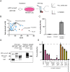
VpdC is allosterically activated by binding to host cell ubiquitin. (A) Schematic overview of the enrichment procedure and identification of host factors bound by VpdC. (B) Graph showing significant hits identified by LC-MS/MS. Proteins with an enrichment ratio (protein molarity in mcVpdC relative to the MD) above 3 are plotted against their quality score (as an indicator of confidence). Ribosomal proteins are shown in blue, while ubiquitin/polyubiquitin is colored in red. (C) Ubiquitin stimulates the PLA2 activity of mcVpdC in vitro. mcVpdC was incubated with ubiquitin, and substrate hydrolysis was determined 40 min after incubation. (D) VpdC directly interacts with ubiquitin through its CTD. Ubiquitin immobilized on agarose beads was incubated with E. coli lysates containing either MBP or the indicated MBP-VpdC variants, and proteins precipitated with the resin were analyzed by immunoblot probed with anti-MBP antibody. (E) PLA2 inhibitors suppress the activity of VpdC. mcVpdC was incubated with ubiquitin in the presence of increasing concentrations of the indicated PLA2 inhibitors, and phospholipid hydrolysis was measured after 40 min. Data were normalized to the DMSO control and are shown as mean ± SD.
Fig. 3.
Fig. 3.
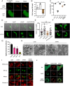
Exacerbated VpdC activity negatively impacts vacuole expansion and host organelle morphology. (A) Translocated VpdC localizes to LCVs. COS-1 cells producing anti-SunTag antibody were challenged for 10 h with Lp02ΔvpdC or Lp03 producing SunTag24x-VpdC. Intracellular bacteria were labeled with anti-Lp antibody, and GFP localization was examined by confocal microscopy. (Scale bar, 5 μm.) Quantifications of SunTag24x-VpdC on LCVs are shown as mean ± SD. Numbers are an average of at least 250 vacuoles from three biological replicates. (B) VpdC is an intermediate/late-stage effector. Expression of the indicated effector-encoding genes was determined at exponential and stationary growth phase by RT-quantitative polymerase chain reactions (qPCR) and normalized to 16S rRNA. The relative fold change in gene expression analyzed with the 2−ΔΔCT method was plotted. Data are shown as mean ± SD in log2 scale. (C) VpdC overproduction attenuates intracellular bacterial growth. Human U937 macrophages were challenged with the indicated bacterial strains, and fold-growth was determined based on colony-forming units (CFU) after 2 h and 48 h in a plating assay. Individual data points are shown with their mean ± SD (***P < 0.001, two-tailed, unpaired t test). (D) Overproduction of VpdC causes vacuole expansion defects. LCV in U937 macrophages were labeled with anti-Lp antibody and examined at 10 hpi. (Scale bar, 5 μm.) (E) Volume measurement of Legionella vacuoles. Human U937 macrophages were challenged with the indicated bacterial strains and immuostained for calnexin as a marker of the ER surrounding LCVs. Z-stack images were acquired by confocal microscopy and used to determine the volume of the vacuoles. (F) PLA2 inhibitors prevent the vacuole expansion defect caused by VpdC overproduction. During infection, 20 μM of the indicated PLA2 inhibitors was added, and vacuoles were examined. (Scale bar, 5 μm.) (G) Quantification of (F) scoring spatially confined vacuoles. Numbers are an average of at least 150 LCVs from three experimental replicates. ***P < 0.001, **P < 0.01. (H) Overproduction of translocated VpdC disrupts the ultrastructure of LCVs and that of surrounding host membranes. Electron micrographs showing LCVs 10 hpi with the indicated L. pneumophila strains. Representative regions of the vacuolar membrane (arrow) and ER tubules or fragments (arrowheads) are magnified in the insets. (Scale bar, 200 nm.) (I) Translocated VpdC causes mitochondria fragmentation during infection. U937 challenged for 10 h with the indicated L. pneumophila strains were stained with Mitotracker-Red, and intracellular bacteria are labeled green. Insets show magnified regions of interest (arrowheads). (Scale bar, 5 μm.) (J) VpdC causes mitochondria fragmentation in transiently transfected COS-1 cells. GFP-VpdC variants were produced for 8 h in transiently transfected COS-1 cells, and mitochondria were labeled by DsRed-Mito. (Scale bar, 5 μm.)
Fig. 4.
Fig. 4.
Lysophospholipid accumulation correlates with membrane morphology changes caused by VpdC. (A) VpdC overproduction induces elevated level of lysophospholipids during infection. Lipids extracted from U937 macrophages infected with Lp02 overproducing VpdC or VpdC(S338A) were quantified. Numbers are an average of three experimental replicates. *** Adjust P < 0.001, ** adjust P < 0.01, * adjust P < 0.05. VpdC preferably hydrolyzed PC (B) and PE (C) with C16 or C18 acyl chain. (D) Exogenous addition of LPC to digitonin-permeabilized COS-1 caused mitochondria fragmentation. Mitochondria were labeled with Mitotracker Red and LPC was conjugated to a green TopFluor fluorophore. LPC-positive and LPC-negative cells are outlined in yellow and white, respectively. Plasma membrane clearly lining the tubules emanating from cells are indicated with arrows. (Scale bar, 5 μm.) (E) Quantification of (D) enumerating cells with fragmented mitochondria. Data are as mean ± SD (n = 3, **P < 0.01, unpaired t test).

References

    1. Dowhan W., “The role of phospholipids in cell function” in Advances in Lipobiology, Gross R. W., Ed. (JAI, 1997), vol. 2, pp. 79–107.
    1. Heringdorf D. M. z., “Lysophospholipids” in Encyclopedia of Molecular Pharmacology, Offermanns S., Rosenthal W., Eds. (Springer, Berlin, Heidelberg, 2008), pp. 710–716, 10.1007/978-3-540-38918-7_90. - DOI
    1. Yeagle P. L., Smith F. T., Young J. E., Flanagan T. D., Inhibition of membrane fusion by lysophosphatidylcholine. Biochemistry 33, 1820–1827 (1994). - PubMed
    1. Chernomordik L. V., Kozlov M. M., Mechanics of membrane fusion. Nat. Struct. Mol. Biol. 15, 675–683 (2008). - PMC - PubMed
    1. Smith G. A., et al. , The two distinct phospholipases C of listeria monocytogenes have overlapping roles in escape from a vacuole and cell-to-cell spread. Infect. Immun. 63, 4231–4237 (1995). - PMC - PubMed

Publication types

LinkOut - more resources