. 2022 Dec 1;109(12):2253-2269.
doi: 10.1016/j.ajhg.2022.11.002. Epub 2022 Nov 21.

A recurrent de novo splice site variant involving DNM1 exon 10a causes developmental and epileptic encephalopathy through a dominant-negative mechanism

Affiliations

A recurrent de novo splice site variant involving DNM1 exon 10a causes developmental and epileptic encephalopathy through a dominant-negative mechanism

Shridhar Parthasarathy et al. Am J Hum Genet. .

Abstract

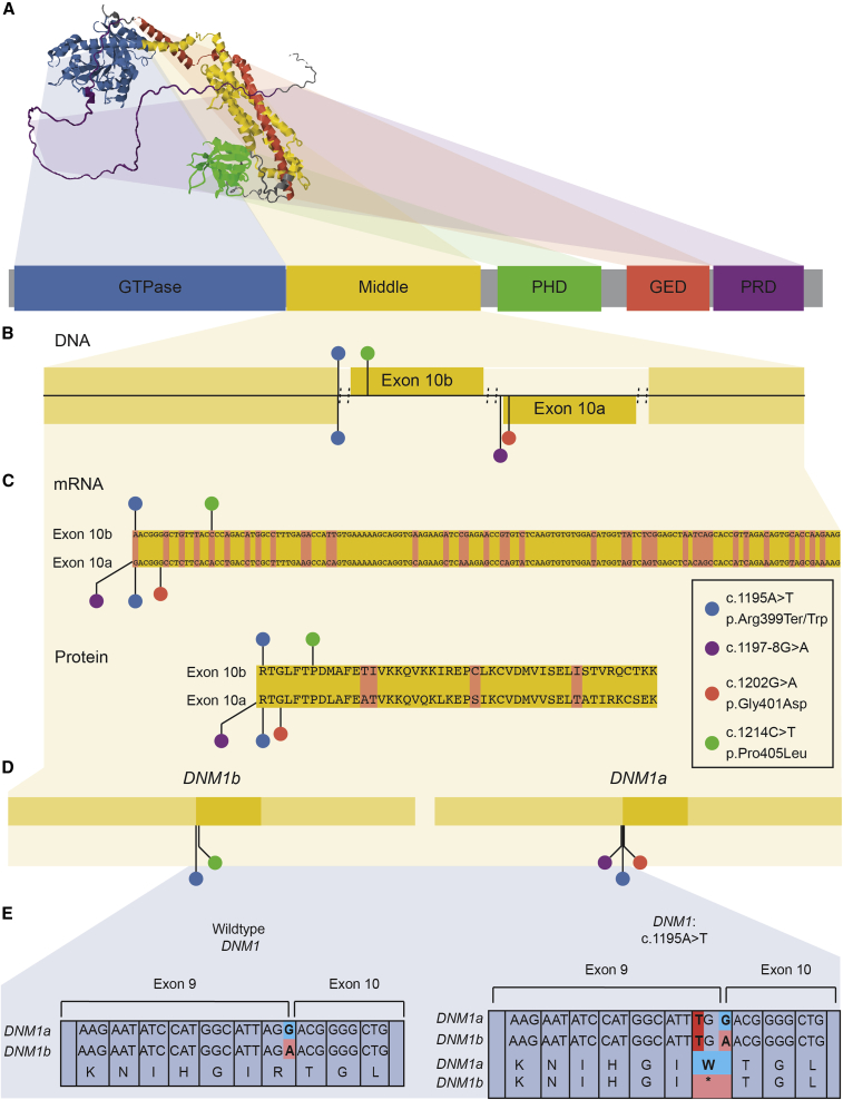
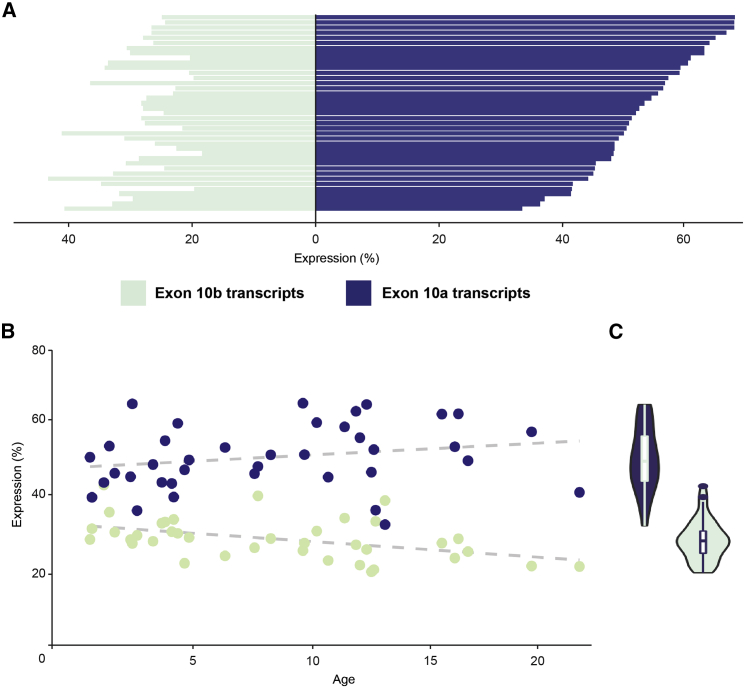
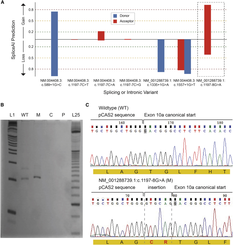
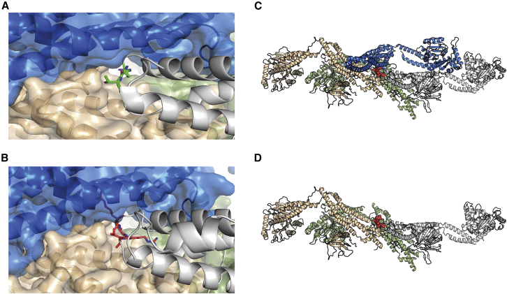
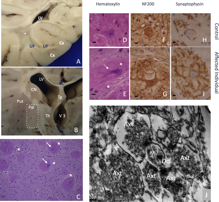
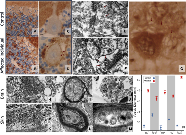
Heterozygous pathogenic variants in DNM1 cause developmental and epileptic encephalopathy (DEE) as a result of a dominant-negative mechanism impeding vesicular fission. Thus far, pathogenic variants in DNM1 have been studied with a canonical transcript that includes the alternatively spliced exon 10b. However, after performing RNA sequencing in 39 pediatric brain samples, we find the primary transcript expressed in the brain includes the downstream exon 10a instead. Using this information, we evaluated genotype-phenotype correlations of variants affecting exon 10a and identified a cohort of eleven previously unreported individuals. Eight individuals harbor a recurrent de novo splice site variant, c.1197-8G>A (GenBank: NM_001288739.1), which affects exon 10a and leads to DEE consistent with the classical DNM1 phenotype. We find this splice site variant leads to disease through an unexpected dominant-negative mechanism. Functional testing reveals an in-frame upstream splice acceptor causing insertion of two amino acids predicted to impair oligomerization-dependent activity. This is supported by neuropathological samples showing accumulation of enlarged synaptic vesicles adherent to the plasma membrane consistent with impaired vesicular fission. Two additional individuals with missense variants affecting exon 10a, p.Arg399Trp and p.Gly401Asp, had a similar DEE phenotype. In contrast, one individual with a missense variant affecting exon 10b, p.Pro405Leu, which is less expressed in the brain, had a correspondingly less severe presentation. Thus, we implicate variants affecting exon 10a as causing the severe DEE typically associated with DNM1-related disorders. We highlight the importance of considering relevant isoforms for disease-causing variants as well as the possibility of splice site variants acting through a dominant-negative mechanism.

Keywords: DNM1; alternative transcripts; developmental and epileptic encephalopathy; dominant negative; dynamin-1; epilepsy; synapse; vesicle fission.


Conflict of interest statement

Declaration of interests The authors declare no competing interests.

Figures

Figure 1
Figure 1
Structure and location of variants in DNM1 (A) Predicted 3D structure of dynamin-1 from AlphaFold and primary sequence of dynamin-1, highlighting distinct domains. (B and C) Subjects’ variants, including the recurrent variant c.1197−8G>A (GenBank: NM_001288739.1), mapped to the dynamin-1 middle domain primary sequence, including alternative exons. The pathogenic variant-rich middle domain includes the alternatively spliced exon 10 of DNM1, with multiple disease-causing variants identified at positions uniquely affecting exon 10a. (D) Subjects’ variants have different effects on each isoform. Variants causing severe disease affect exon 10a in the DNM1a isoform. (E) Exon 10-dependent effects of the c.1195A>T variant. This variant is missense in DNM1a but nonsense in DNM1b; dominant-negative missense, not loss-of-function, variants are the established disease mechanism of DNM1.
Figure 2
Figure 2
Expression profiles of DNM1 transcripts from Ensembl in 39 pediatric brain tissue samples (A) Individual transcript expression, highlighting identifiable transcripts within RefSeq. Bars represent individual samples. (B) Proportional transcript expression for transcripts containing exon 10a and exon 10b versus age, showing age and expression are not strongly correlated. (C) Cumulative expression per individual of full-length DNM1 transcripts containing exon 10a (blue) or exon 10b (green). DNM1a has markedly higher expression than DNM1b in the pediatric brain. Proportional transcript expression was computed by quantifying transcripts in TPM, classifying by 10th exon, and normalizing to the total TPM across all DNM1 transcripts. All data are reported as ratios.
Figure 3
Figure 3
Splicing assay assessing consequence of c.1197−8G>A (GenBank: NM_001288739.1) variant (A) SpliceAI predictions for the consequence of population variants in gnomAD including curated, predicted loss-of-function splicing variants; variants at a position similar to the recurrent disease variant, i.e., c.1197−7 (GenBank: NM:004408.3); and the recurrent disease variant itself. Only the disease variant (c.1197−8G>A [GenBank: NM_001288739]) is strongly predicted to have an effect that may not be loss of function. (B) cDNAs PCR products. L1, 1 Kb plus DNA ladder (Invitrogen); WT, wild-type amplification; M, mutant amplification; C, amplification from HEK293 cells without minigene transfection; P, PCR control without cDNA; L25, 25 bp ladder (Invitrogen). (C) Sanger sequencing of wild-type and mutant PCR products. In red, the insertion of “GTGCAG” from 5ʹ of intron 9 to the mutant transcript, which results in a mutant protein with a cysteine and an arginine (CR) in-frame insertion after Arg399 residue.
Figure 4
Figure 4
Structural modeling of the effects of the c.1197−8G>A (GenBank: NM_001288739.1) variant (A) Mutation site in dynamin-1 corresponding to c.1197−8G>A (GenBank: NM_001288739.1). Green, two residues neighboring the CR insertion. (B) Model of the CR insertion (pink). Given the lack of space to accommodate, the new residues steric clashes would be created, disrupting the interactions to the neighboring protomers. (C and D) Tetrameric organization of DNM1 (PDB ID: 5A3F). The tetramer is formed by two sets of parallel protomers facing in the same direction. Gray and green and blue and orange. When a mutant protomer containing the CR insertion (bottom: gray chain) forms part of the complex, the corresponding parallel protomer (blue) can no longer bind as a result of steric hindrance.
Figure 5
Figure 5
Neuropathological hallmarks of the c.1197−8G>A (GenBank: NM_001288739.1) variant (A and B) Brain macroscopic aspect. (A) White matter displayed severe atrophy with lateral ventriculomegaly, spindly gyri, and extremely thin corpus callosum. A central myelin discoloration was observed consisting of a myelin defect sparing U fiber. (B) Among gray structures, thalamus was severely atrophic associated with a third ventricle distention. Cortical thickness seemed preserved but was difficult to estimate considering the associated severe white matter atrophy. CC, corpus callosum; Cx, cortex; LV, lateral ventricle; Put, putamen; Pal, pallidum; Th, thalamus; Tg, trigone; UF, U fibers; V3, third ventricles, , deep white matter. (C–J) Pallidal synaptic dysplasia. (C) Pallidal glomeruli (star) appeared as rounded formations containing intermingled eosinophilic neurites (arrow). (D–I) After immunohistochemistry and control matching, these glomeruli (E) (star) consisted of exuberant neurites proliferation (arrow) (NF200(+)) that were covered by diffuse synaptic areas (synaptophysin). (D), (F), and (H) are age-matched controls; (F) and (G) are immunohistochemistry with anti-neurofilament 200; (H) and (I) are with anti-synaptophysin; initial magnification ×100. (J) Brain biopsy at pallidum level after electron microscopy conditioning. Initial magnification: ×7,500. Five hyperplastic synaptic terminations (Axt) surrounding one receptive dendrite (Dd). Synaptic vesicles appeared abnormally numerous and packed. M, mitochondria.
Figure 6
Figure 6
Synaptic dysplasia expression in brain and skin biopsy (A–D) Cerebellar cortex revealed an excess of synapses as disorganization of granular glomeruli (arrows); (A) and (B) are with initial magnification ×40 and (C) and (D) are with ×100; (A) and (C) are age-matched controls. (E–H) Synaptic dysplasia consisted of (G) dendrite proliferation (arrows) evident in pallidal glomeruli; (E) and (F) show excess synaptic vesicles in thalamus with abnormal shape, irregular size, densely packed and adherent to synaptic membrane (arrows); (H)–(J) show excess mitochondria in cervical spinal cord axon with abnormal appearance, with crests irregular in size, rounded and disorganized. (E) with initial magnification ×15,000, (F) with ×12,000, (G) with ×100, (H) with ×7,500, (I) with ×12,000, (J) with ×7,500. (K–M) In skin biopsy, most of the observed axons contained heterogenous membranous vesicles that are not pathognomonic features but suggest altered trafficking. However, synaptic areas (axonal zone filled by synaptic vesicles), which are typically difficult to find in skin biopsy, were easily observed in this individual. (K) with initial magnification ×12,000, (L) with ×10,000, (M) with ×15,000. (N) Vesicle diameters in control and affected neuropathological samples from thalamus (Th), cervical spinal cord (SpC), globus pallidus (GP), cerebellum (Cb), and skin. Points and bars are mean ± SEM. Vesicles were significantly enlarged (Mann-Whitney p < 0.001) in the affected individual in all tissue types measured.
Figure 7
Figure 7
Imaging and electroencephalography from individual 1 with the recurrent splice variant c.1197−8G>A (GenBank: NM_001288739.1) (A and B) MRI showing markedly decreased cerebral volume, with deficiency of cerebral white matter. (A) Coronal FLAIR. Slightly dysmorphic lateral ventricles are enlarged, with prominent frontal horns and mild prominence of the temporal horn. (B) Sagittal T1. Small brainstem and small corpus callosum. (C) Abnormal EEG from the same individual with poorly organized, diffusely slow background; nearly continuous focal slowing in the left occipital region; and multi-focal epileptiform discharges.

References

    1. Helbig I., Heinzen E.L., Mefford H.C., International League Against Epilepsy Genetics Commission Genetic literacy series: Primer part 2-Paradigm shifts in epilepsy genetics. Epilepsia. 2018;59:1138–1147. - PubMed
    1. Helbig I., Tayoun A.A.N. Understanding genotypes and phenotypes in epileptic encephalopathies. Mol. Syndromol. 2016;7:172–181. - PMC - PubMed
    1. McTague A., Howell K.B., Cross J.H., Kurian M.A., Scheffer I.E. The genetic landscape of the epileptic encephalopathies of infancy and childhood. Lancet Neurol. 2016;15:304–316. - PubMed
    1. Scheffer I.E., Berkovic S., Capovilla G., Connolly M.B., French J., Guilhoto L., Hirsch E., Jain S., Mathern G.W., Moshé S.L., et al. ILAE classification of the epilepsies: position paper of the ILAE commission for classification and terminology. Epilepsia. 2017;58:512–521. - PMC - PubMed
    1. Helbig K.L., Farwell Hagman K.D., Shinde D.N., Mroske C., Powis Z., Li S., Tang S., Helbig I. Diagnostic exome sequencing provides a molecular diagnosis for a significant proportion of patients with epilepsy. Genet. Med. 2016;18:898–905. - PubMed

Publication types