. 2023 Feb;12(4):e2201897.
doi: 10.1002/adhm.202201897. Epub 2022 Dec 4.

3D-Printed Radiopaque Microdevices with Enhanced Mucoadhesive Geometry for Oral Drug Delivery

Affiliations

3D-Printed Radiopaque Microdevices with Enhanced Mucoadhesive Geometry for Oral Drug Delivery

Tien-Jen Chang et al. Adv Healthc Mater. 2023 Feb.

Abstract

During the past decades, microdevices have been evaluated as a means to overcome challenges within oral drug delivery, thus improving bioavailability. Fabrication of microdevices is often limited to planar or simple 3D designs. Therefore, this work explores how microscale stereolithography 3D printing can be used to fabricate radiopaque microcontainers with enhanced mucoadhesive geometries, which can enhance bioavailability by increasing gastrointestinal retention. Ex vivo force measurements suggest increased mucoadhesion of microcontainers with adhering features, such as pillars and arrows, compared to a neutral design. In vivo studies, utilizing planar X-ray imaging, show the time-dependent gastrointestinal location of microcontainers, whereas computed tomography scanning and cryogenic scanning electron microscopy reveal information about their spatial dynamics and mucosal interactions, respectively. For the first time, the effect of 3D microdevice modifications on gastrointestinal retention is traced in vivo, and the applied methods provide a much-needed approach for investigating the impact of device design on gastrointestinal retention.

Keywords: X-ray imaging; gastrointestinal tracking; microcontainers; microscale 3D printing; mucoadhesion; stereolithography.


Conflict of interest statement

The authors declare no conflict of interest.

Figures

Figure 1
Figure 1
Schematic illustration of the experimental setup. Radiopaque microcontainers were 3D‐printed with three different designs (utilizing microscale 3D printing) and subsequently dosed to rats in gelatin capsules using oral gavage. Their location in the GI tract was determined by planar X‐ray imaging 0.5–3 h after dosing. Further investigation of spatial dynamics and mucosal interactions in the small intestine was carried out using CT scanning and CryoSEM, respectively.
Figure 2
Figure 2
3D‐printed biocompatible microcontainers with enhanced mucoadhesive geometry. A) Illustration of neutral, pillar, and arrow microcontainer design. B) Picture of 3D‐printed microcontainers in a 10 × 10 matrix on a silicon chip. C) Detailed microscope image of 3D‐printed microcontainers. SEM images of D) neutral, E) pillar, and F) arrow microcontainers (Figure S1A–C, Supporting Information, for mass‐production).
Figure 3
Figure 3
Ex vivo mucoadhesion characterization of 3D‐printed microcontainer. A) SEM images of the top side of a i) square, ii) neutral, or iii) arrow microcontainer on the microprobe, and the bottom side of a iv) square, v) neutral, or vi) arrow microcontainer on the microprobe. B) SEM images of the top side of a i) neutral, ii) pillar, or iii) fork microcontainer on the microprobe. C) Mucoadhesive force versus displacement curve while approaching and withdrawing bottom side of an arrow microcontainer to porcine small intestinal tissue. D) Orientation study with normalized mucoadhesion of square, neutral, and arrow microcontainer's top and bottom sides. Mean ± SD, n = 6, Tukey's pairwise test. E) Additive feature study shows normalized mucoadhesion of neutral, pillar, and fork microcontainer's top side. Mean ± SD, n = 6.
Figure 4
Figure 4
Incorporation of BaSO4 into 3D‐printed microcontainers. SEM images of 3D‐printed radiopaque microcontainers with A) neutral, B) pillar, and C) arrow design, respectively (Figure S1D–F, Supporting Information, for mass‐production). D) High contrast SEM image clearly indicating the BaSO4 particles in the pillar design.
Figure 5
Figure 5
EDX analysis and µCT scanning. A) SEM image of a radiopaque arrow microcontainer's surface and EDX analysis, which includes carbon, oxygen, barium, and sulfur. B) µCT scanning showing the homogeneity of BaSO4 nanoparticles in the 3D‐printed radiopaque microcontainers (Figure S3, Supporting Information, for multiple microcontainers).
Figure 6
Figure 6
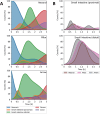
GI retention and transit time of microcontainers. A) Graphs with overlayed fits (from Figure S5, Supporting Information) showing the quantity of microcontainers at specific locations over time for the neutral, pillar, and arrow design, respectively. B) Graphs directly comparing the quantity of microcontainers in the small intestinal sections (proximal and distal) for the three designs.
Figure 7
Figure 7
Spatial dynamics and mucosal interactions of microcontainers. A) CT scan images of neutral, pillar, and arrow microcontainers inside small intestinal pieces, revealing their spatial dynamics. Accompanying CT scans movies are found online (Movies S1–S3, Supporting Information). B,C) CryoSEM images of pillar microcontainers showing their mucosal interactions, such as embedment into the intestinal tissue and the microcontainer orientation.

References

    1. Alqahtani M. S., Kazi M., Alsenaidy M. A., Ahmad M. Z., Front. Pharmacol. 2021, 12, 618411. - PMC - PubMed
    1. Maher S., Brayden D. J., Drug Discovery Today: Technol. 2012, 9, e113.
    1. Atuma C., Strugala V., Allen A., Holm L., Am. J. Physiol. Gastrointest. Liver 2001, 280, G922. - PubMed
    1. Chirra H. D., Shao L., Ciaccio N., Fox C. B., Wade J. M., Ma A., Desai T. A., Adv. Healthcare Mater. 2014, 3, 1648. - PMC - PubMed
    1. Ghosh A., Li L., Xu L., Dash R. P., Gupta N., Lam J., Jin Q., Akshintala V., Pahapale G., Liu W., Sarkar A., Rais R., Gracias D. H., Selaru F. M., Sci. Adv. 2020, 6, 4133. - PMC - PubMed

Publication types

MeSH terms