Review
. 2023 Jan;39(1):15-30.
doi: 10.1016/j.tig.2022.10.006. Epub 2022 Nov 19.

Mitochondria and G-quadruplex evolution: an intertwined relationship

Affiliations
Review

Mitochondria and G-quadruplex evolution: an intertwined relationship

Vinodh J Sahayasheela et al. Trends Genet. 2023 Jan.

Abstract

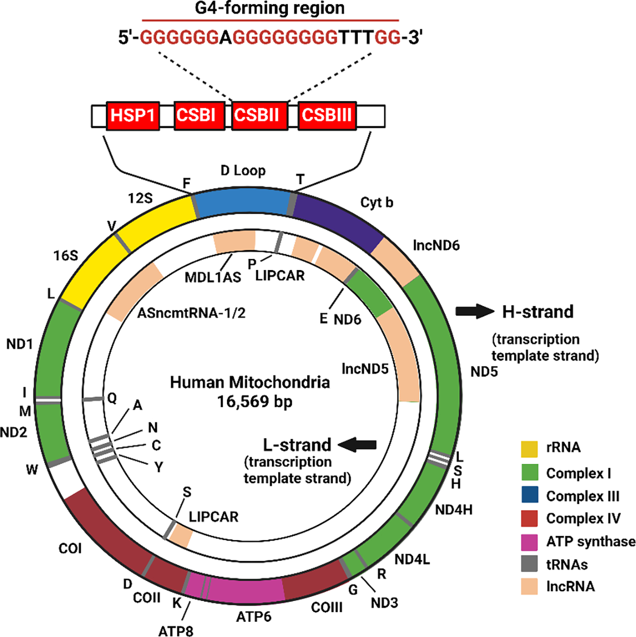
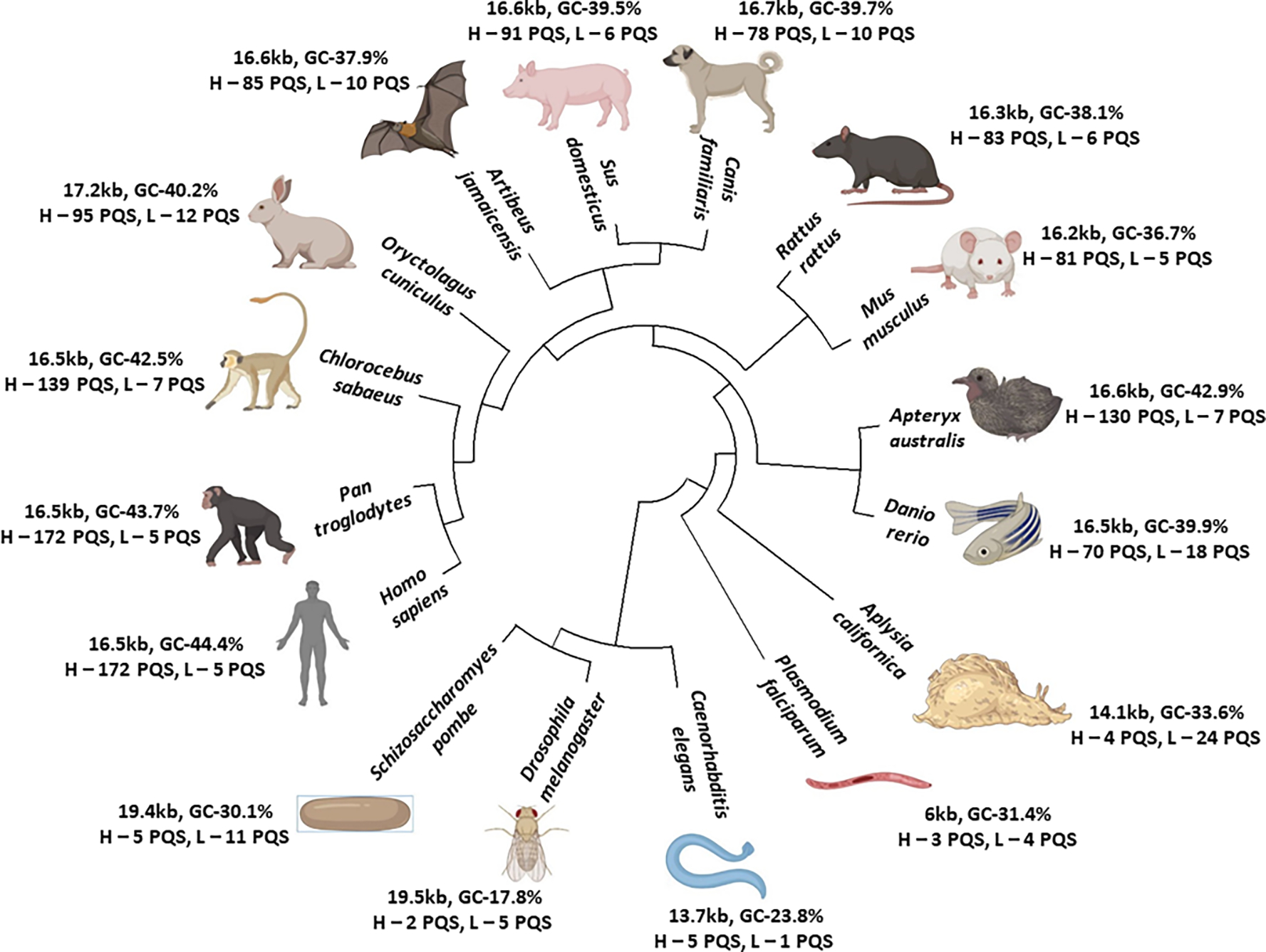
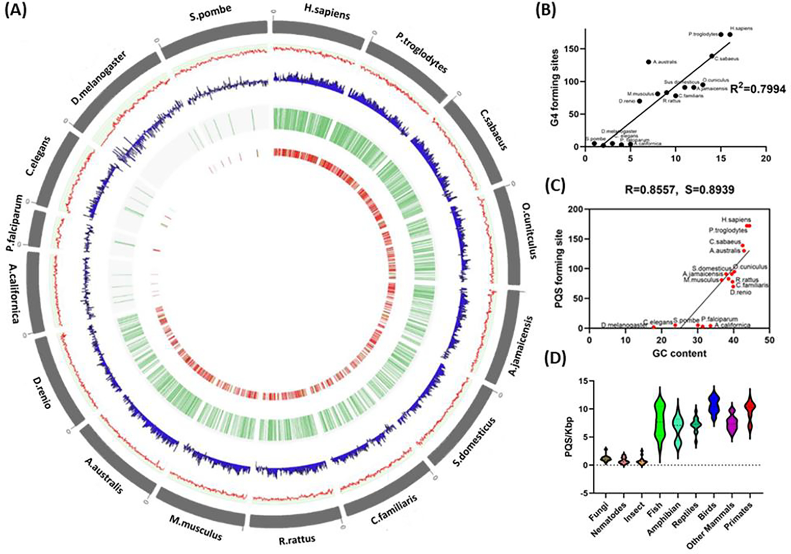
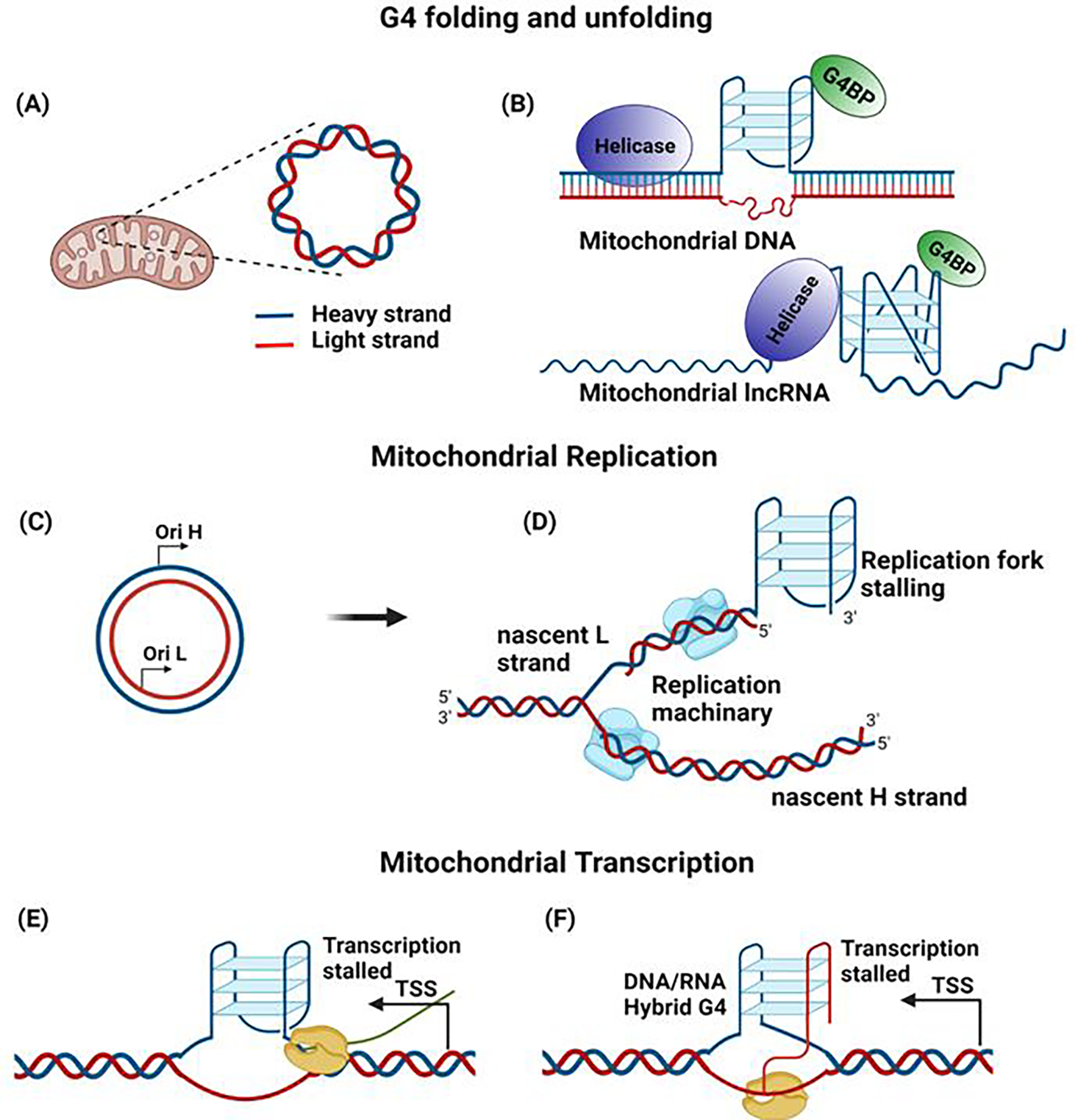
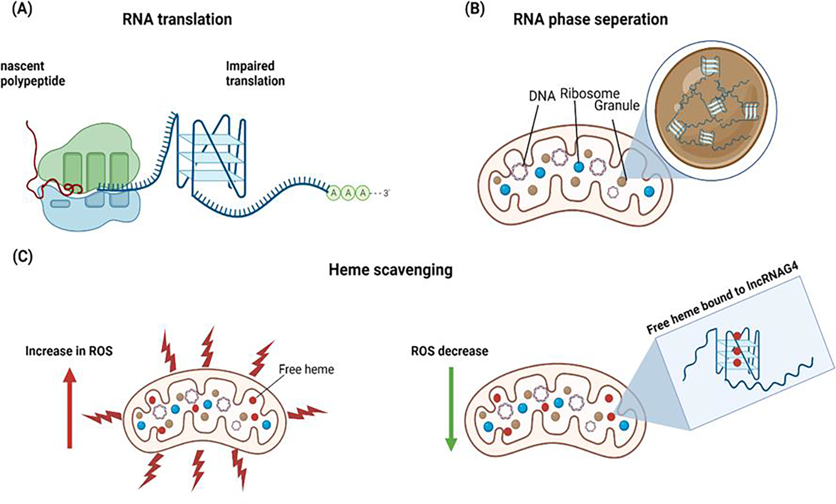
G-quadruplexes (G4s) are non-canonical structures formed in guanine (G)-rich sequences through stacked G tetrads by Hoogsteen hydrogen bonding. Several studies have demonstrated the existence of G4s in the genome of various organisms, including humans, and have proposed that G4s have a regulatory role in various cellular functions. However, little is known regarding the dissemination of G4s in mitochondria. In this review, we report the observation that the number of potential G4-forming sequences in the mitochondrial genome increases with the evolutionary complexity of different species, suggesting that G4s have a beneficial role in higher-order organisms. We also discuss the possible function of G4s in mitochondrial (mt)DNA and long noncoding (lnc)RNA and their role in various biological processes.

Keywords: G-quadruplexes; evolution; long noncoding RNA; mitochondria.


Conflict of interest statement

Declaration of interests The authors declare no conflicts of interest.

Figures

Figure 1.
Figure 1.
G-quadruplex structure and topologies (A) The nucleotide sequence of the G-4 motif, where N denotes the loop sequences. (B) A planar guanine tetrad formed by Hoogsteen bonds and stabilized by metal cation M+. (C) Schematic representation of some topologies of G4.
Figure 2.
Figure 2.
Map of human mitochondrial DNA. The highlighted region in the D loop indicates the conserved block sequence II (CSBII), G4-forming sequence, which is highly conserved in most vertebrates.
Figure 3.
Figure 3.
The phylogeny tree of 16 species together with their mitochondrial genome size. The GC content and the number of potential G4-forming sequences (PQS) in the heavy (H) and light (L) strands is denoted.
Figure 4.
Figure 4.
(A) Circos plot of the representative mitochondrial genomes. The outer circle represents the organism and its mtDNA in gray followed by the GC content in the form of a line plot in red, further followed by GC skew in blue. The next circle indicates the localization of the G4 plotted in the form of tiles in green, and the inner circle represents the score of the G4-forming regions plotted in the form of a heatmap. b) The GC content of the mitochondrial genomes of the 16 species. (B) Potential G4-forming motifs in the mitochondrial heavy strand of the 16 species. The x-axis represents the ranking of organisms based on their complexity by phylogeny analysis, from lower- to higher-order organisms. (C) The scatter plot indicates the PQS-forming sequence for all species on the y-axis with respect to GC content on the x-axis. The R and S values at the top denote the Pearson correlation coefficient (R) and the Spearman correlation coefficient (S). (D) The frequency of PQS-forming sites, relative to genome length expressed as PQS/kbp in the different taxonomy sub-groups.
Figure 5
Figure 5. Role of G4 in mtDNA.
(A) Mitochondrial DNA is represented as an H strand and an L strand, based on guanine content. (B) The presence of proteins that can bind and stabilize G4 (G4BP-G4 binding protein), as well as helicases that can unwind G4, can influence the G4 in mitochondria. (C) The mitochondrial replication origin in the H and L strand (OH & OL) initiates DNA replication in the respective strands. (D) The formation of G4 can influence mitochondrial DNA replication by stalling the process. (E) The presence of G4 in the transcription start site (TSS) can block the progression of mtRNA polymerase, resulting in altered transcription. (F) The formation of hybrid DNA:RNA G4 in the CSBII region terminates the transcription.
Figure 6 –
Figure 6 –. Role of G4 in RNA.
(A) Formation of RNA G-quadruplex can impair the translation of mRNA rich in guanine. (B) The lncRNA with G-quadruplex structures can trigger phase separation and contribute to RNA granules. The lncRNA can scavenge the free heme in the mitochondria and reduce reactive oxygen species (ROS), thereby reducing oxidative stress.

References

    1. Bochman ML et al. (2012) DNA secondary structures: stability and function of G-quadruplex structures. Nat. Rev. Genet. 13, 770–780 - PMC - PubMed
    1. Gellert M et al. (1962) Helix formation by guanylic acid. Proc. Natl. Acad. Sci. U. S. A. 48, 2013–2018 - PMC - PubMed
    1. Bang I (1910) Untersuchungen über die Guanylsäure. Biochemische Zeitschrift 26, 293–311
    1. Burge S et al. (2006) Quadruplex DNA: sequence, topology and structure. Nucleic Acids Res. 34, 5402–5415 - PMC - PubMed
    1. Lombardi EP and Londoño-Vallejo A (2020) A guide to computational methods for G-quadruplex prediction. Nucleic Acids Res. 48, 1603. - PMC - PubMed

Publication types