. 2022 Nov 21;13(1):7137.
doi: 10.1038/s41467-022-34059-1.

Alveolar cell fate selection and lifelong maintenance of AT2 cells by FGF signaling

Affiliations

Alveolar cell fate selection and lifelong maintenance of AT2 cells by FGF signaling

Douglas G Brownfield et al. Nat Commun. .

Abstract

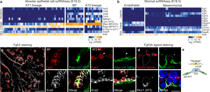
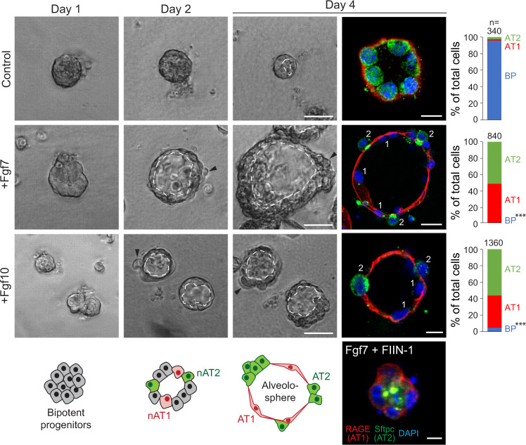
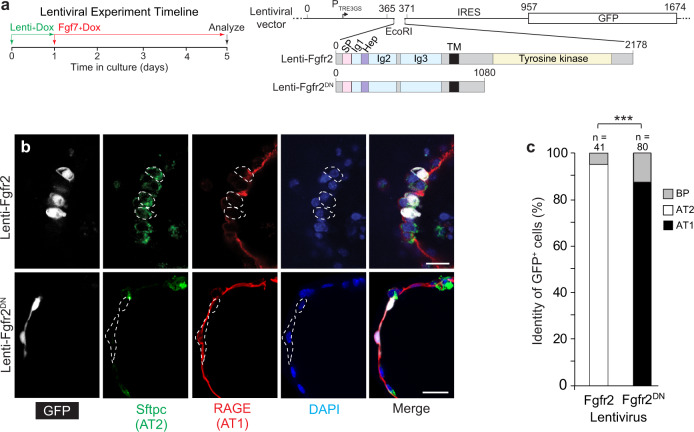
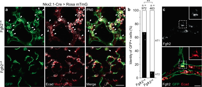
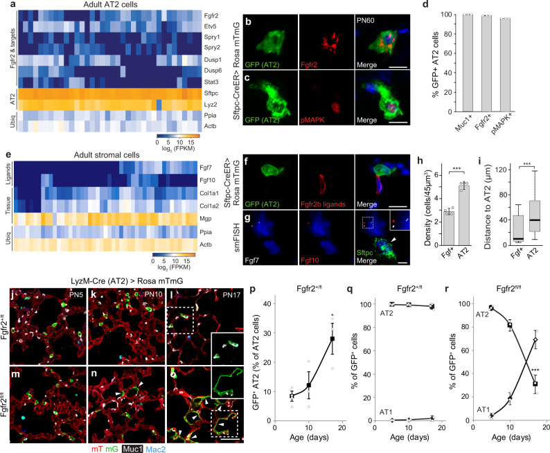
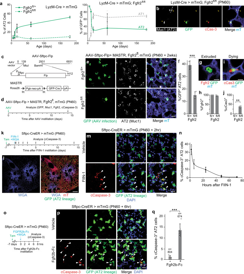
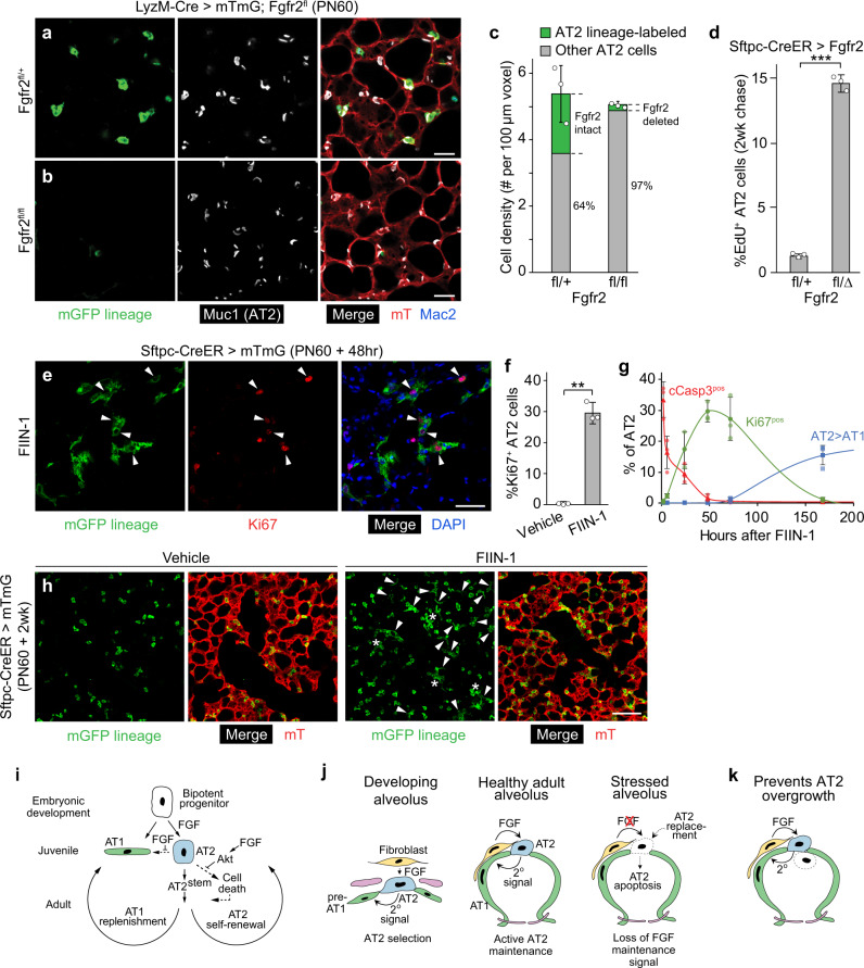
The lung's gas exchange surface is comprised of alveolar AT1 and AT2 cells that are corrupted in several common and deadly diseases. They arise from a bipotent progenitor whose differentiation is thought to be dictated by differential mechanical forces. Here we show the critical determinant is FGF signaling. Fgfr2 is expressed in the developing progenitors in mouse then restricts to nascent AT2 cells and remains on throughout life. Its ligands are expressed in surrounding mesenchyme and can, in the absence of exogenous mechanical cues, induce progenitors to form alveolospheres with intermingled AT2 and AT1 cells. FGF signaling directly and cell autonomously specifies AT2 fate; progenitors lacking Fgfr2 in vitro and in vivo exclusively acquire AT1 fate. Fgfr2 loss in AT2 cells perinatally results in reprogramming to AT1 identity, whereas loss or inhibition later in life triggers AT2 apoptosis and compensatory regeneration. We propose that Fgfr2 signaling selects AT2 fate during development, induces a cell non-autonomous AT1 differentiation signal, then continuously maintains AT2 identity and survival throughout life.


Conflict of interest statement

The authors declare no competing interests.

Figures

Fig. 1
Fig. 1. Expression of Fgfr2 and its ligands during alveolar differentiation.
a Expression of Fgfr2 and the four other receptor genes most selectively expressed in alveolar type 2 (AT2) cell lineage (“Receptors”) from single-cell RNA sequencing (scRNAseq) analysis of distal (alveolar) epithelial cells from embryonic day 18.5 (e18.5) mouse lung. Cells (columns) are arranged in developmental pseudotime (BP, bipotent progenitors, center; AT1 lineage to the left; AT2 lineage to the right) determined as described in ref. by expression of alveolar lineage markers like the four shown (“Lineage”) including two that restrict from BP to AT2 lineage (BP/AT2, Sftpc) or to AT1 lineage (BP/AT1, Rage). (Note due to the very high expression of Sftpc in bipotent progenitors, there is a temporal lag in the extinction of the transcripts in newly-differentiating AT1 cells and thus some nascent AT1 cells co-express Sftpc and Rage.) Ubiq, ubiquitously-expressed control genes. Heat map, mRNA expression level. b Expression of Fgfr2b ligand genes in distal (alveolar) endothelium and mesenchyme from scRNAseq of e18.5 lung. c e17.5 lung immunostained for Fgfr2 (red), bipotent progenitor (BP) and AT1 marker podoplanin (Pdpn, green), and epithelial marker E-cadherin (E-cad, white). Boxed regions, close-ups, and split channels at the right of the bipotent progenitor (BP) and developing AT2 lineage cell (AT2 lin). Note in BP close-up BPs are cuboidal, Pdpn-expressing cells. In AT2 lin closeup, Fgfr2 (red) remains on in developing AT2 cells (arrowhead) but downregulated in neighboring developing AT1 cells, and although BP/AT1 marker (Pdpn) is still detected it too will soon be downregulated as developing AT2 cell matures (see Supplementary Fig. 1a). Bars, 50 µm (left panel), 10 µm (close-ups). Biological triplicates were analyzed. d e17.5 lung stained with the Fgfr2 (isoform iiib) ligand-binding domain fused to human IgG1 domain to show Fgfr2b ligands (red), and co-stained for Pdpn (green), and AT2 marker mucin1 (Muc1, white). Fgfr2b ligands are detected in nearby mesenchymal cells (asterisk) and diffusely around developing alveolar epithelial cells. Bar, 10 µm. Repeated in biological triplicate. e Schematic showing inferred signaling from Fgf ligand-expressing mesenchymal cell (fibroblast) to nearby Fgfr2-expressing nascent AT2 cell during alveologenesis. pre-AT1, nascent AT1 cells that downregulated Fgfr2. All experiments were repeated at least three times.
Fig. 2
Fig. 2. Effect of Fgfr2 ligands on purified alveolar epithelial progenitors in culture.
Phase images (left panels) and immunostains (right) of epithelial progenitors purified from tips of e16.5 lungs and cultured in Matrigel for the period indicated with media alone (Control, top panels) or media supplemented every two days with Fgf7 (50 ng/ml, middle panels) or Fgf10 (100 ng/ml) and HSPG (100 ng/ml, lower panels). Note an increase in luminal surface area (dashed lines) and basal extrusion (arrowheads) in cultures with Fgf ligands, and differentiation into intermingled cuboidal AT2 (Sftpcpos, green) and squamous AT1 cells (RAGEpos, red) at day 4 (“alveolospheres”). In control cultures lacking FGFs (top row) or treated both with Fgf7 and the Fgfr inhibitor FIIN-1 (bottom panel), cells remained bipotent progenitors throughout, as shown by co-expression of both markers. nAT1, nascent AT1 cell; nAT2, nascent AT2 cell. Bars, 50 µm (left panels and right +Fgf7 panel), 10 µm (right top and +Fgf10 panels), and 10 µm (Fgf7 + FIIN-1 panel). Co-treatment with both Fgf7 and Fgf10 (with HSPG) gave similar results as each treatment alone. Quantification at right gives the percent of each cell type (BP), AT1, AT2) in the culture at day 4 determined by immunostaining (n = 348 cells scored for control, 843 for Fgf7-treatment, and 1365 for Fgf10-treatment in experimental triplicate). ***p = 7.7 × 10−28 for Fgf7-treatment and 6.4 × 10−26 for Fgf10-treatment (Student’s two-sided t-test), BP abundance of either Fgf treatment condition vs control.
Fig. 3
Fig. 3. Fgfr2 signaling controls AT2 fate selection cell autonomously in culture.
a Experiment timeline and structures of lentiviral vectors co-expressing wild-type Fgfr2 (Lenti-Fgfr2) or dominant negative Fgfr2 lacking the tyrosine kinase domain (Lenti-Fgfr2DN) and GFP. Numbers above the vector indicate nucleotide position and above Fgfr2 structure indicate amino acid residue; colors highlight functional domains in Fgfr2. TRE tetracycline responsive element, IRES internal ribosome entry site, GFP green fluorescent protein, SP signal peptide, Ig immunoglobulin domain, Hep heparin sulfate binding domain, TM transmembrane domain, DN dominant negative. b e16.5 alveolar progenitors were mosaically infected (less than 1% of the cultured cells were infected) with the lentiviral vectors indicated at the time of cell plating in doxycycline-containing media (100 ng/ml) for 24 h, then cultured with Fgf7 and doxycycline as in Fig. 2 for 4 days and immunostained for markers indicated. Note that GFPpos cells in top panel with Fgfr2 signaling promoted cell autonomously by expression of wild-type Fgfr2 became Sftpcpos RAGEneg cuboidal AT2 cells, whereas GFPpos cells in the bottom panel with Fgfr2 signaling cell autonomously inhibited by expression of Fgfr2DN became Sftpcneg RAGEpos squamous AT1 cells. See Supplementary Fig. 4A for images of rare infected cells that did not acquire the predominant fate. Bars, 20 µm. c Quantification of b. Sftpcpos RAGEpos cuboidal cells were scored as bipotent progenitors (BP). n number of GFPpos cells scored in three experiments. ***p = 4.0 × 10−13 for Lenti-Fgfr2 and 2.2 × 10−16 for Lenti-Fgfr2DN (chi-squared). All experiments were repeated at least three times.
Fig. 4
Fig. 4. Cell-autonomous requirement of Fgfr2 for AT2 fate selection in vivo.
a Alveolar region of lungs from Nkx2.1-Cre; Rosa26-mTmG; Fgfr2fl/+ (upper panels) or Nkx2.1-Cre; Rosa26-mTmG; Fgfr2fl/delta (lower) mice at postnatal day 0 with Cre expressed in developing lung epithelial cells to delete conditional Fgfr2 allele (Fgfr2fl) and activate a farnesylated GFP protein reporter, which targets all membranes including cytoplasmic vesicles (mTmG, green). Lungs were immunostained for GFP, E-cadherin (E-cad), and AT2 marker Muc1 as indicated. Note mosaic GFP expression showing Cre is active in some but not all alveolar epithelial cells, and that Fgfr2 is required cell autonomously for AT2 cell development because GFPpos control cells carrying wild-type Fgfr2 allele (Fgfr2fl/+, upper panels) became either cuboidal Muc1pos AT2 cells (arrowheads) or squamous AT1 cells, whereas GFPpos cells lacking Fgfr2 (Fgfr2fl/delta, lower panels) almost exclusively became AT1 cells. Bar, 50 µm. b Quantification of a. n number of GFPpos cells scored in three lungs. **p = 0.0022 (Student’s two-sided t-test, data as mean ± SD). c Close-up of a rare GFPpos AT2 cell (boxed) from Nkx2.1-Cre; Rosa26-mTmG; Fgfr2fl/delta lung, as in bottom panels of a, stained for Fgfr2 and markers indicated. Note that Fgfr2 has not yet been lost from the GFPpos cell, implying the recent deletion of conditional allele Fgfr2fl and perdurance of Fgfr2 that promoted AT2 fate selection. Bar, 10 µm. Stain repeated in biological triplicate. All experiments were repeated at least three times.
Fig. 5
Fig. 5. Fgfr2 pathway prevents AT2 reprogramming to AT1 after birth.
a scRNAseq of adult AT2 cells. Expression of Fgfr2, downstream transcription factor Etv5, and target genes indicates pathway remains on after birth. AT2, AT2 markers; Ubiq, ubiquitously-expressed genes. b, c AT2 in adult (PN60) Sftpc-CreER;Rosa26-mTmG mouse tamoxifen-induced to label AT2 (GFP) and co-stained for Fgfr2 (b) or phosphorylated MAP kinase (pMAPK) to show pathway activity (c). Blue, DAPI counterstain. Bars, 10 µm. d Quantification of b, c. Nearly all labeled cells (GFP+) express AT2 marker (Muc1) and Fgfr2, and show Fgfr2 activity (pMAPK+). n = 300 GFP+ cells scored (three animals). e Fgfr2 ligand expression (scRNAseq) in lung stromal cells. f AT2 in adult Sftpc-CreER;Rosa26-mTmG mouse tamoxifen-induced to label AT2 (GFP+) and co-stained for Fgfr2 (isoform iiib) ligand-binding domain to show Fgfr2 ligands (ligands identified in Supplementary Fig. 2). Blue, DAPI. Bar, 10 µm. g smFISH (Fgf7, Fgf10, Sftpc) in adult (PN60) lung. Cell neighboring AT2 co-expresses Fgf7 (white dots) and Fgf10 (red dots) (enlarged in inset). Blue, DAPI; arrowhead, AT2. Bar, 5 µm. h, i Quantification of g showing the density of Fgf-expressing and AT2 cells (h, ***p = 4.8 × 10−5, Student’s two-sided t-test, data shown as mean ± SD) and distance between them (i, ***p = 2.9 × 10−50, Mann Whitney, boxplot shows minimum, maximum, first quartile, second quartile (median), and third quartile). n = 182 AT2, 103 Fgf+ cells scored (four animals). (Values indicate Fgf availability and how many AT2 it supports, and how far each Fgf-expressing cell must extend (or ligand diffuse) for support). jo Alveolar region of LyzM-Cre;Rosa26-mTmG;Fgfr2fl/+ (jl) or LyzM-Cre;Rosa26-mTmG;Fgfr2fl/fl (mo) mice (ages indicated) immunostained for Cre reporter (mTmG), AT2 (Muc1), and macrophage (Mac2, distinguishes GFP-labeled macrophages) markers. LyzM-Cre becomes active in AT2 in early postnatal life, activating the GFP reporter and deleting Fgfr2fl. Control GFPpos AT2 cells carry a wild-type Fgfr2 allele (Fgfr2fl/+, jl) and remain AT2 (inset in l), whereas those lacking Fgfr2 (Fgfr2fl/fl, mo) convert to AT1 (arrowheads, inset in o). Bar, 50 µm. pr Quantification (mean ± SD) of jo showing increasing Cre activity in AT2 in early postnatal life (p) and identities of GFPpos cells in control (q) and following Fgfr2 loss (r). n = 300 GFPpos cells scored (three animals) per timepoint/condition; *p = 0.01 for (p) and ***p = 0.0002 for (r) (Student’s two-sided t-test, time series end vs. start).
Fig. 6
Fig. 6. Fgfr2 signaling continuously prevents adult AT2 apoptosis.
a (Left) Time course showing the proportion of AT2 (Muc1+) cells labeled with GFP in control (LyzM-Cre; Rosa26-mTmG Fgfr2fl/+) or Fgfr2 deleted (LyzM-Cre; Rosa26-mTmG; Fgfr2fl/fl) lungs in postnatal life (control, extension of Fig. 5p, mean values ± SD). No further accumulation of GFPpos AT2 occurs in Fgfr2 deleted lungs (dashed line) after PN20, despite continued LyzM-Cre activity (AT2 labeling) apparent in control (solid line). n = 3955 AT2 (3 animals/genotype). (Right) Time course showing identities of GFPpos cells following Fgfr2 loss (extension of Fig. 5r). After a rapid increase in AT1 due to AT2 reprogramming in the juvenile period (through PN20), no further accumulation of GFPpos AT1 or AT2 occurs. n = 300 GFPpos cells scored (3 animals/condition and timepoint, mean ± SD). b Close-up of the alveolar region in adult (PN60) LyzM-Cre;Rosa26-mTmG;Fgfr2fl/fl mouse stained for apoptosis marker cleaved Caspase-3 (cCaspase-3). Note GFPpos AT2 deleted for Fgfr2 undergoing apoptosis (asterisk) but not unlabeled control AT2 nearby (arrowhead). Bar, 10 µm. cf MASTR system for the conditional, complete deletion of Fgfr2 in AT2. c AAV-Sftpc-Flp virus (top) expresses Flp from Sftpc promoter. MASTR allele (bottom), following Flp-mediated deletion (at frt, triangles) of Pgk-neo-pA cassette, constitutively expresses GFP-Cre fusion. d Scheme. Two weeks after AAV-Sftpc-Flp instillation into lungs of adult Fgfr2fl/fl;Rosa26mTmG/MASTR mice to induce constitutive GFP-Cre and complete deletion of Fgfr2fl/fl in AT2, lungs were immunostained (GFP, Muc1, Fgfr2, cCaspase-3). e Alveolar regions from Fgfr2fl/+ control (top) or Fgfr2fl/fl (bottom) mice treated as in (d). Note the reduction in GFPpos AT2 (Muc1pos) (bottom). Bar, 25 µm. f Quantification of AT2 loss in (e). n = 890 Muc1pos cells scored/condition (three experiments, mean ± SD). g, h Quantification (h) of Fgfr2 immunostaining (n = 100 GFPpos cells scored/condition, three experiments, mean ± SD). Nearly all (96%) GFPpos AT2 remaining after MASTR-mediated Fgfr2fl/fl deletion (e, f) have not fully lost Fgfr2. Micrograph (g) shows an example of rare GFPposFgfr2neg AT2, which were all rounded and extruded into the lumen. Bar, 10 µm. i, j Quantification (j) of cCaspase-3 immunostaining (n = 470 GFPpos cells scored/condition, mean ± SD). Note that 36% of GFPpos AT2 cells remaining after MASTR-mediated Fgfr2fl/fl deletion were cCaspase-3pos, indicating apoptosis initiated (micrograph, i). Bar, 10 µm. kn Acute inhibition of Fgfr2 signaling by FIIN-1. k Scheme for co-instillation of FIIN-1 and fluorescent marker WGA-405 into lungs of adult SftpcCreER/+;Rosa26mTmG/+ mice tamoxifen(Tam)-treated to label AT2 then analyzed over 2 weeks. Bar, 200 µm. l Lung regions exposed to FIIN-1 (WGA-labeled, left) and shown merged (right) with mTmG fluorescence (AT2 lineage GFP-labeled, other cells tdTomato (mT)-labeled). Bar, 250 µm. m WGA-labeled alveolar regions exposed 2 h to the vehicle (top) or FIIN-1 (bottom). FIIN-1-exposed AT2 cells (GFPpos) undergo apoptosis (cCaspase-3pos, arrowheads). Bar, 20 µm. n Kinetics of AT2 apoptosis in (m). In vehicle control, <2.5% of AT2 were cCaspase-3pos (n = 650 cells scored/condition, three experiments, mean ± SD). No conversion of labeled AT2 to AT1 was detected during this period (n = 390 cells scored/condition, three experiments) (see also Suppl. Fig. 7). oq Effect of acute Fgfr2 inhibition in AT2 cells by Fgfr2iiib-Fc protein. o Scheme. After Tam-induction of adult SftpcCreER/+;Rosa26mTmG/+ mice to GFP-label AT2, Fgfr2iiib-Fc (or vehicle) and WGA-405 were co-instilled and lungs subsequently stained for cCaspase-3. p WGA-labeled alveolar regions from vehicle control (top) and Fgfr2iiib-Fc (bottom) instilled lungs. Bar, 25 µm. q Quantification of p. n, cells scored, six experiments (mean ± SD). ***p = 4.3 × 10−4 for (f), **p = 3.4 × 10−3 for (j), and ***p = 2.5 × 10−4 for (q) (Student’s two-sided t-test) (panels f, j, q).
Fig. 7
Fig. 7. Robust regeneration following focal AT2 loss.
a, b Alveolar regions of adult control LyzM-Cre;Rosa26-mTmG;Fgfr2fl/+ (a) and AT2 conditional deletion LyzM-Cre;Rosa26-mTmG;Fgfr2fl/fl (b) mice immunostained as indicated. Note loss in b of lineage-labeled AT2 but preserved alveolar architecture. Mac2, macrophage marker. Bars, 50 µm. c Quantification of a, b. AT2 density is maintained in Fgfr2fl/fl deletion by the compensatory expansion of unlabeled AT2 (with unrecombined Fgfrfl alleles). n = 300 cells scored/condition (six animals, mean ± SD); p = 0.65 (not significant, Student’s two-sided t-test) for density difference. d AT2 proliferation in control adult Sftpc-CreER;Rosa26-mTmG;Fgfr2fl/+ and AT2 conditional Fgfr2 deletion Sftpc-CreER;Rosa26-mTmG;Fgfr2fl/delta by EdU labeling for 2 weeks following tamoxifen. n = 400 AT2 cells scored/condition (six lungs, mean ± SD). ***p = 5.4 × 10−6 (Student’s two-sided t-test). e Alveolar region of adult Sftpc-CreER;Rosa26-mTmG tamoxifen-induced to label AT2 and instilled with Fgfr inhibitor FIIN-1 then stained 48 h later for Ki67 and markers indicated. Arrowheads, proliferating AT2 (Ki67pos,GFPpos). Bar, 50 µm. f Quantification of e. n = 800 AT2 cells scored/condition (six animals, mean ± SD). **p = 2.6 × 10−3 (Student’s two-sided t-test). g Time course of AT2 apoptosis (cCasp3pos), compensatory AT2 proliferation (Ki67pos), and conversion to AT1 (Pdpnpos) after FIIN-1 instillation in adult SftpcCreER/+;Rosa26mTmG/+ lungs (see Fig. 6k–n) and subsequent staining (AT2 lineage mGFP, cCasp3, Ki67, Pdpn). mGFPpos (AT2 lineage) cells were scored in instilled (WGApos) regions; uninstilled regions and vehicle-instilled lungs showed no changes. n = 300 mGFPpos cells scored per stain/timepoint (21 animals, mean ± SD). h Vehicle and FIIN-1-treated alveolar regions 2 weeks after instillation. Note clusters of lineage-labeled AT2 (asterisks) and conversion to AT1 (arrowheads) in FIIN-1 lung, and overall restoration of the alveolar structure. Bar, 100 µm. Stain repeated in biological triplicate. i Sequential roles of Fgf/Fgfr2 pathway in alveolus: AT2 selection and 2o induction of AT1 (embryo), AT2 fate consolidation and prevention of reprogramming to AT1 (juvenile), and AT2 survival (adult, mediated by Akt). AT2 loss triggers regeneration by AT2stem. j Source and dynamics of FGF signaling. After AT2 selection in development (left), a 2° signal induces other progenitors to AT1 fate. FGF signaling remains active in adults, maintaining AT2 (middle). Loss of FGF in the stressed alveolus (right) triggers AT2 apoptosis and rapid replacement. k FGF signaling in adults might prevent AT2 overgrowth/tumors by depriving daughters (dashed) moving away from the FGF source of survival signal. All experiments were repeated at least three times.

References

    1. Bertalanffy FD, Leblond CP. Structure of respiratory tissue. Lancet. 1955;269:1365–1368. doi: 10.1016/S0140-6736(55)93164-0. - DOI - PubMed
    1. Barnes PJ, et al. Chronic obstructive pulmonary disease. Nat. Rev. Dis. Prim. 2015;1:15076. doi: 10.1038/nrdp.2015.76. - DOI - PubMed
    1. Martinez FJ, et al. Idiopathic pulmonary fibrosis. Nat. Rev. Dis. Prim. 2017;3:17074. doi: 10.1038/nrdp.2017.74. - DOI - PubMed
    1. Siegel R, Naishadham D, Jemal A. Cancer statistics, 2013. CA Cancer J. Clin. 2013;63:11–30. doi: 10.3322/caac.21166. - DOI - PubMed
    1. Desai TJ, Brownfield DG, Krasnow MA. Alveolar progenitor and stem cells in lung development, renewal and cancer. Nature. 2014;507:190–194. doi: 10.1038/nature12930. - DOI - PMC - PubMed

Publication types