

Peripapillary and Subfoveal Choroidal Thickness in Retinal Vein Occlusions
- PMID: 36415602
- PMCID: PMC9675997
- DOI: 10.2147/OPTH.S379373
Peripapillary and Subfoveal Choroidal Thickness in Retinal Vein Occlusions
Abstract
Purpose: This work aimed to longitudinally assess the peripapillary (PPCT) and subfoveal (SFCT) choroidal thickness (CT), in patients diagnosed with central (CRVO) or branch retinal vein occlusions (BRVO), correlating SFCT with central macular thickness (CMT) and PPCT with peripapillary retinal nerve fiber layer thickness (pRNFL).
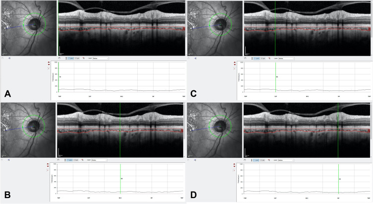
Patients and methods: This was a retrospective longitudinal study of 71 eyes from 71 patients with treatment-naïve retinal vein occlusion (24 CRVO and 40 BRVO). Spectral-domain optical coherence tomography (SD-OCT, Spectralis HRA-OCT, Heidelberg) was used to measure PPCT, SFCT, pRNFL and CMT of the affected and fellow eyes at baseline (acute phase) and at 3 and 9 months post anti-VEGF treatment. IBM SPSS Statistics version 27.0 (IBM Corp., Armonk, NY, USA) was used for statistical analysis. A p-value ≤0.05 was considered statistically significant.
Results: Affected eyes presented a thicker baseline PPCT and SFCT compared to their fellow eyes both in CRVO and BRVO (p < 0.05). Both groups presented a significant decrease of PPCT in the affected eyes at 3 months compared to baseline (p < 0.05). At 9 months, compared to 3 months, PPCT remained stable (p > 0.05). Similarly, affected eyes' SFCT significantly decreased at 3 months (p < 0.05) in both groups. At 9 months, compared to 3 months, SFCT decreased in the CRVO patients (p = 0.047) but remained stable in the BRVO patients (p = 0.850). No correIations between SFCT and CMT were seen at any timepoint in both groups (p > 0.05). PPCT correlates with pRNFL in CRVO at 3 months, although no other correlations were found during the follow-up. In BRVO, PPCT did not show any significant correlation with pRNFL.
Conclusion: Both in CRVO and BRVO eyes, PPCT and SFCT at diagnosis are significantly thicker compared to the fellow eye, suggesting a possible increase in CT immediately after the occlusion, which is followed by a decrease at an early follow-up stage.
Keywords: biomarkers; choroidal thickness; optical coherence tomography; retinal vein occlusions; vascular retinal diseases.
© 2022 Moleiro et al.
Conflict of interest statement
Dr Susana Penas reports personal fees from Alimera, Bayer, Novartis, and Roche, outside the submitted work. The author reports no other conflicts of interest in this work.
Figures

References
LinkOut - more resources
Full Text Sources
