

Didymin, a natural flavonoid, relieves the progression of myocardial infarction via inhibiting the NLR family pyrin domain containing 3 inflammasome
- PMID: 36416076
- PMCID: PMC9704078
- DOI: 10.1080/13880209.2022.2148170
Didymin, a natural flavonoid, relieves the progression of myocardial infarction via inhibiting the NLR family pyrin domain containing 3 inflammasome
Abstract
Context: Globally, the morbidity and mortality of cardiovascular diseases remain high. Didymin, a flavonoid glycoside, has long been used as a dietary antioxidant.
Objective: To determine the role of didymin in myocardial infarction (MI), and its possible myocardial protective mechanism.
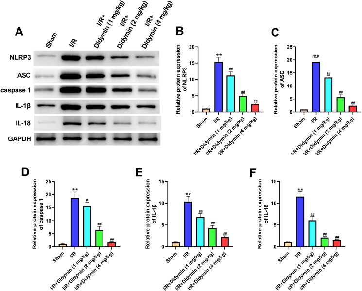
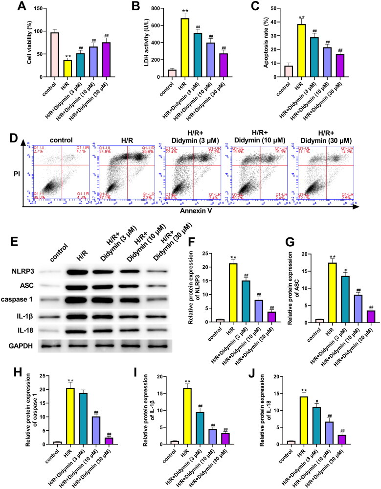
Materials and methods: C57/BL6 mice (aged 6-8 weeks, n = 40) were divided into five groups: sham group, ischaemia-reperfusion (I/R) group, I/R + didymin (1 mg/kg) group, I/R + didymin (2 mg/kg) group and I/R + didymin (4 mg/kg) group. Didymin was administered intragastrically daily before I/R for 5 consecutive days. H9C2 cells were divided into five groups: control group, H/R group, H/R + didymin (3 μM) group, H/R + didymin (10 μM) group and H/R + didymin (30 μM) group. H9C2 cells were treated with didymin for 24 h before hypoxia/reoxygenation (H/R).
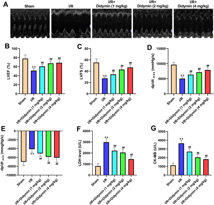
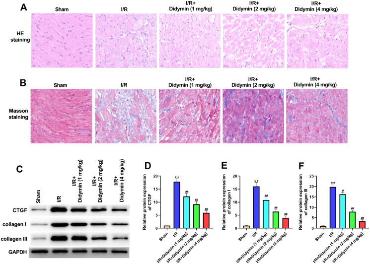
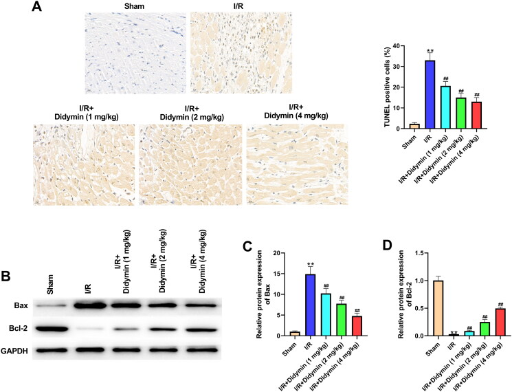
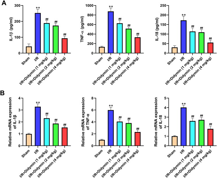
Results: In vivo, didymin reduced the pathological damage and fibrosis of myocardial tissues, decreased the levels of lactate dehydrogenase, creatine kinase, connective tissue growth factor, collagen I and collagen III. Moreover, didymin reduced myocardial apoptosis, inhibited NLRP3, ASC and caspase-1 expression, and alleviated the inflammatory response. In vitro, didymin reduced MI, apoptosis, inflammation and the levels of NLRP3, ASC and caspase-1 in H9C2.
Discussion and conclusions: Didymin prevented the deterioration of MI by inhibiting NLRP3 inflammasome in vivo and in vitro, and may be a potential natural drug for the treatment of MI. Our study provides the scientific basis for further research of didymin.
Keywords: Ischaemia–reperfusion; NOD-like receptor protein 3; fibrosis.
Conflict of interest statement
The authors declare no conflict of interest
Figures
Similar articles
-
The NLRP3 inflammasome is up-regulated in cardiac fibroblasts and mediates myocardial ischaemia-reperfusion injury.Cardiovasc Res. 2013 Jul 1;99(1):164-74. doi: 10.1093/cvr/cvt091. Epub 2013 Apr 10. Cardiovasc Res. 2013. PMID: 23580606
-
The protective effect of Luteolin on myocardial ischemia/reperfusion (I/R) injury through TLR4/NF-κB/NLRP3 inflammasome pathway.Biomed Pharmacother. 2017 Jul;91:1042-1052. doi: 10.1016/j.biopha.2017.05.033. Epub 2017 May 15. Biomed Pharmacother. 2017. PMID: 28525945
-
Lipopolysaccharide (LPS) Aggravates High Glucose- and Hypoxia/Reoxygenation-Induced Injury through Activating ROS-Dependent NLRP3 Inflammasome-Mediated Pyroptosis in H9C2 Cardiomyocytes.J Diabetes Res. 2019 Feb 17;2019:8151836. doi: 10.1155/2019/8151836. eCollection 2019. J Diabetes Res. 2019. PMID: 30911553 Free PMC article.
-
NLRP3 Inflammasome in Cardioprotective Signaling.J Cardiovasc Pharmacol. 2019 Oct;74(4):271-275. doi: 10.1097/FJC.0000000000000696. J Cardiovasc Pharmacol. 2019. PMID: 31356546 Review.
-
Cell-Specific Roles of NLRP3 Inflammasome in Myocardial Infarction.J Cardiovasc Pharmacol. 2019 Sep;74(3):188-193. doi: 10.1097/FJC.0000000000000709. J Cardiovasc Pharmacol. 2019. PMID: 31356542 Review.
Cited by
-
Unveiling the geroprotective potential of Monarda didyma L.: insights from in vitro studies and a randomized clinical trial on slowing biological aging and improving quality of life.Geroscience. 2025 Jun;47(3):4253-4290. doi: 10.1007/s11357-025-01580-2. Epub 2025 Mar 10. Geroscience. 2025. PMID: 40064804 Free PMC article. Clinical Trial.
-
Research Progress on the Pharmacodynamic Mechanisms of Sini Powder against Depression from the Perspective of the Central Nervous System.Medicina (Kaunas). 2023 Apr 10;59(4):741. doi: 10.3390/medicina59040741. Medicina (Kaunas). 2023. PMID: 37109699 Free PMC article. Review.
-
Cellular and Molecular Bases for the Application of Polyphenols in the Prevention and Treatment of Cardiovascular Disease.Diseases. 2025 Jul 15;13(7):221. doi: 10.3390/diseases13070221. Diseases. 2025. PMID: 40710011 Free PMC article. Review.
-
Combined prediction value of coronary plaque burden, serum creatinine, MLR and NLR in reinfarction risk after PCI in middle-aged and elderly patients with myocardial infarction.Eur J Med Res. 2025 Jun 24;30(1):522. doi: 10.1186/s40001-025-02804-z. Eur J Med Res. 2025. PMID: 40555986 Free PMC article.
-
Metabolic-Associated Fatty Liver Disease: The Influence of Oxidative Stress, Inflammation, Mitochondrial Dysfunctions, and the Role of Polyphenols.Pharmaceuticals (Basel). 2024 Oct 10;17(10):1354. doi: 10.3390/ph17101354. Pharmaceuticals (Basel). 2024. PMID: 39458995 Free PMC article. Review.
References
-
- Aghaei M, Motallebnezhad M, Ghorghanlu S, Jabbari A, Enayati A, Rajaei M, Pourabouk M, Moradi A, Alizadeh AM, Khori V.. 2019. Targeting autophagy in cardiac ischemia/reperfusion injury: a novel therapeutic strategy. J Cell Physiol. 234(10):16768–16778. - PubMed
-
- Ali MY, Zaib S, Rahman MM, Jannat S, Iqbal J, Park SK, Chang MS.. 2019. Didymin, a dietary citrus flavonoid exhibits anti-diabetic complications and promotes glucose uptake through the activation of PI3K/Akt signaling pathway in insulin-resistant HepG2 cells. Chem Biol Interact. 305:180–194. - PubMed
-
- Bagheri F, Khori V, Alizadeh AM, Khalighfard S, Khodayari S, Khodayari H.. 2016. Reactive oxygen species-mediated cardiac-reperfusion injury: mechanisms and therapies. Life Sci. 165:43–55. - PubMed
-
- Bajpai G, Bredemeyer A, Li W, Zaitsev K, Koenig AL, Lokshina I, Mohan J, Ivey B, Hsiao H-M, Weinheimer C, et al. . 2019. Tissue resident CCR2– and CCR2+ cardiac macrophages differentially orchestrate monocyte recruitment and fate specification following myocardial injury. Circ Res. 124(2):263–278. - PMC - PubMed
MeSH terms
Substances
LinkOut - more resources
Full Text Sources
Medical
Miscellaneous