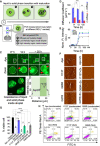
A Zn-dependent structural transition of SOD1 modulates its ability to undergo phase separation
- PMID: 36416085
- PMCID: PMC9841336
- DOI: 10.15252/embj.2022111185
A Zn-dependent structural transition of SOD1 modulates its ability to undergo phase separation
Abstract
The misfolding and mutation of Cu/Zn superoxide dismutase (SOD1) is commonly associated with amyotrophic lateral sclerosis (ALS). SOD1 can accumulate within stress granules (SGs), a type of membraneless organelle, which is believed to form via liquid-liquid phase separation (LLPS). Using wild-type, metal-deficient, and different ALS disease mutants of SOD1 and computer simulations, we report here that the absence of Zn leads to structural disorder within two loop regions of SOD1, triggering SOD1 LLPS and amyloid formation. The addition of exogenous Zn to either metal-free SOD1 or to the severe ALS mutation I113T leads to the stabilization of the loops and impairs SOD1 LLPS and aggregation. Moreover, partial Zn-mediated inhibition of LLPS was observed for another severe ALS mutant, G85R, which shows perturbed Zn-binding. By contrast, the ALS mutant G37R, which shows reduced Cu-binding, does not undergo LLPS. In addition, SOD1 condensates induced by Zn-depletion exhibit greater cellular toxicity than aggregates formed by prolonged incubation under aggregating conditions. Overall, our work establishes a role for Zn-dependent modulation of SOD1 conformation and LLPS properties that may contribute to amyloid formation.
Keywords: SOD1; aggregation; amyotrophic lateral sclerosis (ALS); liquid-liquid phase separation (LLPS); molecular dynamics (MD) simulation.
© 2022 The Authors.
Figures

Human SOD1 amino acid sequence comprised 8‐beta sheets (highlighted in blue) connected by seven loop regions (in red); Zn‐binding loop IV and electrostatic loop VII highlighted in purple.
Cartoon representation of SOD1 monomer highlighting loops IV and VII (Coulombic surface coloring: red = negative, white = neutral, blue = positive).
Schematic representation of SOD1 variants used in the study. WTSOD12SH is the disulfide‐reduced holo form with both metal ion cofactors Zn and Cu. ApoSOD12SH is the disulfide‐reduced de‐metalated form of WTSOD12SH lacking both metal cofactors. ZnSOD12SH and CuSOD12SH are the only Zn‐ and Cu‐bound forms of the disulfide‐reduced monomer.

SOD12SH undergoes LLPS on the removal of metal ion cofactors.
DIC microscopic image of WTSOD12SH diluted to 100 μM in HEPES buffer, pH 7.4 and 100 mM NaCl, and incubated 37°C, 180 RPM for 30 min shows no droplet formation; scale bar: 100 μm; inset shows Alexa Fluor 488 maleimide‐labeled WTSOD12SH; scale bar: 5 μm.
Solution turbidity plot (absorbance at 600 nm) shows that WTSOD12SH (at concentrations ranging from 25 to 200 μM does not undergo LLPS in the presence or absence of heparin, LLPS inducer; data are represented as mean ± SD (from three biological replicates).
DIC microscopic image of ApoSOD12SH under condensate inducing conditions (37°C, 180 RPM) at a concentration of 100 μM incubated 100 mM NaCl; scale bar:100 μm (inset on bottom left shows Alexa Fluor 488 maleimide‐labeled protein condensates; scale bar: 5 μm.
Fluorescent microscopic images of Alexa Fluor 488 maleimide‐labeled ApoSOD12SH condensates diluted in HEPES buffer, pH 7.4 and 100 mM NaCl and incubated (180 RPM, 37°C) in the absence (left) and presence (right) of heparin at 30 min time point; scale bar: 100 μm.
Solution turbidity measurements with ApoSOD12SH (absorbance at 600 nm) show that while in the absence of heparin the turbidity increases with time, the presence of heparin results in faster LLPS; data are represented as mean ± SD (from three biological replicates).
Comparison of ApoSOD12SH droplet numbers per microscopic field of view (view area 0.001963 mm2) calculated from five different images of droplets incubated with and without heparin at 2 h time point. Data are represented as mean ± SD. Statistical significance was established using an unpaired nonparametric t‐test (****P < 0.0001).
Liquid nature of ApoSOD12SH showing droplet fusion and surface wetting; scale bar: 5 μm.
Amplitude normalized FCS curves of ApoSOD12SH (in blue) of dilute phase (DP) and condensed phase (CP). Intense color indicates CP and light variant indicates DP. The increase in τD (as shown by the arrow) translates to a decrease in (D; please refer to equation (2) in Materials and Methods section).
Scheme depicting that presence of Zn and not Cu in a preincubation mixture inhibits LLPS.
DIC microscopic images of ApoSOD12SH in presence of Zn (top) showing no droplet formation and Cu (below) showing the presence of droplets; scale bar: 100 μm.
Solution turbidity plot (absorbance at 600 nm) of ApoSOD12SH subjected to increasing concentrations of Cu and Zn; turbidity decreases in a dose‐dependent manner for Zn while no significant change is observed in presence of Cu; data are represented as mean ± SD (from three biological replicates).

DIC microscopic images of I113T SOD12SH (top) and G85R SOD12SH (bottom) droplets incubated in the absence and presence of Zn and Cu, respectively (scale bar: 100 μm); insets on bottom left show Alexa Fluor 488 maleimide‐labeled protein condensates for I113T SOD12SH and G85R SOD12SH, respectively; scale bar: 5 μm. Droplet dissolution was observed for I113T SOD12SH on the addition of Zn while droplets persisted on the addition of Cu. Red arrowhead indicates fusion of liquid droplets.
Amplitude normalized FCS curves of I113T SOD12SH (in green) and G85R SOD12SH (in gray) of dilute phase (DP) and condensed phase (CP) showing an increase in diffusion time from DP to CP. The intense color indicates CP, and light variant indicates DP.
On metal cofactor removal, ApoG37R SOD12SH formed condensates, which ceased to exist when protein was incubated with Zn, while condensates persisted on Cu addition (left to right); scale bar: 100 μm.

Scheme shows Zn‐dependent disorder to order transition in monomeric SOD1.
Plot of the loge of the hydrodynamic radius (r H) versus the loge of the number of residues in the polypeptide chain. The line fitted to these data for the native folded proteins (yellow dashed) has a slope of 0.29 ± 0.02 and a y‐axis intercept of 1.56 ± 0.1, while the other fitted to the chemically denatured protein (gray dashed) data has a slope of 0.57 ± 0.02 and a y‐axis intercept of 0.79 ± 0.07. Literature data have been used for folded and chemically denatured proteins while we employed FCS to calculate the r H of all SOD12SH variants with (solid shapes) and without Zn (hollow shapes). The hollow gray circle of G85R SOD12SH is directly under hollow green I113T SOD12SH in the log–log plot.
Variation of diffusion coefficients of de‐metalated protein variants determined from FCS measurement with increasing Zn concentration (inset shows a comparison of binding constants between mutants and Zn; ApoSOD12SH and ApoG37R SOD12SH has a higher binding affinity to Zn than ApoI113T SOD12SH and ApoG85R SOD12SH). Data are represented as mean ± SD (from three biological replicates).
Disordered/ extended conformation content decreases in ApoSOD12SH and ApoI113T SOD12SH with the addition of Zn calculated from FTIR spectra; data are represented as mean ± SD (from three biological replicates).

- A, B
(A) Per‐residue RMSF profiles of ApoSOD12SH & its mutants computed from 5 μs simulations performed for each variant. (B) Per‐residue RMSF profiles of ApoSOD12SH, ApoSOD1S‐S, ZnSOD1S‐S, and ZnSOD12SH computed from 5 μs simulations performed for each variant. Error bars in A and B correspond to standard errors, which are estimated by blocks averaging over 10 (500 ns each) blocks.
- C
Salt‐bridge analysis of ApoG85R SOD12SH (dist. cutoff < 0.5 nm = salt bridge).
- D
Snapshots from the ApoG85R SOD12SH trajectory showing the formation of non‐native salt bridges, which are likely to be detrimental for Zn‐binding.
- E
Density profile of ApoSOD12SH and its loop‐deletion variants computed from the CG slab simulations. The condensed phase is centered in the middle of the simulation box.
- F
Slab snapshots from CG simulations of ApoSOD12SH and its variants at 275 K (blue beads represent loop IV, red beads represent loop VII, and orange beads represent the other domains).
- G
Pairwise intermolecular contact formation (normalized by maximum probability) in the condensed phase as a function of residue index.
- H
Saturation concentration measured in the CG phase coexistence simulations for ApoSOD12SH and its variants including ALS mutants at 275 K. The error bars correspond to SD, which are estimated from block averages over four blocks.

Cartoon scheme showing the maturation of protein droplets with time (region1: diffused region outside droplet, region 2: low‐intensity portion inside the droplet, region 3: highly intense portion inside droplet appears during maturation) at different time points.
Diffusion coefficient of regions 1 and 2 (within droplet) was measured using point FCS as shown in the scheme.
Upper panel shows fluorescence images of the maturation of Alexa Fluor 488‐labeled ApoSOD12SH droplets with time, samples were incubated at 37°C up to ~ 25 h. Images are taken at indicated time point: 0* hour (the time after incubation required for droplet formation), 12 h, and > 24 h; images below show magnified scale bar: 5 μm. The middle panel shows magnified view of the marked region from corresponding images on the upper panel. Red arrowhead indicates matured (solid) portion inside the droplet. Lower panel shows the coexistence of liquid and solid phases inside droplet; scale bar: 10 μm. Intensity plot (bottom right) shows the distribution of high‐intensity region within matured droplet.
Diffusion coefficient obtained from point FCS measurement at different regions of the droplet during maturation course; 0* (x‐axis label) indicates the time after incubation required for droplet formation. Data are shown mean ± SD (from three biological replicates). Statistical significance was measured using paired t‐test; ns denotes nonsignificant; **P < 0.01; ***P < 0.001.
ThT fluorescence assay plot showing aggregation kinetics of all SOD12SH variants. ApoSOD12SH, I113T SOD12SH, and G85R SOD12SH show high ThT fluorescence.
AFM micrographs of ApoSOD12SH, I113T SOD12SH, and G85R SOD12SH show fibrillar aggregates after prolonged incubation at 37°C, 180 RPM in absence of Zn (left panel); AFM micrographs of ApoSOD12SH, I113T SOD12SH, and G85R SOD12SH incubated with Zn (at 1:5 protein:Zn molar ratio) show the presence of oligomers for ApoSOD12SH, I113T SOD12SH and short fibrils for G85R SOD12SH (right panel); scale bar: 200 nm.
Cell proliferation of SHSY5Y human neuroblastoma cells assessed by MTT assay post 12 h treatment with 5 μM ApoSOD12SH, I113T SOD12SH, and G85R SOD12SH condensates and aggregates (fibrils). Cell culture medium (DMEM) was used as negative control and untreated SHSY5Y cells were used as a positive control. Data are shown as mean ± SEM (from three biological replicates). P values were determined by two‐tailed unpaired t tests; *P < 0.05, **P < 0.01; ***P < 0.001, ****P < 0.0001.
Dot plots showing flow cytometric analysis of annexin V and propidium iodide (PI) staining of apoptotic SHSY5Y neuroblastoma cells following a 12 h treatment with 5 μM ApoSOD12SH condensates, I113T SOD12SH condensates, G85R SOD12SH condensates, ApoSOD12SH fibrils, I113T SOD12SH fibrils, and G85R SOD12SH fibrils, respectively (see Materials and Methods for details). High cytotoxicity was observed for SOD12SH condensates.

Similar articles
-
A liquid-to-solid phase transition of Cu/Zn superoxide dismutase 1 initiated by oxidation and disease mutation.J Biol Chem. 2023 Feb;299(2):102857. doi: 10.1016/j.jbc.2022.102857. Epub 2022 Dec 31. J Biol Chem. 2023. PMID: 36592929 Free PMC article.
-
The metal cofactor zinc and interacting membranes modulate SOD1 conformation-aggregation landscape in an in vitro ALS model.Elife. 2021 Apr 7;10:e61453. doi: 10.7554/eLife.61453. Elife. 2021. PMID: 33825682 Free PMC article.
-
Exploring the cause of aggregation and reduced Zn binding affinity by G85R mutation in SOD1 rendering amyotrophic lateral sclerosis.Proteins. 2017 Jul;85(7):1276-1286. doi: 10.1002/prot.25288. Epub 2017 Apr 7. Proteins. 2017. PMID: 28321933
-
SOD1 in neurotoxicity and its controversial roles in SOD1 mutation-negative ALS.Adv Biol Regul. 2016 Jan;60:95-104. doi: 10.1016/j.jbior.2015.10.006. Epub 2015 Oct 31. Adv Biol Regul. 2016. PMID: 26563614 Review.
-
Does wild-type Cu/Zn-superoxide dismutase have pathogenic roles in amyotrophic lateral sclerosis?Transl Neurodegener. 2020 Aug 19;9(1):33. doi: 10.1186/s40035-020-00209-y. Transl Neurodegener. 2020. PMID: 32811540 Free PMC article. Review.
Cited by
-
Metallothionein 1X is a tumor suppressor gene and inhibits oxidative stress and metastasis in renal cell carcinoma.Discov Oncol. 2025 Aug 13;16(1):1545. doi: 10.1007/s12672-025-02949-7. Discov Oncol. 2025. PMID: 40802017 Free PMC article.
-
Parkinson-like wild-type superoxide dismutase 1 pathology induces nigral dopamine neuron degeneration in a novel murine model.Acta Neuropathol. 2025 Mar 5;149(1):22. doi: 10.1007/s00401-025-02859-6. Acta Neuropathol. 2025. PMID: 40042537 Free PMC article.
-
Membraneless organelles in health and disease: exploring the molecular basis, physiological roles and pathological implications.Signal Transduct Target Ther. 2024 Nov 18;9(1):305. doi: 10.1038/s41392-024-02013-w. Signal Transduct Target Ther. 2024. PMID: 39551864 Free PMC article. Review.
-
Small molecules in regulating protein phase separation.Acta Biochim Biophys Sin (Shanghai). 2023 Jun 12;55(7):1075-1083. doi: 10.3724/abbs.2023106. Acta Biochim Biophys Sin (Shanghai). 2023. PMID: 37294104 Free PMC article.
-
PICNIC accurately predicts condensate-forming proteins regardless of their structural disorder across organisms.Nat Commun. 2024 Dec 11;15(1):10668. doi: 10.1038/s41467-024-55089-x. Nat Commun. 2024. PMID: 39663388 Free PMC article.
References
-
- Andersen PM (2006) Amyotrophic lateral sclerosis associated with mutations in the CuZn superoxide dismutase gene. Curr Neurol Neurosci Rep 6: 37–46 - PubMed
-
- Anderson JA, Glaser J, Glotzer SC (2020) HOOMD‐blue: a python package for high‐performance molecular dynamics and hard particle Monte Carlo simulations. Comput Mater Sci 173: 109363
Publication types
MeSH terms
Substances
Grants and funding
LinkOut - more resources
Full Text Sources
Miscellaneous

