. 2023 Jan 31;147(5):388-408.
doi: 10.1161/CIRCULATIONAHA.122.059062. Epub 2022 Nov 23.

Macrophage-Derived 25-Hydroxycholesterol Promotes Vascular Inflammation, Atherogenesis, and Lesion Remodeling

Affiliations

Macrophage-Derived 25-Hydroxycholesterol Promotes Vascular Inflammation, Atherogenesis, and Lesion Remodeling

Alberto Canfrán-Duque et al. Circulation. .

Abstract

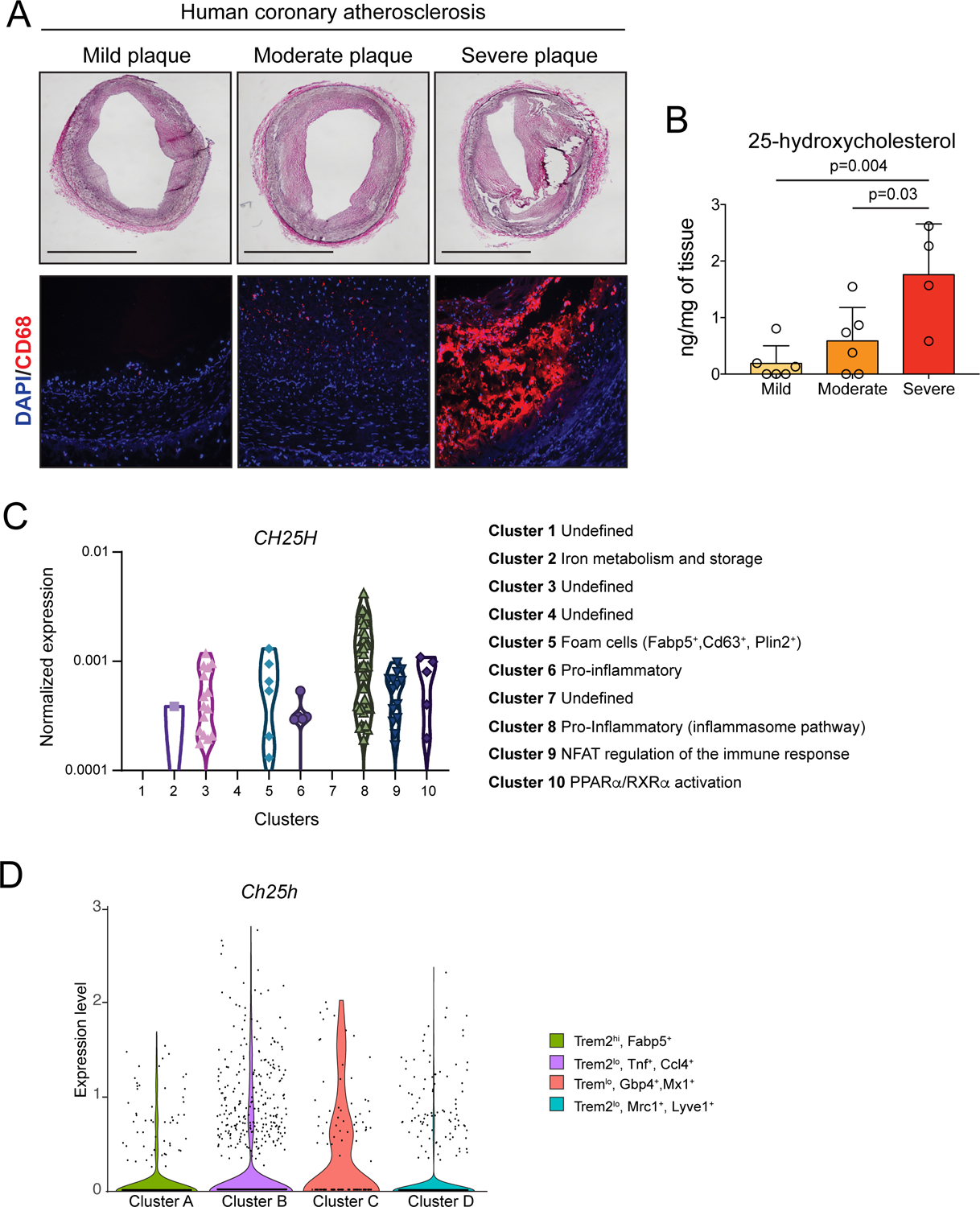
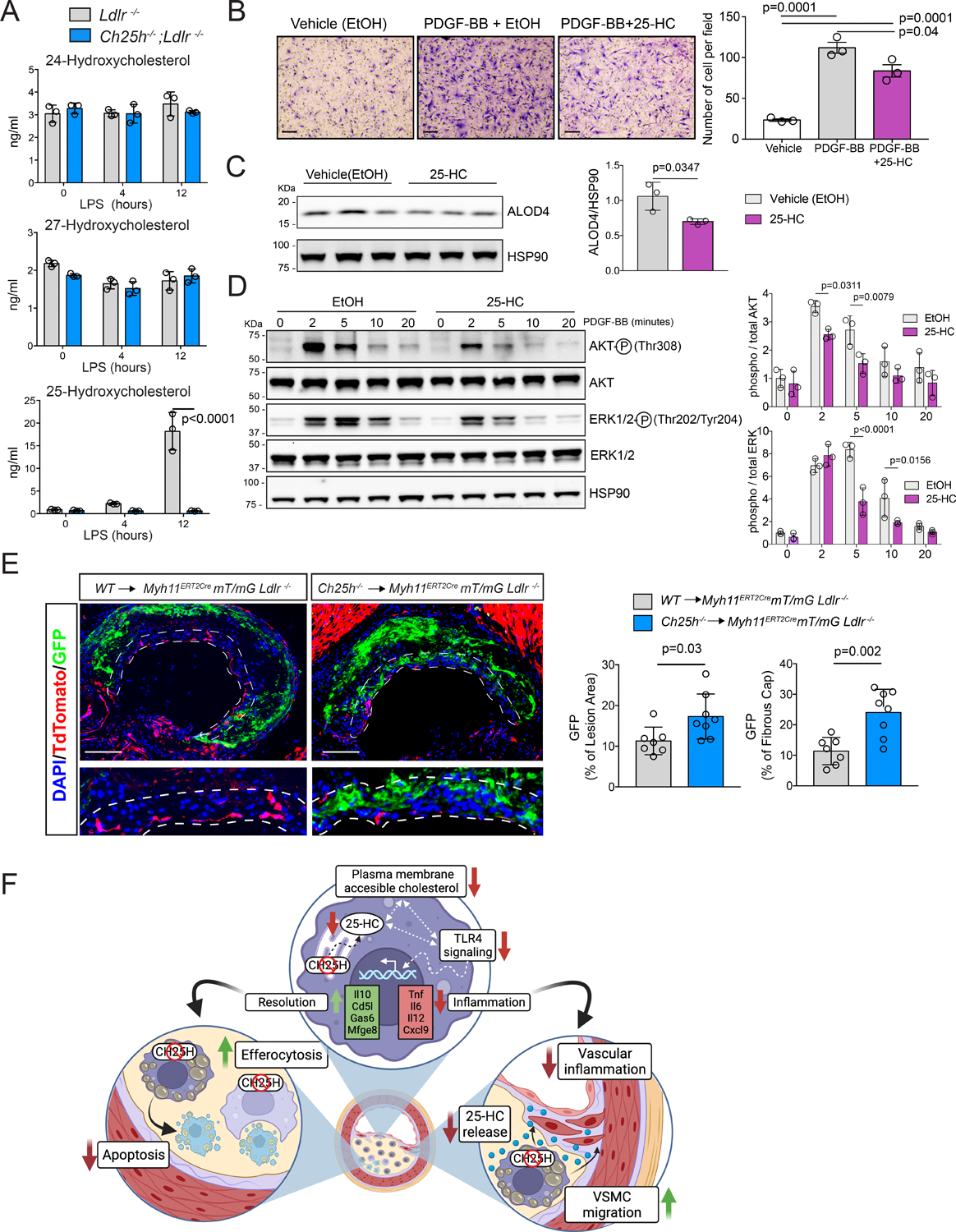
Background: Cross-talk between sterol metabolism and inflammatory pathways has been demonstrated to significantly affect the development of atherosclerosis. Cholesterol biosynthetic intermediates and derivatives are increasingly recognized as key immune regulators of macrophages in response to innate immune activation and lipid overloading. 25-Hydroxycholesterol (25-HC) is produced as an oxidation product of cholesterol by the enzyme cholesterol 25-hydroxylase (CH25H) and belongs to a family of bioactive cholesterol derivatives produced by cells in response to fluctuating cholesterol levels and immune activation. Despite the major role of 25-HC as a mediator of innate and adaptive immune responses, its contribution during the progression of atherosclerosis remains unclear.

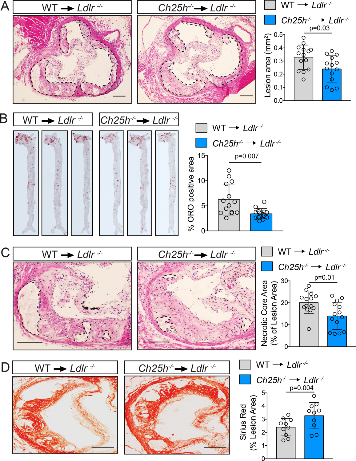
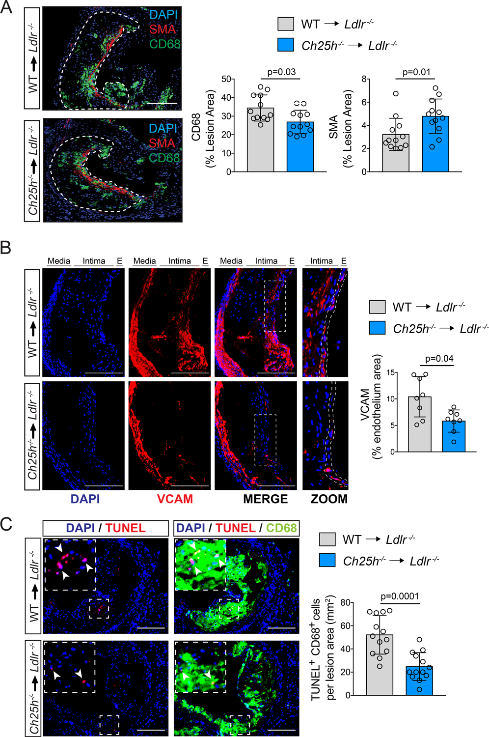
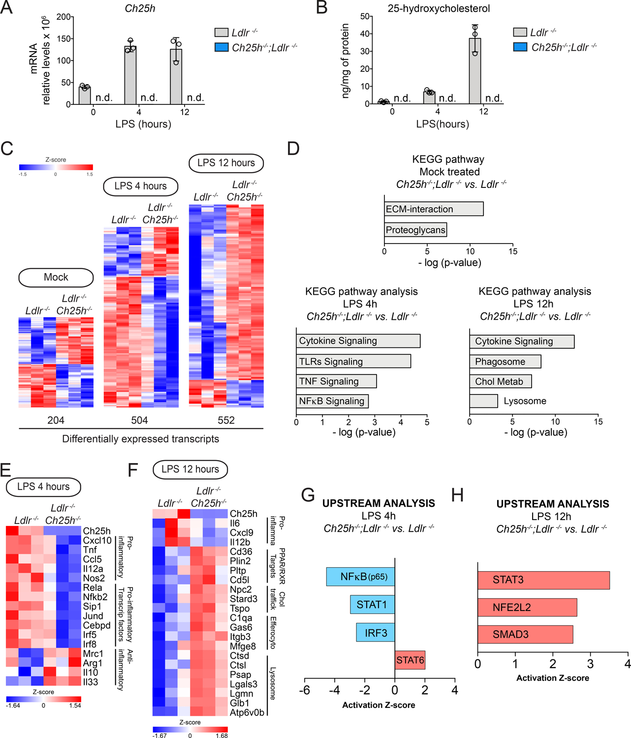
Methods: The levels of 25-HC were analyzed by liquid chromatography-mass spectrometry, and the expression of CH25H in different macrophage populations of human or mouse atherosclerotic plaques, respectively. The effect of CH25H on atherosclerosis progression was analyzed by bone marrow adoptive transfer of cells from wild-type or Ch25h-/- mice to lethally irradiated Ldlr-/- mice, followed by a Western diet feeding for 12 weeks. Lipidomic, transcriptomic analysis and effects on macrophage function and signaling were analyzed in vitro from lipid-loaded macrophage isolated from Ldlr-/- or Ch25h-/-;Ldlr-/- mice. The contribution of secreted 25-HC to fibrous cap formation was analyzed using a smooth muscle cell lineage-tracing mouse model, Myh11ERT2CREmT/mG;Ldlr-/-, adoptively transferred with wild-type or Ch25h-/- mice bone marrow followed by 12 weeks of Western diet feeding.

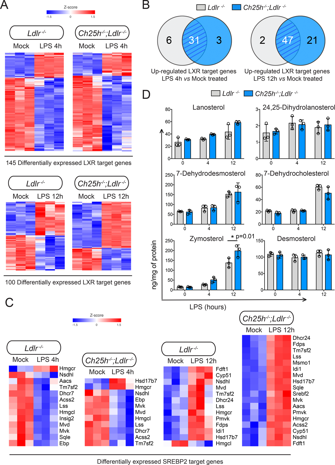
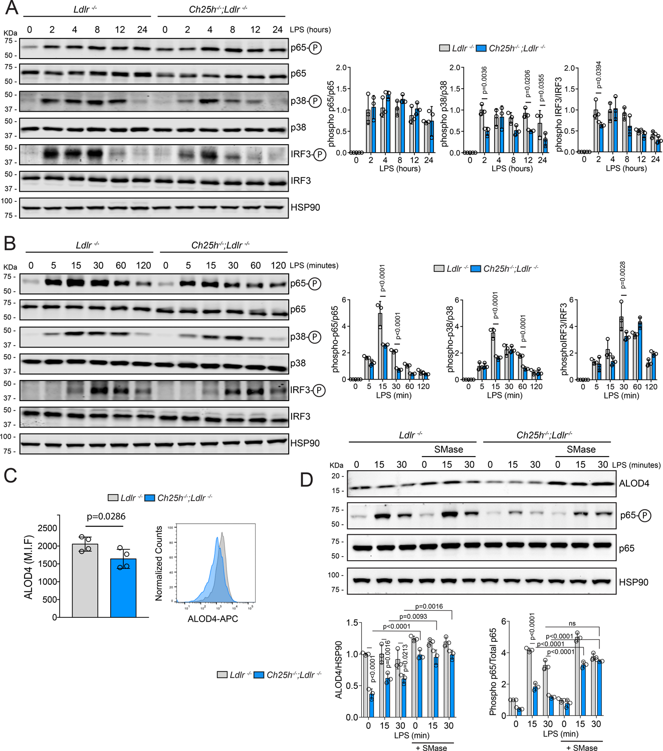
Results: We found that 25-HC accumulated in human coronary atherosclerotic lesions and that macrophage-derived 25-HC accelerated atherosclerosis progression, promoting plaque instability through autocrine and paracrine actions. 25-HC amplified the inflammatory response of lipid-loaded macrophages and inhibited the migration of smooth muscle cells within the plaque. 25-HC intensified inflammatory responses of lipid-laden macrophages by modifying the pool of accessible cholesterol in the plasma membrane, which altered Toll-like receptor 4 signaling, promoted nuclear factor-κB-mediated proinflammatory gene expression, and increased apoptosis susceptibility. These effects were independent of 25-HC-mediated modulation of liver X receptor or SREBP (sterol regulatory element-binding protein) transcriptional activity.

Conclusions: Production of 25-HC by activated macrophages amplifies their inflammatory phenotype, thus promoting atherogenesis.

Keywords: atherosclerosis; inflammation; macrophages; oxysterol.


Conflict of interest statement

Disclosures

None

Figures

Figure 1.
Figure 1.. 25-hydroxycholesterol accumulates in human coronary atherosclerotic plaque and CH25H is highly express in human and mouse inflammatory plaque macrophages.
A, Representative hematoxylin, and eosin (H&E) staining (upper panels) and CD68 (macrophage marker) immunofluorescence (lower panels) of atherosclerotic plaques isolated from human coronary arteries. Scale bar: 1 mm. B, 25-hydroxycholesterol quantification in atherosclerotic plaques isolated from human coronary arteries. Data were analyzed by one-way ANOVA, post-hoc Bonferroni’s multiple comparison test (n= 6 mild plaques, 6 moderate plaques and 4 severe plaques). C, Violin plots showing CH25H expression among different macrophages clusters presents in human plaque tissue. D, Violin plot showing the expression of Ch25h withing the indicated macrophage clusters from leukocyte isolated from Ldlr−/− aortic tissue of mice fed a WD for 12 weeks.
Figure 2.
Figure 2.. Ch25h deficiency in hematopoietic cells protects against atherosclerosis.
Analysis of Ldlr−/− mice transplanted with WT or Ch25h−/− bone marrow and fed for 12 weeks on a WD. A, C, Representative histological analysis of cross sections of the aortic sinus stained with hematoxylin and eosin (H&E). Dashed lines delimit the plaque area (A) and show the edge of the developing necrotic core (C). Quantification of plaque size (A) and necrotic core (C) are shown on the right panels (mean ±SD, n=14 mice per group). B, Representative en face ORO staining of aortas. Quantification of the ORO positive area is shown in the right panel and represents the mean ±SD (n=14 mice per group). D, Representative histological analysis of cross sections of the aortic sinus stained with Picrosirius Red. Quantification of the Picrosirius Red positive area is shown at the right panel and represents the mean ±SD (n=10 mice per group). All data were analyzed by Mann-Whitney non-parametric test. Scale bar: 100 μm.
Figure 3.
Figure 3.. Ch25h deficiency in hematopoietic cells reduces vascular inflammation and promotes plaque stability.
Analysis of Ldlr−/− mice transplanted with WT or Ch25h−/− bone marrow and fed for 12 weeks on a WD. A, Representative histological analysis of cross sections of the aortic sinus stained with CD68 or smooth muscle actin (SMA). Dashed lines delimit the plaque area (A) Quantification of the CD68 or SMA positive area is shown on the right panels and represents the mean ±SD (n=12 mice per group). B, Representative histological analysis of cross sections of the aortic sinus stained with VCAM and DAPI. Quantification of the VCAM positive endothelium area is shown in the right panel and represents the mean ±SD (n=8 mice per group). In the enlarged images the dashed lines show how the endothelium area was delimited for quantification. C, Representative histological analysis of cross sections of the aortic sinus stained with CD68 and TUNEL. Quantification of the CD68-positive cells with TUNEL-positive nuclei is shown in the right panel and represents the mean ±SD (n=13 mice per group). DAPI was used to stain the nuclei. All data were analyzed by Mann-Whitney non-parametric test. Scale bar: 100 μm.
Figure 4.
Figure 4.. 25-hydroxycholesterol in lipid-laden macrophages promotes an inflammatory gene expression profile.
Analysis of lipid-laden TG-EPM isolated from Ldlr−/− or Ch25h−/−;Ldlr−/− mice (n=3 mice per genotype), treated with LPS (100 ng/ml) for 4 or 12 hours. A, Ch25h relative mRNA expression levels in where n.d. means not detected B, LC/MS-MS quantification of 25-hydroxycholesterol, in where n.d. means not detected. C, Heatmaps of differentially expressed genes. A default P-value ≤ 0.05 was considered statistically significant with a fold-change ≥ 1.5 for up-regulated transcripts or ≤ −1.5 for down-regulated transcripts. D, Kyoto Encyclopedia of Gene and Genomes (KEGG) pathway enrichment analysis of differentially expressed genes at different time points. E & F, Heatmaps of representative genes differentially expressed involved in inflammation and resolution of inflammation. A default P-value ≤ 0.05 was considered statistically significant with a fold-change ≥ 1.5 for up-regulated transcripts or ≤ −1.5 for down-regulated transcripts. G & H, Upstream analysis using Ingenuity Pathway Analysis Software of Transcription factors in the above indicated macrophages treated with 100ng/ml LPS for 4 hours (G) or 12 hours (H). Transcription factors predicted to be activated (z-score > 0) appear in red and those that were predicted to be inactive (z-score < 0) appear in blue.
Figure 5.
Figure 5.. 25-hydroxycholesterol in lipid-laden macrophages is not necessary for LXR or SREBP transcriptional regulation.
Analysis of lipid-laden TG-EPM isolated from Ldlr−/− or Ch25h−/−;Ldlr−/− mice (n=3 mice per genotype), treated with LPS (100 ng/ml) for 4 or 12 hours. A, Heatmaps showing LXR target genes differentially expressed in response to LPS treatment for 4 or 12 hours respectively. A default P-value ≤ 0.05 was considered statistically significant with a fold-change ≥ 1.5 for up-regulated transcripts or ≤ −1.5 for down-regulated transcripts. B, Venn diagrams depicting the overlap of upregulated LXR target genes. C, Heatmaps illustrating the effect of LPS treatment for 4 or 12 hours on SREBP2. A default P-value ≤ 0.05 was considered statistically significant with a fold-change ≥ 1.5 for up-regulated transcripts or ≤ −1.5 for down-regulated transcripts. D, Lipidomic analysis of sterol biosynthetic intermediates in macrophages isolated and treated as above. Data were analyzed by two-way ANOVA, post-hoc Bonferroni’s multiple comparison test (n=3 mice per genotype).
Figure 6.
Figure 6.. Ch25h deficiency in macrophage reduces LPS-induced apoptosis independently of inflammasome activation but increases macrophages efferocytotic activity.
A, Representative Western blot analysis of cleaved and total caspase-3 in Ldlr−/− or Ch25h−/−;Ldlr−/− lipid-laden TG-EPM treated with LPS (100 ng/ml) at the indicated times. HSP90 was used as a loading control. Relative protein quantification by band densitometry is shown in the right panel. Data were analyzed by two-way ANOVA, post-hoc Bonferroni’s multiple comparison test (n=3). n.d, not detectable. B, Representative Western blot analysis of caspase-1 and IL-1β as a read out of inflammasome activation of Ldlr−/− or Ch25h−/−;Ldlr−/− lipid-laden TG-EPM with LPS (100 ng/ml) for 8 hours. ATP (5 mM) was added the last 30 minutes and prior to samples collection. Relative protein quantification by band densitometry is shown in the right panels. β-Actin was used as a loading control. Data were analyzed by two-way ANOVA, post-hoc Bonferroni’s multiple comparison test (n=3). n.d. not detectable. C, Representative images of the in vitro engulfment of CellTracker Deep Red labeled apoptotic Jurkat cells by lipid-laden TG-EPM from Ldlr−/− or Ch25h−/−;Ldlr−/− mice (n=14 mice per group). Data were acquired by Amnis Imagestream-X MarkII Imaging Flow Cytometer. Right panel shows efferocytosis quantification as phagocytic index, which is the number of apoptotic cells (red) ingested in 1 h per F4/80-positive macrophage (green) × 100. Significance was determined by Mann-Whitney non-parametric test. D, Representative images of the in situ efferocytosis assay. Apoptotic bodies were identified as TUNEL-positive nuclei (white arrow heads) and macrophages as CD68 positive cells (asteriscs). Free apoptotic bodies were those TUNEL-positive nuclei that do not overlap with CD68 (upper panels), whereas TUNEL-positive nuclei that do overlap with CD68 (lower panels) are considered phagocytosed apoptotic bodies. Right panel shows the quantification of the phagocytosed versus free apoptotic bodies ratio (n= 13 mice per group). Data were analyzed by Mann-Whitney non-parametric test. Scale bar: 10 μm.
Figure 7.
Figure 7.. 25-hydroxycholesterol alters TLR4 downstream signaling by reducing the accessible cholesterol in the plasma membrane.
A-B, Representative Western blot of phospho-p65, p65, phospho-p38, p38, phospho-IRF3, IRF3 or HSP90 in Ldlr−/− or Ch25h−/−;Ldlr−/− lipid-laden TG-EPM treated with LPS (100 ng/ml) at the indicated time points. HSP90 was used as loading control. Quantification of the relative phosphorylation is showing at the right panels. Data were analyzed by two-way ANOVA, post-hoc Bonferroni’s multiple comparison test (n=3). C, Quantification of ALOD4 PM binding, in lipid-laden TG-EPM from Ldlr−/− or Ch25h−/−;Ldlr−/− mice, by flow cytometry. Representative histogram is showing in the bottom panel. Data are average of the mean of Median Intensity Fluorescence (M.I.F.) in arbitrary units (a.u.) and analyzed by Mann-Whitney non-parametric test (n=4). D, Representative Western blot of phospho-p65, p65, ALOD4 or HSP90 in lipid-laden TG-EPM from Ldlr−/− or Ch25h−/−;Ldlr−/− treated with LPS (100 ng/ml) in the presence or absence of Sphingomyelinase (200 mU/ml) at the indicated time points. Quantification of ALOD4 and the relative p65 phosphorylation is showed in the right panel. HSP90 was used as loading control. Data were analyzed by two-way ANOVA, post-hoc Bonferroni’s multiple comparison test (n=3).
Figure 8.
Figure 8.. 25-hydroxycholesterol secreted by macrophages inhibits vascular smooth muscle cells migration.
A, LC/MS-MS quantification of several oxysterols presents in the media of cultured lipid-laden TG-EPM from Ldlr−/− or Ch25h−/−;Ldlr−/− (n=3 mice per genotype) treated with LPS (100 ng/ml) for 4 or 12 hours. Data were analyzed by two-way ANOVA, post-hoc Bonferroni’s multiple comparison test. B, SMC migration in the presence of PDGF-BB (10 ng/ml) or PDGF-BB plus 25-HC (5μM). 5 images were taken randomly per trans-well. Quantification is showing in the right panel and represent the mean ±SEM of 3 independent experiments. Data were analyzed by one-way ANOVA, post-hoc Bonferroni’s multiple comparison test. Scale bar: 100 μm. C, Western blot analysis of ALOD4 binding into the PM in SMC treated with ethanol (EtOH) or 25-hydroxycholesterol (25-HC) at a concentration of 5μM for 2 hours. Quantification of ALOD4 is showed in the right panel. Data were analyzed by Mann-Whitney non-parametric test. D, Representative Western blot of phospho-AKT, AKT, phospho-ERK1/2, ERK1/2 or HSP90 in SMC pre-treated with EtOH or 25-HC (5μM) for 2 hours and then stimulated with PDGF-BB (10 ng/ml) for the indicated times. Quantification of the relative AKT and ERK1/2 phosphorylation is showed in the right panel. Data were analyzed by two-way ANOVA, post-hoc Bonferroni’s multiple comparison test (n=3). E, Representative histological analysis of cross sections of the aortic sinus stained with eGFP and Tdtomato of Myh11CRE;mT/mG; Ldlr−/− mice transplanted with bone marrow from WT or Ch25h−/− donor mice and fed for 12 weeks a WD. Dashed lines define the fibrous cap area used for quantification. Scale bar: 100 μm. Quantification of the eGFP positive area within the whole plaque or the fibrous cap are shown at the right panels and represents the mean ±SD (n=8 mice per group) DAPI was used to stain the cell nucleus. All data were analyzed by Mann-Whitney non-parametric test. F, Proposed working model of 25-HC in macrophages in the context of atherosclerosis. Macrophages-derived 25-HC accelerates atherosclerosis progression and promotes plaque instability. Mechanistically we found that lack of 25-HC synthesis favors a reduction of accessible cholesterol in the PM, what diminishes pro-inflammatory response initiated by pattern recognition receptors in lipid-laden macrophages and promotes a reprogramming into a more pro-resolving phenotype (up). This pro-resolving phenotype is characterized by a better efferocytotic capacity and a lower susceptibility to stress-associated apoptosis in hypercholesterolemic macrophages Ch25h deficient (right). Additionally, the lack of macrophage-derived 25-HC released allows the migration of tunica media smooth muscle cells into the intima to form the fibrous cap (left).

References

    1. Cushing SD, Berliner JA, Valente AJ, Territo MC, Navab M, Parhami F, Gerrity R, Schwartz CJ, Fogelman AM. Minimally modified low density lipoprotein induces monocyte chemotactic protein 1 in human endothelial cells and smooth muscle cells. Proc Natl Acad Sci U S A. 1990;87:5134–5138. doi: 10.1073/pnas.87.13.5134 - DOI - PMC - PubMed
    1. Hurt-Camejo E, Camejo G, Rosengren B, Lopez F, Ahlstrom C, Fager G, Bondjers G. Effect of arterial proteoglycans and glycosaminoglycans on low density lipoprotein oxidation and its uptake by human macrophages and arterial smooth muscle cells. Arterioscler Thromb. 1992;12:569–583. doi: 10.1161/01.atv.12.5.569 - DOI - PubMed
    1. Skalen K, Gustafsson M, Rydberg EK, Hulten LM, Wiklund O, Innerarity TL, Boren J. Subendothelial retention of atherogenic lipoproteins in early atherosclerosis. Nature. 2002;417:750–754. doi: 10.1038/nature00804 - DOI - PubMed
    1. Robbins CS, Hilgendorf I, Weber GF, Theurl I, Iwamoto Y, Figueiredo JL, Gorbatov R, Sukhova GK, Gerhardt LM, Smyth D, et al. Local proliferation dominates lesional macrophage accumulation in atherosclerosis. Nat Med. 2013;19:1166–1172. doi: 10.1038/nm.3258 - DOI - PMC - PubMed
    1. Stewart CR, Stuart LM, Wilkinson K, van Gils JM, Deng J, Halle A, Rayner KJ, Boyer L, Zhong R, Frazier WA, et al. CD36 ligands promote sterile inflammation through assembly of a Toll-like receptor 4 and 6 heterodimer. Nat Immunol. 2010;11:155–161. doi: 10.1038/ni.1836 - DOI - PMC - PubMed

Publication types