. 2022 Dec 16;66(7):821-830.
doi: 10.1042/EBC20220044.

Protein interactions: anything new?

Affiliations

Protein interactions: anything new?

Susana Barrera-Vilarmau et al. Essays Biochem. .

Abstract

How do proteins interact in the cellular environment? Which interactions stabilize liquid-liquid phase separated condensates? Are the concepts, which have been developed for specific protein complexes also applicable to higher-order assemblies? Recent discoveries prompt for a universal framework for protein interactions, which can be applied across the scales of protein communities. Here, we discuss how our views on protein interactions have evolved from rigid structures to conformational ensembles of proteins and discuss the open problems, in particular related to biomolecular condensates. Protein interactions have evolved to follow changes in the cellular environment, which manifests in multiple modes of interactions between the same partners. Such cellular context-dependence requires multiplicity of binding modes (MBM) by sampling multiple minima of the interaction energy landscape. We demonstrate that the energy landscape framework of protein folding can be applied to explain this phenomenon, opening a perspective toward a physics-based, universal model for cellular protein behaviors.

Keywords: energy landscape framework; fuzziness; higher-order assembly; liquid-liquid phase separation; protein-protein interactions.


Conflict of interest statement

The authors declare that there are no competing interests associated with the manuscript.

Figures
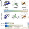
Figure 1
Figure 1. Protein interactions range from ordered to disordered binding modes
(A) Structural order and contact patterns of the bound complex. In ordered binding modes, represented by the complex of p53 tumor suppressor (blue) with Mdm2 (gray, PDB: 2MWY, [12]), the binding interface is well-defined, the bound conformations exhibit limited mobility. Ordered binding modes are characterized by a well-defined contact pattern between specific residues. In disordered binding modes, represented by the complex formed between Gcn4 transcription factor (orange) and Med15 (gray, PDB: 2LPB [18]), the binding interface is conformationally heterogeneous as either or both partners are disordered in the bound complex. Disordered binding modes are generated by alternative contact patterns among the same set of residues. In context-dependent binding modes, represented by the complex formed between p53 (green) and HMGB1 (gray, PDB: 2LY4, [11]) the degrees of disorder are modulated by the cellular or experimental conditions. In context-dependent binding modes, some contacts are well-defined, whereas others are variable and depend on the conditions. The upper panels show the structure of the bound complex, while the lower panels display the residues, which are in contact in at least one model of the ensemble. (B) Heterogeneity of contact patterns. The variability of interactions can be quantified by the contact frequency, defined as the fraction of models in which the given residue is observed to form a contact (defined by https://getcontacts.github.io). The color scale ranges from more stable (blue) to more transient (light orange) interactions corresponding to highly or sparsely populated contacts. The interface residues of p53 exhibit more stable, well-defined contacts in complex with Mdm2, while more variable contacts with HMGB1. In contrast, Gcn4 forms contacts through any of its residues with a low frequency, leading to contact pattern variability and disordered binding modes.
Figure 2
Figure 2. Protein interactions sample multiplicity of binding modes (MBM)
Protein interactions with low MBM sample only one binding mode (unimodal), which can be ordered (blue, p53/Mdm2 complex [12]) or disordered (orange, Gcn4/Med15 complex [18]). Context-dependent protein interactions have high MBM, as they sample multiple binding modes (multimodal) in between ordered (blue) and disordered (orange) modes (p53/HMGB1 complex [11]). The distributions show the frequencies of different binding modes, which can be sampled under different cellular conditions or partners. These can be evaluated using the computational methods described in Ref. [38].
Figure 3
Figure 3. The droplet landscape indicates an impact of ALS-associated mutations on the multiplicity of binding modes (MBM)
The droplet landscape represents the change in the cellular state as a function of MBM (x-axis) and droplet-promoting probability pDP (y-axis) in ALS-associated mutations (empty symbols) versus the wild-type sequence (filled symbols). On the droplet landscape, ALS-associated mutations of droplet-forming proteins, such as FUS G156E [49], G187S [50] (red circle); TDP-43 A321V [51], A315T [52] (cyan cross); TIA-1 E384K [53] (blue diamond); hnRNPA2 D214V [54], D290V [55,56] (orange triangle); UBQLN2 P506T [57] (purple square) are shown. As compared with the wild-type sequences (filled symbols), ALS mutations (open symbols) significantly impact MBM (shift to right), while exhibiting a negligible decrease in droplet-promoting probability (shift upward), which is proportional to the increase in ordered binding modes. Data are derived from Ref. [46]. The MBM values were derived from the binding mode entropy [38] and were normalised to [0,1].
Figure 4
Figure 4. Energetic frustration in specific protein complexes
Structures for translation initiation factor 2 (eIF2g) subunit gamma in complex with alpha and beta (PDB: 3CW2 [66], upper left) and in complex with a substrate-mimicking small molecule (PDB 3I1F; lower right). The disordered binding region, which undergoes templated folding, is shown with the yellow backbone on the insets. Contact patterns (3CW2 upper triangle; and 3I1F lower triangle) show the local frustration patterns of the protein, with the minimally frustrated interactions in green, the neutral shown in gray, and highly frustrated interactions in red. The contact patterns indicate that both complexes are energetically suboptimal, yet the pattern of frustrated contacts are different, enabling specific associations. This figure was adapted from Ref. [37], and is reproduced with permission. Copyright 2021, American Chemical Society.

References

    1. Wu H. and Fuxreiter M. (2016) The structure and dynamics of higher-order assemblies: amyloids, signalosomes, and granules. Cell 165, 1055–1066 10.1016/j.cell.2016.05.004 - DOI - PMC - PubMed
    1. Lyon A.S., Peeples W.B. and Rosen M.K. (2021) A framework for understanding the functions of biomolecular condensates across scales. Nat. Rev. Mol. Cell Biol. 22, 215–235 10.1038/s41580-020-00303-z - DOI - PMC - PubMed
    1. Boeynaems S., Alberti S., Fawzi N.L., Mittag T., Polymenidou M., Rousseau F.et al. . (2018) Protein phase separation: a new phase in cell biology. Trends Cell Biol. 28, 420–435 10.1016/j.tcb.2018.02.004 - DOI - PMC - PubMed
    1. Banani S.F., Afeyan L.K., Hawken S.W., Henninger J.E., Dall'Agnese A., Clark V.E.et al. . (2022) Genetic variation associated with condensate dysregulation in disease. Dev. Cell. 57, 1776e8–1788e8 10.1016/j.devcel.2022.06.010 - DOI - PMC - PubMed
    1. Vendruscolo M. and Fuxreiter M. (2022) Protein condensation diseases: therapeutic opportunities. Nat. Commun. 13, 5550 10.1038/s41467-022-32940-7 - DOI - PMC - PubMed

Publication types