. 2022 Dec 8;14(12):mfac090.
doi: 10.1093/mtomcs/mfac090.

Exploring the K isotope composition of Göttingen minipig brain regions, and implications for Alzheimer's disease

Affiliations

Exploring the K isotope composition of Göttingen minipig brain regions, and implications for Alzheimer's disease

Brandon Mahan et al. Metallomics. .

Abstract

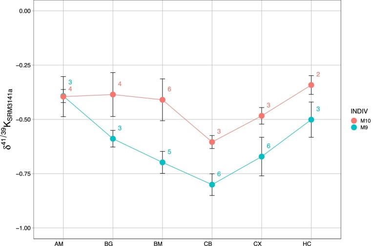
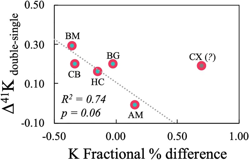
Natural stable metal isotopes have shown utility in differentiation between healthy and diseased brain states (e.g. Alzheimer's disease, AD). While the AD brain accumulates some metals, it purges others, namely K (accompanied by increased serum K, suggesting brain-blood transferal). Here, K isotope compositions of Göttingen minipig brain regions for two AD models at midlife are reported. Results indicate heavy K isotope enrichment where amyloid beta (Aβ) accumulation is observed, and this enrichment correlates with relative K depletion. These results suggest preferential efflux of isotopically light K+ from the brain, a linkage between brain K concentrations and isotope compositions, and linkage to Aβ (previously shown to purge cellular brain K+). Brain K isotope compositions differ from that for serum and brain K is much more abundant than in serum, suggesting that changes in brain K may transfer a measurable K isotope excursion to serum, thereby generating an early AD biomarker.

Keywords: Alzheimer's disease, brain potassium; isotope geochemistry; isotope metallomics; neurodegeneration, porcine model.


Conflict of interest statement

The authors declare no conflicts of interest.

Figures

Graphical Abstract
Graphical Abstract
Potassium isotope compositions of minipig brain regions indicate heavy K isotope enrichment coincident with amyloid beta accumulation, suggesting a mechanistic linkage and potential for downstream signal transferal to blood serum (i.e. noninvasive diagnostic potential).
Fig. 1
Fig. 1
Anatomical schematic of the Göttingen minipig brain with K isotope ratios—δ41K—for each region. Values in parenthesis are calculated heavy K isotope enrichment in porcine model M10 (APP/PS1) relative to M9 (PS1 only) (i.e. δ41K offset between APP/PS1 and PS1 animals, respectively). See Table 1 and Fig. 2 for associated analytical uncertainties.
Fig. 2
Fig. 2
Spider plot of K isotope results highlighting the general ∼0.20‰ increase in δ41K for the younger double-transgenic animal (M10) relative to the older single-transgenic animal (M9) (except for the amygdala). AM—amygdala, BG—basal ganglia, BM—brainstem, CB—cerebellum, CX—cerebral cortex, and HC—hippocampus. Numbers next to data points indicate number of replicate analyses; analytical uncertainties represented by 2σ ± brackets for each sample (determined from replicate analyses).
Fig. 3
Fig. 3
Collated isotopic data to date for minipig brain regions from this work and Mahan et al. and Higgins et al. (Note: data for δ41K are indicative only, as isotopic composition varies considerably both within and between animal models.)
Fig. 4
Fig. 4
Plots of δ41K for the single- and double-transgenic AD model animals in the current work as a function of concentration for biologically relevant elements (biometals and P) and δ44Ca (color coded as mentioned earlier). Reproducibility of measurements via Q–ICP–MS was better than 5% (RSD) for all elements, and symbols encompass analytical error; analytical uncertainties for K isotope measurements in Table 2. Statistically significant relations were determined for Mg(*), P(**), Fe(*), and Cu(*). The increasing power of the statistical significance is defined as: P ≤ 0.05 (*); P ≤ 0.01, **; P ≤ 0.001, ***; and P ≤ 0.0001, ****.
Fig. 5
Fig. 5
Statistical results for dependence of metal concentrations and δ44Ca as a function of δ41K (unpaired t-tests in GraphPad Prism with 2-tailed P-values).
Fig. 6
Fig. 6
Heavy K isotope enrichment (high δ41K) in the double-transgenic model (M10) relative to the single-transgenic model (M9), expressed as Δ41Kdouble–single41KM10–δ41KM9), as a function of the % difference in K concentration in the same reference frame (percentage depletion in M10 relative to M9). All brain regions fall on a discernible trendline (except for the cerebral cortex, CX), suggesting a mechanistic linkage between brain K depletion (associated with AD) and δ41K. At present, the reason for the excursion of the cerebral cortex off this trendline is unknown.

References

    1. Adlard P. A., Bush A. I., Metals and Alzheimer's disease, J. Alzheimers Dis., 2006, 10 (2-3), 145–163. - PubMed
    1. Li Y., Jiao Q., Xu H., Du X., Shi L., Jia F., Jiang H., Biometal Dyshomeostasis and toxic metal accumulations in the development of Alzheimer's disease, Front. Mol. Neurosci., 2017, 10, 339. - PMC - PubMed
    1. Roberts B. R., Doecke J. D., Rembach A., Yevenes L. F., Fowler C. J., McLean C. A., Lind M., Volitakis I., Masters C. L., Bush A. I., Hare D. J., group Ar., Rubidium and potassium levels are altered in Alzheimer's disease brain and blood but not in cerebrospinal fluid, Acta Neuropathol. Commun., 2016, 4 (1), 119. - PMC - PubMed
    1. Balter V., Zazzo A., Moloney A. P., Moynier F., Schmidt O., Monahan F. J., Albarede F., Bodily variability of zinc natural isotope abundances in sheep, Rapid Commun. Mass Spectrom., 2010, 24 (5), 605–612. - PubMed
    1. Balter V., Lamboux A., Zazzo A., Télouk P., Leverrier Y., Marvel J., Moloney A. P., Monahan F. J., Schmidt O., Albarède F., Contrasting Cu, Fe, and Zn isotopic patterns in organs and body fluids of mice and sheep, with emphasis on cellular fractionation, Metallomics, 2013, 5 (11), 1470–1482. - PubMed

Publication types