Glioblastoma PET/MRI: kinetic investigation of [18F]rhPSMA-7.3, [18F]FET and [18F]fluciclovine in an orthotopic mouse model of cancer
- PMID: 36416908
- PMCID: PMC9931868
- DOI: 10.1007/s00259-022-06040-z
Glioblastoma PET/MRI: kinetic investigation of [18F]rhPSMA-7.3, [18F]FET and [18F]fluciclovine in an orthotopic mouse model of cancer
Abstract
Purpose: Glioblastoma multiforme (GBM) is the most common glioma and standard therapies can only slightly prolong the survival. Neo-vascularization is a potential target to image tumor microenvironment, as it defines its brain invasion. We investigate [18F]rhPSMA-7.3 with PET/MRI for quantitative imaging of neo-vascularization in GBM bearing mice and human tumor tissue and compare it to [18F]FET and [18F]fluciclovine using PET pharmacokinetic modeling (PKM).
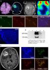
Methods: [18F]rhPSMA-7.3, [18F]FET, and [18F]fluciclovine were i.v. injected with 10.5 ± 3.1 MBq, 8.0 ± 2.2 MBq, 11.5 ± 1.9 MBq (n = 28, GL261-luc2) and up to 90 min PET/MR imaged 21/28 days after surgery. Regions of interest were delineated on T2-weighted MRI for (i) tumor, (ii) brain, and (iii) the inferior vena cava. Time-activity curves were expressed as SUV mean, SUVR and PKM performed using 1-/2-tissue-compartment models (1TCM, 2TCM), Patlak and Logan analysis (LA). Immunofluorescent staining (IFS), western blotting, and autoradiography of tumor tissue were performed for result validation.
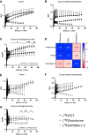
Results: [18F]rhPSMA-7.3 showed a tumor uptake with a tumor-to-background-ratio (TBR) = 2.1-2.5, in 15-60 min. PKM (2TCM) confirmed higher K1 (0.34/0.08, p = 0.0012) and volume of distribution VT (0.24/0.1, p = 0.0017) in the tumor region compared to the brain. Linearity in LA and similar k3 = 0.6 and k4 = 0.47 (2TCM, tumor, p = ns) indicated reversible binding. K1, an indicator for vascularization, increased (0.1/0.34, 21 to 28 days, p < 0.005). IFS confirmed co-expression of PSMA and tumor vascularization. [18F]fluciclovine showed higher TBR (2.5/1.8, p < 0.001, 60 min) and VS (1.3/0.7, p < 0.05, tumor) compared to [18F]FET and LA indicated reversible binding. VT increased (p < 0.001, tumor, 21 to 28 days) for [18F]FET (0.5-1.4) and [18F]fluciclovine (0.84-1.5).
Conclusion: [18F]rhPSMA-7.3 showed to be a potential candidate to investigate the tumor microenvironment of GBM. Following PKM, this uptake was associated with tumor vascularization. In contrast to what is known from PSMA-PET in prostate cancer, reversible binding was found for [18F]rhPSMA-7.3 in GBM, contradicting cellular trapping. Finally, [18F]fluciclovine was superior to [18F]FET rendering it more suitable for PET imaging of GBM.
Keywords: Amino acid transport; Glioblastoma; Neo-vascularization; PET/MRI; PSMA.
© 2022. The Author(s).
Conflict of interest statement
The authors declare no competing interests.
Figures


Comment on
-
Quantification of O-(2-[18F]fluoroethyl)-L-tyrosine kinetics in glioma.EJNMMI Res. 2018 Jul 31;8(1):72. doi: 10.1186/s13550-018-0418-0. EJNMMI Res. 2018. PMID: 30066053 Free PMC article.
Similar articles
-
A comparison study of dynamic [18F]Alfatide II imaging and [11C]MET in orthotopic rat models of glioblastoma.J Cancer Res Clin Oncol. 2024 Apr 22;150(4):208. doi: 10.1007/s00432-024-05688-4. J Cancer Res Clin Oncol. 2024. PMID: 38647690 Free PMC article.
-
Prognostic impact of postoperative, pre-irradiation (18)F-fluoroethyl-l-tyrosine uptake in glioblastoma patients treated with radiochemotherapy.Radiother Oncol. 2011 May;99(2):218-24. doi: 10.1016/j.radonc.2011.03.006. Epub 2011 Apr 16. Radiother Oncol. 2011. PMID: 21497925
-
More advantages in detecting bone and soft tissue metastases from prostate cancer using 18F-PSMA PET/CT.Hell J Nucl Med. 2019 Jan-Apr;22(1):6-9. doi: 10.1967/s002449910952. Epub 2019 Mar 7. Hell J Nucl Med. 2019. PMID: 30843003
-
18F-Fluciclovine (18F-FACBC) PET/CT or PET/MRI in gliomas/glioblastomas.Ann Nucl Med. 2020 Feb;34(2):81-86. doi: 10.1007/s12149-019-01426-w. Epub 2019 Nov 26. Ann Nucl Med. 2020. PMID: 31773466
-
Approaches to PET Imaging of Glioblastoma.Molecules. 2020 Jan 28;25(3):568. doi: 10.3390/molecules25030568. Molecules. 2020. PMID: 32012954 Free PMC article. Review.
Cited by
-
Radiosynthesis and preclinical evaluation of a 68Ga-labeled tetrahydroisoquinoline-based ligand for PET imaging of C-X-C chemokine receptor type 4 in an animal model of glioblastoma.EJNMMI Radiopharm Chem. 2024 Aug 20;9(1):61. doi: 10.1186/s41181-024-00290-y. EJNMMI Radiopharm Chem. 2024. PMID: 39162901 Free PMC article.
-
A comparison study of dynamic [18F]Alfatide II imaging and [11C]MET in orthotopic rat models of glioblastoma.J Cancer Res Clin Oncol. 2024 Apr 22;150(4):208. doi: 10.1007/s00432-024-05688-4. J Cancer Res Clin Oncol. 2024. PMID: 38647690 Free PMC article.
-
Evaluating the Potential of PSMA Targeting in CNS Tumors: Insights from Large-Scale Transcriptome Profiling.Cancers (Basel). 2025 Apr 6;17(7):1239. doi: 10.3390/cancers17071239. Cancers (Basel). 2025. PMID: 40227800 Free PMC article.
References
-
- ENCR E. Eurocim version 4.0, European incidence database, V 2.2 (1999) Lyon, ENCR. ENCR; 2001.
-
- Stupp R, Tonn J-C, Brada M, Pentheroudakis G. High-grade malignant glioma: ESMO Clinical Practice Guidelines for diagnosis, treatment and follow-up. Ann Oncol [Internet]. 2010;21:v190–3. Available from: https://www.sciencedirect.com/science/article/pii/S0923753419396358. - PubMed
-
- Stupp R, Mason WP, van den Bent MJ, Weller M, Fisher B, Taphoorn MJB, et al. Radiotherapy plus concomitant and adjuvant temozolomide for glioblastoma. N Engl J Med. Massachusetts Medical Society; 2005;352:987–96. - PubMed
Publication types
MeSH terms
Substances
LinkOut - more resources
Full Text Sources
Medical
Miscellaneous

