Review
. 2023 Mar;242(3):510-524.
doi: 10.1111/joa.13796. Epub 2022 Nov 23.

Anatomical study of the red flour beetle using synchrotron radiation X-ray phase-contrast micro-tomography

Affiliations
Review

Anatomical study of the red flour beetle using synchrotron radiation X-ray phase-contrast micro-tomography

Maria Luigia Vommaro et al. J Anat. 2023 Mar.

Abstract

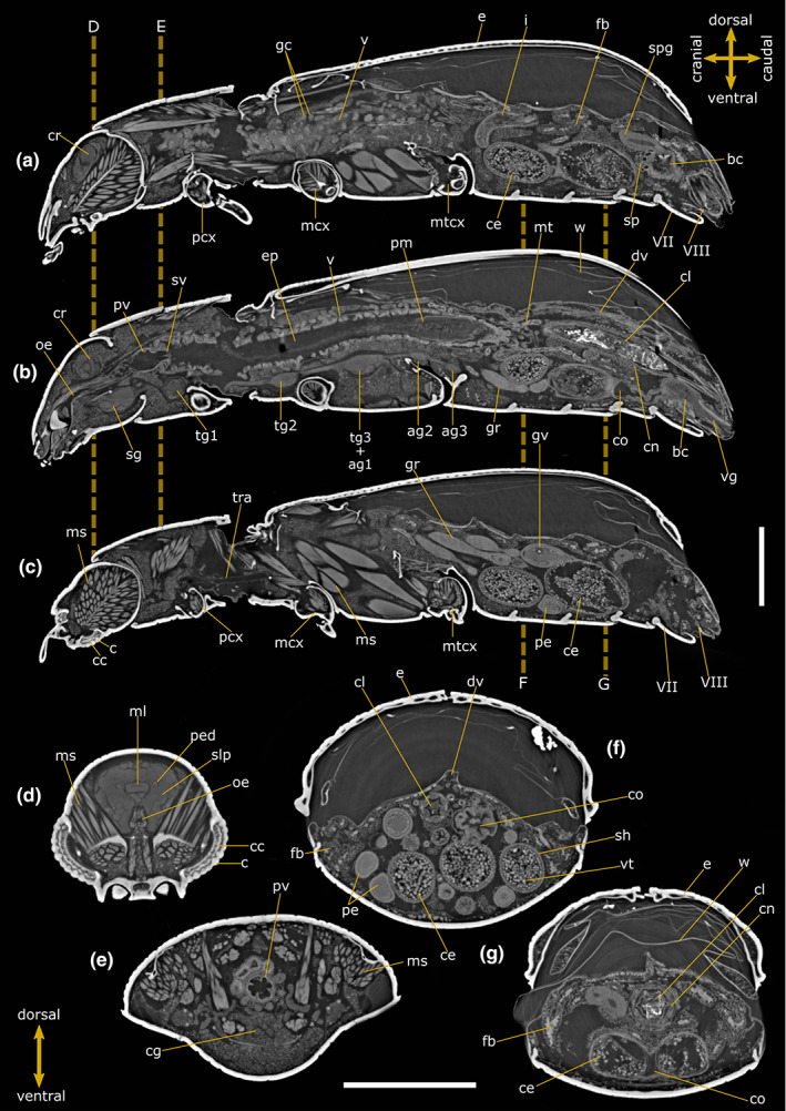
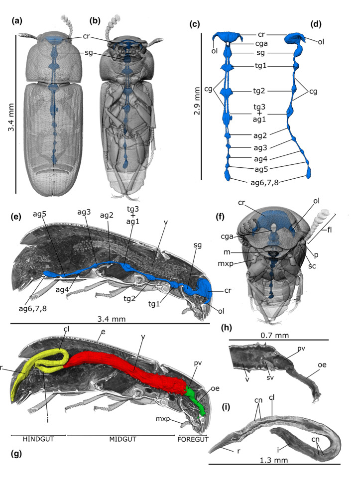
Synchrotron X-ray phase-contrast microtomography (SR-PhC micro-CT) is well established, fast and non-destructive imaging technique for data acquisition that is currently being used to obtain new insights into insect anatomy and function in physiological, morphological and phylogenetic studies. In this study, we described in situ the internal organs of the red flour beetle Tribolium castaneum Herbst 1797, a widespread pest of cereals and stored food causing serious damage to the human economy. Two-dimensional virtual sections and volumetric reconstructions of the nervous, alimentary and reproductive systems were carried out in both sexes. The results provided a comprehensive overview of the morphological characteristics of this species, such as the different maturation stages of ovarioles and the realistic location, size and shape of internal organs. Given the great interest in this model species in experimental biology and forensic entomology, complete knowledge of the general anatomy is required for future functional applications in pest control and experimental studies. In addition, this study confirms SR-PhC micro-CT as a powerful and innovative tool in entomology, particularly suitable for small species and chitinized structures that are difficult to analyse using conventional dissection and histological methods.

Keywords: 3D rendering; abdominal gland; brain; cryptonephridial system; image segmentation; microtomography.


Conflict of interest statement

The authors declare that they have no conflicts of interest

Figures

FIGURE 1
FIGURE 1
Internal anatomy of Tribolium castaneum female. Two‐dimensional PhC micro‐CT‐based longitudinal (a–c) and cross (d–g) sections. Dashed lines indicate the levels of head (d), pronotum (e) and abdominal (f, g) cross‐sections. VII: Abdominal sternite VII; VIII: Abdominal sternite VIII; ag 2–3: Abdominal ganglia 2 and 3; bc: Bursa copulatrix; c: Cornea; cc: Crystalline cones; ce: Chorionic egg; cg: Connective of ganglia; cl: Colon; cn: Cryptonephridial system; co: Common oviduct; cr: Cerebrum; dv: Dorsal vessel; e: Elytrae; ep: Ectoperitrophic space; fb: Fat bodies; gc: Gastric caeca; gr: Germarium; gv: Germinal vesicle; i: Ileum; mcx: Mesocoxae; ms: Muscle; ml: Median lobe; mt: Malpighian tubules; mtcx: Metacoxae; oe: Oesophagus; pcx: Procoxae; pe: Pre‐vitellogenic egg; ped: Peduncle; pm: Peritrophic matrix; pv: Proventriculus; sg: Suboesophagean ganglion; sh: Epithelial sheath; slp: Superior lateral protocerebrum; spg: Spermathecal gland; sp: Spermatheca; sv: Stomodeal valve; tg1‐2: Thoracic ganglia 1 and 2; tg3 + ag1: Complex of thoracic ganglion 3 and abdominal ganglion 1; tra: Tracheae; v: Ventriculus; vg: Vagina; vt: Vitellum; w: Membranous wings. Scale bars: 500 μm (a–g).
FIGURE 2
FIGURE 2
Internal anatomy of Tribolium castaneum male. Two‐dimensional PhC micro‐CT‐based longitudinal (a–c) and cross (d–g) sections. Dashed lines indicate the levels of head (d), pronotum (e) and abdominal (f, g) cross‐sections. VIII: Abdominal sternite VIII; IX: Abdominal sternite IX; ae: Aedeagus; agl: Accessory glands; c: Cornea of compound eye; cc: Crystalline cones; cg: Connective of ganglia; cl: Colon; cn: Cryptonephridial system; cr: Cerebrum; dd: Deferent duct; dv: Dorsal vessel; e: Elytrae; ed: Ejaculatory duct; ep: Ectoperitrophic space; fb: Fat bodies; gc: Gastric caeca; i: Ileum; lb: Lobe of testis; m: Mandible; mcx: Mesocoxae; ml: Median lobe; ms: Muscle; mt: Malpighian tubules; mcx: Mesocoxae; mtcx: Metacoxae; mx: Maxilla; mxp: Maxillary palp; oe: Oesophagus; ol: Optical lobe; p: Pedicel; pcx: Procoxae; ped: Peduncle; py: Pyloric valve; pm: Peritrophic matrix; pv: Proventriculus; r: Rectum; rb: Rhabdoms; sc: Scape of antennae; sg: Subesophagean ganglion; slp: Superior lateral protocerebrum; sv: Stomodeal valve; t: Testis; tg1‐2: Thoracic ganglia 1 and 2; tg3 + ag1: Complex of thoracic ganglion 3 and abdominal ganglion 1; tr: Tritocerebrum; v: Ventriculus; w: Membranous wings. Scale bars: 500 μm (a–g).
FIGURE 3
FIGURE 3
PhC micro‐CT 3D rendering and reconstructions of the nervous (a–f) and alimentary (g–i) systems in Tribolium castaneum male. Localization of nervous system in the transparent rendered body, dorsal (a), ventral (b) and frontal (f) view. Dorsal (c) and lateral (d) view of the segmented nervous system. (e) Longitudinal section of the body showing in situ localization of the cerebrum (cr) connected to the ganglia through the connectives (cg). (g) alimentary canal reconstruction showed in a longitudinal sectioned volume of the rendered body. Detailed rendering images of the foregut (h) and hindgut (i). ag2‐5: Abdominal ganglia 2–5; ag6, 7, 8: Terminal abdominal ganglia fused to form a large caudal ganglion; cga: Circumesophageal connectives loop around the digestive system to link the brain (cr) and subesophageal complex together; cl: Colon; cn: Cryptonephridial system; fl: Antennal flagellum; i: Ileum; m: Mandible; mxp: Maxillary palp; oe: Oesophagus; ol: Optical lobe; p: Pedicel of antenna; pv: Proventriculus; r: Rectum; sc: Scape of antenna; sg: Subesophagean ganglion; sv: Stomodeal valve; tg1‐2: Thoracic ganglia 1–2; tg3 + ag1: Complex of thoracic ganglion 3 and abdominal ganglion 1; v: Ventriculus.
FIGURE 4
FIGURE 4
PhC micro‐CT 3D rendering and reconstructions of reproductive system in Tribulium castaneum female. Volume reconstruction of right (blue) and left (light blue) ovaries (ov) showed in the transparent rendered body, dorsal (a) and ventral (B). (c) Segmentation of ovaries showing six ovarioles (ovl) and the apical filaments (f) connecting the gonads to the sospensorium. (d) Reconstruction of the ovaries showed in a longitudinal sectioned volume of the rendered body. (e, f) Longitudinal and lateral rendering of telotrophic ovarioles showing the germinarium (gr) end vitellarium (vr). (g, h) Frontal sections of the abdomen showing in situ oviduct and ovarioles at different level of egg maturation (H) and a detail of the spermatheca (sp) and spermathecal gland (spg) (g). VII: Abdominal sternite VII; VIII: Abdominal sternite VIII; bc: Bursa copulatrix; ce: Chorionic egg; co: Common oviduct; e: Elytrae; gr: Germinarium; gv: Germinal vesicle; nc: Nurse cells; pe: Pre‐vitellogenic egg; sh: Epithelial sheath; spg: Spermathecal gland; sp: Spermatheca; v: Ventriculus; vg: Vagina; vr: Vitellarium; vt: Vitellum.
FIGURE 5
FIGURE 5
PhC micro‐CT 3D rendering and reconstructions of reproductive system of Tribolium castaneum male. Right (blue) and left (light blue) testis (t) in rendering image of beetle's body, dorsal (a) and ventral (b) view. (c) Volumetric segmentation of testis. (d) Longitudinal section transparent rendered of the body showing in situ testis. (e) External view of testis showing six lobes (lb). Frontal (f) and sagital (g) sections of the abdominal cavity –showing in situ left and right testis (t), accessory glands (agl) and aedeagus (ae). (h) Longitudinal section of the testis, showing different compartmentalization of spermatic cystis at different degrees of maturation. VIII: Abdominal sternite VIII; IX: Abdominal sternite IX; ae: Aedeagus; agl: Accessory glands; dd: Deferent duct; e: Elytrae; ed: Ejaculatory duct; fo: Foramen of aedeagus; lb: Testis lobes; r: Rectum; t: Testis; v: Ventriculus.
FIGURE 6
FIGURE 6
PhC micro‐CT‐based images of T. castaneum female. Two‐dimensional cross (a, b) sections and 3D rendering of the abdomen (c) showing immature ovarioles and abdominal exocrine glands (re). ag2: Abdominal ganglion 2; bc: Bursa copulatrix; cd: Collective duct of abdominal glands; cl: Colon; cn: Cryptonephridial system; dv: Dorsal vessel; e: Elytrae; fb: Fat bodies; gr: Germarium; gv: Germinal vesicle; i: Ileum; pe: Pre‐vitellogenic egg; r: Rectum; v: Ventriculus; w: Membranous wings. Scale bars: 500 μm (a, b).

Similar articles

Cited by

References

    1. Adamski, Z. , Bufo, S.A. , Chowański, S. , Falabella, P. , Lubawy, J. , Marciniak, P. et al. (2019) Beetles as model organisms in physiological, biomedical and environmental studies—a review. Frontiers in Physiology, 10, 1–22. Available from: 10.3389/fphys.2019.00319 - DOI - PMC - PubMed
    1. Alba‐Alejandre, I. , Alba‐Tercedor, J. & Hunter, W.B. (2020) Anatomical study of the female reproductive system and bacteriome of Diaphorina citri Kuwayama, (Insecta: Hemiptera, Liviidae) using micro‐computed tomography. Scientific Reports, 10(1), 1–14. Available from: 10.1038/s41598-020-64132-y - DOI - PMC - PubMed
    1. Alba‐Alejandre, I. , Alba‐Tercedor, J. & Vega, F.E. (2019) Anatomical study of the coffee berry borer (Hypothenemus hampei) using micro‐computed tomography. Scientific Reports, 9(1), 1–16. Available from: 10.1038/s41598-019-53537-z - DOI - PMC - PubMed
    1. Almeida Rossi, C. , Roat, T.C. , Tavares, D.A. , Cintra‐Socolowski, P. & Malaspina, O. (2013) Effects of sublethal doses of imidacloprid in malpighian tubules of africanized Apis mellifera (hymenoptera, Apidae). Microscopy Research and Technique, 76(5), 552–558. - PubMed
    1. Ameen, M.U. & Rahman, M.F. (1973) Larval and adult digestive tracts of Tribolium castaneum (Herbst) (Coleoptera: Tenebrionidae). International Journal of Insect Morphology and Embryology, 2(2), 137–152. Available from: 10.1016/0020-7322(73)90014-7 - DOI

Publication types