. 2022 Nov 29;119(48):e2202934119.
doi: 10.1073/pnas.2202934119. Epub 2022 Nov 23.

A gut microbial metabolite of dietary polyphenols reverses obesity-driven hepatic steatosis

Affiliations

A gut microbial metabolite of dietary polyphenols reverses obesity-driven hepatic steatosis

Lucas J Osborn et al. Proc Natl Acad Sci U S A. .

Abstract

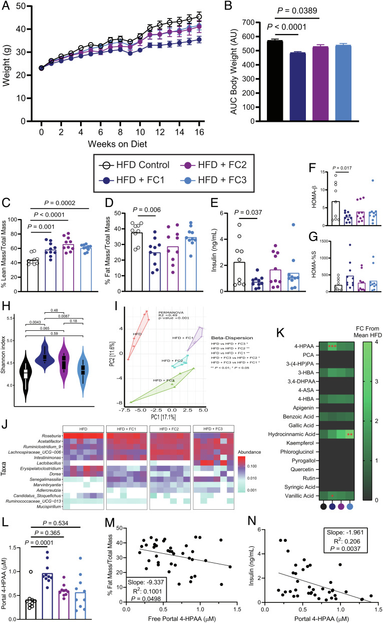
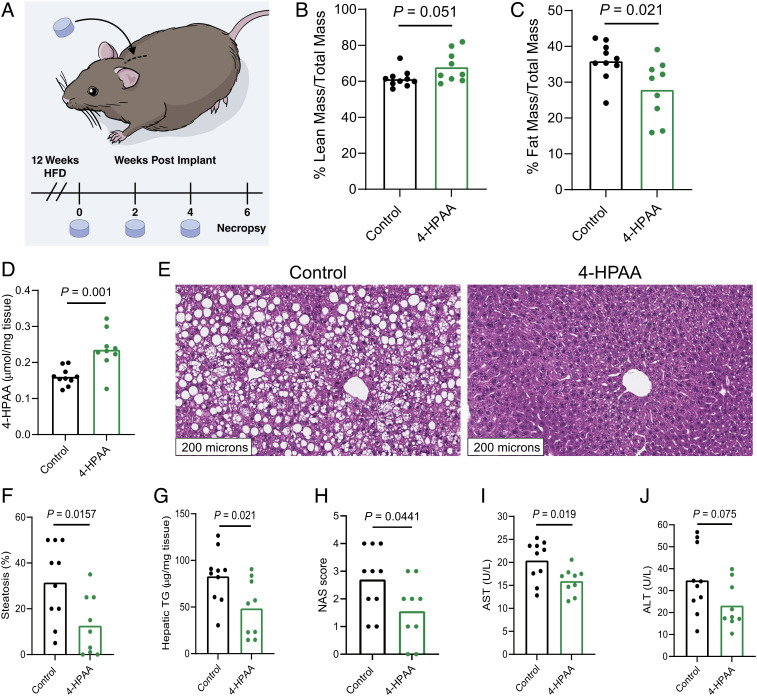
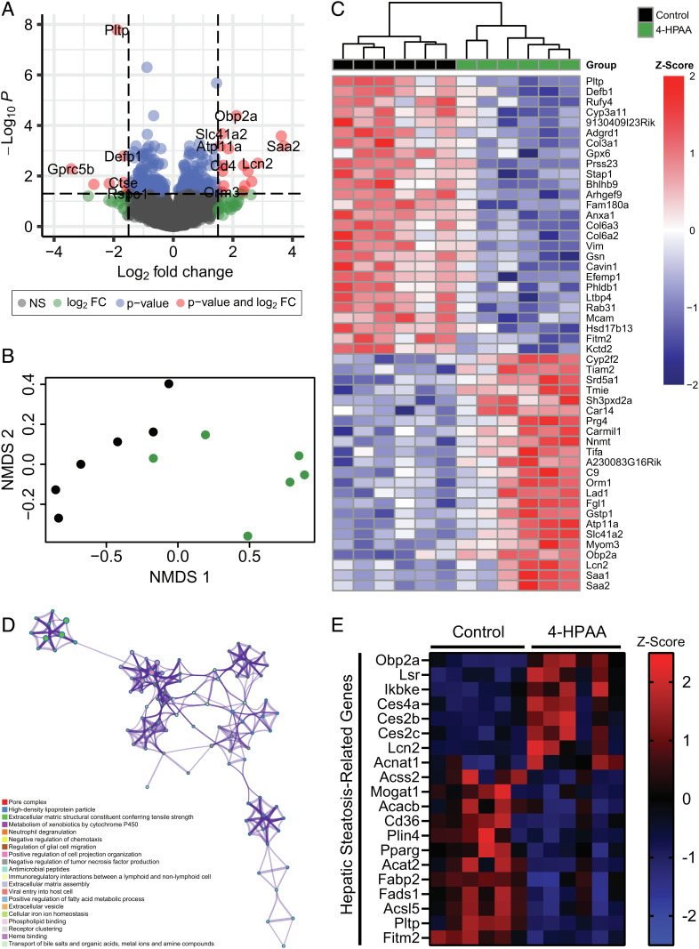
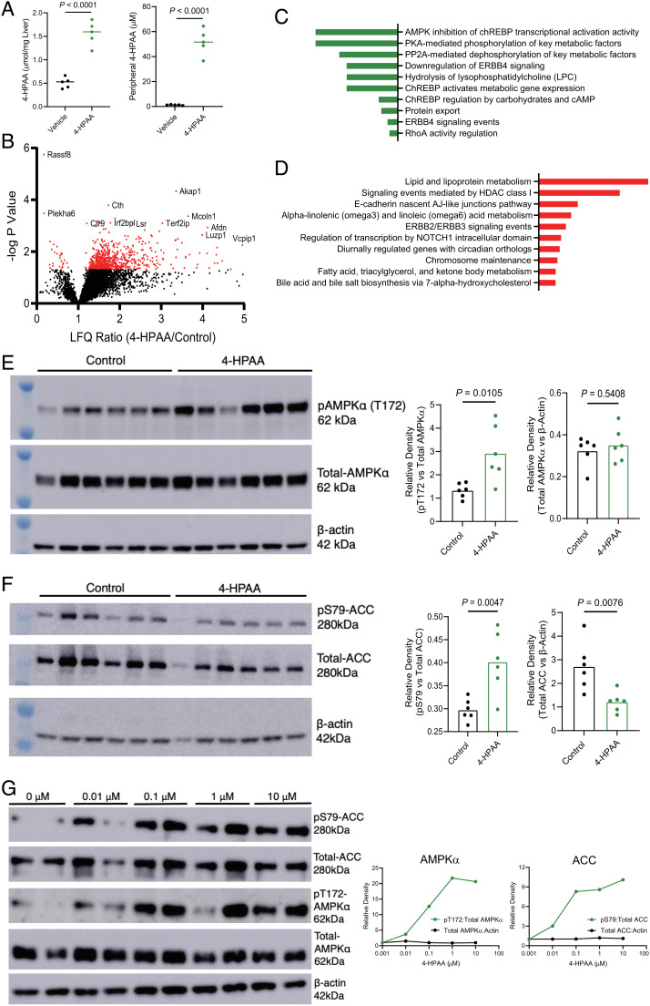
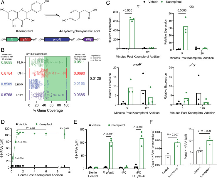
The molecular mechanisms by which dietary fruits and vegetables confer cardiometabolic benefits remain poorly understood. Historically, these beneficial properties have been attributed to the antioxidant activity of flavonoids. Here, we reveal that the host metabolic benefits associated with flavonoid consumption hinge, in part, on gut microbial metabolism. Specifically, we show that a single gut microbial flavonoid catabolite, 4-hydroxyphenylacetic acid (4-HPAA), is sufficient to reduce diet-induced cardiometabolic disease (CMD) burden in mice. The addition of flavonoids to a high fat diet heightened the levels of 4-HPAA within the portal plasma and attenuated obesity, and continuous delivery of 4-HPAA was sufficient to reverse hepatic steatosis. The antisteatotic effect was shown to be associated with the activation of AMP-activated protein kinase α (AMPKα). In a large survey of healthy human gut metagenomes, just over one percent contained homologs of all four characterized bacterial genes required to catabolize flavonols into 4-HPAA. Our results demonstrate the gut microbial contribution to the metabolic benefits associated with flavonoid consumption and underscore the rarity of this process in human gut microbial communities.

Keywords: AMP-activated protein kinase; cardiometabolic disease; flavonoid metabolism; gut microbiome; obesity.


Conflict of interest statement

The authors declare a competing interest. J.C. is a Scientific Advisor for Seed Health, Inc. Z.W. and S.L.H. report being named as co-inventor on pending and issued patents held by the Cleveland Clinic relating to cardiovascular diagnostics and therapeutics. S.L.H. reports being a paid consultant for Procter & Gamble, and Zehna Therapeutics, having received research funds from Procter & Gamble, Roche Diagnostics and Zehna therapeutics, and being eligible to receive royalty payments for inventions or discoveries related to cardiovascular diagnostics or therapeutics from Cleveland Heart Lab, a fully owned subsidiary of Quest Diagnostics, Zehna Therapeutics and Procter & Gamble. The other authors declare they have no competing interests.

Figures

Fig. 1.
Fig. 1.
Flavonoid composites reduce HFD-induced obesity and alter the cecal microbiome. (A) Body weights of 6-wk-old male C57BL/6 mice fed a high fat control diet, or the same diet supplemented with 1% w/w FC1, FC2, or FC3 for 16 wk; n = 9–10 per group, error bars represent SEM. (B) Mean cumulative area under the curve (AUC) for body weights after 16 wk, error bars represent SEM. (C) and (D) Lean and fat mass after 16 wk as measured by EchoMRI and normalized to total body mass, n = 9–10 per group. (E) Plasma insulin after 16 wk on either control or experimental diets following a 4-h fast, n = 9–10 per group. (F) Homeostatic model assessment of β-cell function (HOMA-β), n = 9–10 per group. (G) Percent insulin sensitivity after 16 wk, n = 9–10 per group. (H) Shannon alpha diversity estimates for cecal microbiomes based on 16S rRNA profiles of all the four groups. Statistical analysis was performed via ANOVA. (I) Principal component analysis (PCA) plot based on the Bray–Curtis dissimilarity index between the cecal 16S rRNA profile of all the four groups. Statistical analysis was performed with PERMANOVA where R2 values are noted for comparisons with significant P-values and stand for percentage variance explained by the variable of interest. (J) Heatmap of the significantly differentially abundant bacterial taxa across the cecal 16S rRNA profiles of all the four groups. n = 6 for all 16S rRNA sequencing analyses. (K) Heatmap of portal plasma flavonoids and microbial flavonoid catabolites measured by LC-MS/MS, n = 9–10 per group. Statistical analysis was performed with one-way ANOVA with Tukey’s multiple comparison test. (L) Portal plasma concentration of the microbial flavonol catabolite 4-HPAA measured by LC-MS/MS. (M) Simple linear regression analysis of fat mass proportion and portal plasma 4-HPAA. (N) Simple linear regression analysis of terminal plasma insulin and portal plasma 4-HPAA. P values shown were calculated using one-way ANOVA with Dunnett’s multiple comparisons test; n = 9–10 per group. Individual points represent individual mice, and bars represent group means.
Fig. 2.
Fig. 2.
4-HPAA is sufficient to reverse HFD-induced hepatic steatosis and liver injury. (A) 4-wk-old male C57BL/6 mice were fed a HFD for 12 wk to induce obesity. After the induction of diet-induced obesity, mice were randomly assigned to receive a subcutaneously implanted control scaffold pellet, or a pellet releasing 350 µg 4-HPAA per day for 2 wk. New pellets were implanted every 2 wk for a total of 6 wk. Lean mass (B) and fat mass (C) were measured by Echo MRI, n = 9–10 per group. (D) 4-HPAA accumulates in the liver as measured by LC-MS/MS, n = 9–10 per group. (EG) After a total of 18 wk of HFD feeding, H&E-stained sections of the liver revealed profound hepatic steatosis in scaffold control mice, while 4-HPAA-treated mice had marked reversal of steatosis and triglyceride deposition, n = 6–10 per group. (H) NAFLD activity score (NAS) incorporating histologic assessments of steatosis, hepatocellular ballooning, and inflammation. (I and J) Quantification of liver injury biomarkers in peripheral plasma, n = 9–10 per group. Statistical analysis was performed using unpaired two-tailed Student’s t test. Individual points represent individual mice, and bars represent group means. (Fig. 2A reprinted with permission, Cleveland Clinic Foundation ©2022. All Rights Reserved.)
Fig. 3.
Fig. 3.
4-HPAA influences the hepatic transcriptome. (A) Volcano plot of RNA-Seq hepatic transcriptome from control and 4-HPAA-treated mice after 6 wk. (B) NMDS of hepatic RNA-Seq data representing the distribution of samples in space based on the top 500 differentially expressed genes by adjusted P value and log2 fold change between control (black) and 4-HPAA-treated (green) mice. (C) Heatmap of differentially expressed genes with hierarchical clustering arranged by adjusted P value with Z-score values scaled by row. (D) Metascape-enriched ontology clusters based on the top 100 most significant differentially expressed genes between control and 4-HPAA-treated mice. Each ontology term is represented by a circle node where the size of the node is proportional to the number of genes that fall into that ontology assignment. (E) Heatmap representing the Z-score of genes implicated in hepatic lipid metabolism and subsequent related disorders in control and 4-HPAA-treated mice. A sample size of n = 6 per group was used for all RNA-Seq-related analyses.
Fig. 4.
Fig. 4.
Untargeted phosphoproteomic analysis reveals that 4-HPAA activates the AMPK pathway and downstream effectors to reduce de novo hepatic lipogenesis. (A) 4-HPAA concentration in the liver and peripheral plasma, 10 min after animals received an intraportal injection of 150 µg 4-HPAA or saline control. (B) Volcano plot of label-free quantitation (LFQ) ratio vs. −log P value for 4-HPAA/control hepatic phosphoproteomic analysis following portal vein injection of 4-HPAA or a saline vehicle control. (C) Pathway enrichment analysis of all hepatic phosphopeptides down-regulated following 4-HPAA/control portal vein injections. (D) Pathway enrichment analysis of top 125 up-regulated hepatic phosphopeptides following portal vein injection of 4-HPAA or a saline vehicle control, n = 5 per group for A–D. (E) Western blot analysis of pAMPKα (T172), total AMPKα, and β-actin with densitometric quantification, n = 6 per group. (F) Western blot analysis of pACC (S79), total ACC, and β-actin with densitometric quantification, n = 6 per group. (G) Primary murine hepatocytes were treated with either DMSO vehicle control (0 μM) or 0.01, 0.1, 1, and 10 μM 4-HPAA for 30 min at which point the protein expression of pACC (S79), total ACC, pAMPKα (T172), total AMPKα, and β-actin was measured via western blot analysis with densitometric quantification, with dots representing the mean of two biological replicates. Statistical analysis for panels A and B was performed using unpaired two-tailed Student’s t test. Individual points represent individual mice, and bars represent group means.
Fig. 5.
Fig. 5.
Flavonol- and flavone-catabolizing genes are rarely found in human microbiomes. (A) Graphical depiction of the complete bacterial gene set required to catabolize the flavonol kaempferol into 4-HPAA. (B) Computational screening of 1,899 assemblies of metagenomic data derived from human fecal samples representing over 1,300 human subjects for each of the bacterial genes required to catabolize flavonols/flavones. Assemblies with >40% coverage to the parent Flavonifractor plautii YL31 gene of interest were considered present. (C) Relative expression of F. plautii flavonol-catabolizing genes as measured by RT-qPCR following the addition of a vehicle control or kaempferol over time, n = 2–3 per group. (D) F. plautii rapidly converts kaempferol into 4-HPAA as measured by LC-MS/MS, n = 3 per group. (E) F. plautii converts kaempferol into 4-HPAA after 24 h of anaerobic incubation in vitro and retains its function when added to a nonconverting human fecal community (hFC). (F) Portal and cecal 4-HPAA concentrations in mice monocolonized with F. plautii after oral gavage with kaempferol or saline control. Individual points represent technical replicates in panels C, D, and E. Individual points in panel F represent biological replicates, with bars representing group means. Statistical analysis for panels C and E was performed using two-way ANOVA with Tukey’s correction for multiple comparisons (n = 3 per group). Statistical analysis for panel D was performed using a two-tailed unpaired t test with Holm–Šídák correction for multiple comparisons (n = 3 per group), and for panel F with a two-tailed Student’s t test (n = 3 per group).

Similar articles

Cited by

References

    1. Snetselaar L. G., Jesus J. M. D., DeSilva D. M., Stoody E. V., Dietary guidelines for Americans, 2020-2025, Nutr. Today 56, 287–295 (2021). - PMC - PubMed
    1. Manus M. B., Evolutionary mismatch. Evol. Med. Public Health 2018, 190–191, (2018). - PMC - PubMed
    1. Pontzer H., Wood B. M., Raichlen D. A., Hunter-gatherers as models in public health. Obesity Rev. 19, 24–35 (2018). - PubMed
    1. David L. A., et al. , Diet rapidly and reproducibly alters the human gut microbiome. Nature 505, 559–563 (2014). - PMC - PubMed
    1. Wang Z., et al. , Gut flora metabolism of phosphatidylcholine promotes cardiovascular disease. Nature 472, 57–63 (2011). - PMC - PubMed

Publication types