. 2022 Nov 29;119(48):e2202580119.
doi: 10.1073/pnas.2202580119. Epub 2022 Nov 23.

Oligodendrocyte precursor cells ingest axons in the mouse neocortex

Affiliations

Oligodendrocyte precursor cells ingest axons in the mouse neocortex

JoAnn Buchanan et al. Proc Natl Acad Sci U S A. .

Abstract

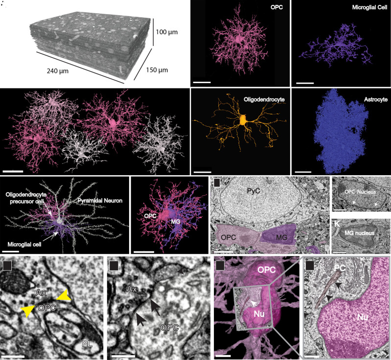
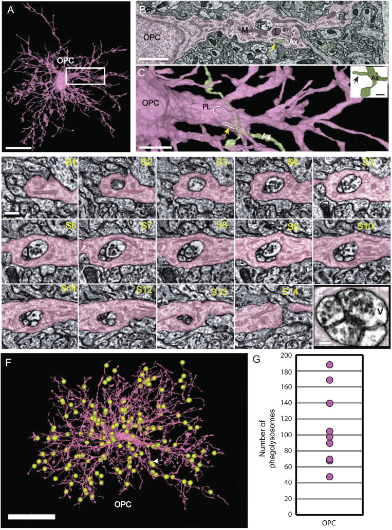
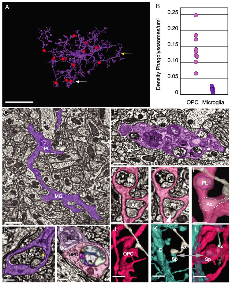
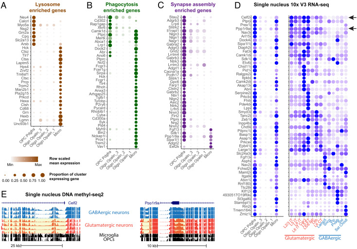
Neurons in the developing brain undergo extensive structural refinement as nascent circuits adopt their mature form. This physical transformation of neurons is facilitated by the engulfment and degradation of axonal branches and synapses by surrounding glial cells, including microglia and astrocytes. However, the small size of phagocytic organelles and the complex, highly ramified morphology of glia have made it difficult to define the contribution of these and other glial cell types to this crucial process. Here, we used large-scale, serial section transmission electron microscopy (TEM) with computational volume segmentation to reconstruct the complete 3D morphologies of distinct glial types in the mouse visual cortex, providing unprecedented resolution of their morphology and composition. Unexpectedly, we discovered that the fine processes of oligodendrocyte precursor cells (OPCs), a population of abundant, highly dynamic glial progenitors, frequently surrounded small branches of axons. Numerous phagosomes and phagolysosomes (PLs) containing fragments of axons and vesicular structures were present inside their processes, suggesting that OPCs engage in axon pruning. Single-nucleus RNA sequencing from the developing mouse cortex revealed that OPCs express key phagocytic genes at this stage, as well as neuronal transcripts, consistent with active axon engulfment. Although microglia are thought to be responsible for the majority of synaptic pruning and structural refinement, PLs were ten times more abundant in OPCs than in microglia at this stage, and these structures were markedly less abundant in newly generated oligodendrocytes, suggesting that OPCs contribute substantially to the refinement of neuronal circuits during cortical development.

Keywords: axonal pruning; engulfment; oligodendrocyte precursor cells; phagolysosomes.


Conflict of interest statement

The authors declare a competing interest. T.M. and H.S.S. disclose financial interests in Zetta AI LLC.

Figures

Fig. 1.
Fig. 1.
Distinct structural features of OPCs in the developing visual cortex. (A) TEM reconstruction of 100-µm3 volume of layer 2/3 mouse visual cortex (P36). (B) 3D reconstructions of a subset of OPCs in P36 dataset, showing discrete territories and tiling. (Scale bar, 30 µm.) (C) 3D rendering of an OPC from P36 dataset showing extensive ramifications emanating from the cell soma. (D) 3D rendering of a microglial cell shows its thicker, less branched processes and elongated and flattened soma. (E) 3D rendering of a mature myelinating oligodendrocyte in P36 dataset has a smooth and ovoid-shaped soma. Note, only the soma and cytoplasmic processes without myelin sheaths are shown. (F) 3D rendering of an astrocyte in P36 dataset showing its densely packed cytoplasmic protrusions. (Scale bars, C–F, 20 µm.) (G) 3D rendered pyramidal neuron (white) with an OPC (pink) and a microglial cell (MG, purple) both in satellite positions. (Scale bar, 30 µm.) (Movie S1) (H) 3D rendering of the same two glial cells in (G) shows their close association and intermingling of branches. (Scale bar, 20 µm.) (I) Ultrathin section slice through an OPC soma (pink) and microglial cell soma (purple). (Scale bar, 3 μm.) (J) Ultrathin section slice of an OPC nucleus with dense rim of heterochromatin and ruffled edge (white arrows). (Scale bar, 1.5 μm.) (K) Ultrathin section slice of a microglial nucleus showing its dense heterochromatin throughout. (Scale bar, 1.5 μm.) (L and M) Axons (Ax) making synaptic contacts (yellow and black arrows) with OPC processes. A phagolysosome (PL) is nearby in L. (Scale bar, 300 nm.) (N) The soma of an OPC (deep pink) bears a primary cilium (white arrow) adjacent to the nucleus (Nu). (Scale bar, 3 µm.) (O) Ultrathin slice of the boxed area in N showing the primary cilium (PC) (black arrows) close to the OPC nucleus (Nu, dark pink). (Scale bar, 750 nm.)
Fig. 2.
Fig. 2.
Phagolysosomes (PLs) are abundant in OPC processes and contain vesicles. (A) 3D rendering of OPC with numerous ramified branches. Boxed area same as micrograph shown in (B) (Scale bar 15µm.) (B) OPC branch contains PLs, lysosomes (L), and mitochondria (M), and an ingested axon (yellow arrow) (Ax). (Scale bar 1.5 µm.) (C) 3D rendering of same OPC in (B) in pink and axon (Ax) (green) arrow (yellow) points to piece of ingested axon outlined in white and PL (white oval). (Scale bar 2 µm.) Inset shows 3D rendering of the same green axon (Ax) bouton with small, ingested protrusion outlined in slice view (yellow arrow). (Scale bar 500 nm.) (D) Twelve ultrathin 40-nm serial sections through a phagolysosome show the distribution of 40-nm vesicles within the organelle. Nine of the twelve sections have vesicles inside the phagolysosome. (Scale bar 300 nm.) (E) The last panel shows high magnification of vesicles inside phagolysosome. (Scale bar 150 nm.) (F) 3D reconstruction of an OPC from the P36 dataset. Yellow spheres represent manual annotations of the 187 PLs found in this OPC. White arrow points to location of PL in serial section Fig. 4D. (Scale bar, 20 μm.) (original data in http://microns-explorer.org/phagolysosomes/opc). (G) Plot showing the number of PLs in nine OPCs in P36 dataset. See Movie S2.
Fig. 3.
Fig. 3.
Axon engulfment by OPCs. (A) OPC process in pink ingests an excitatory axon bouton (gray) at its tip. (Scale bar, 500 nm.) (B) 3D reconstruction reveals a small excitatory axon fragment (Ax) encapsulated within the OPC (pink) at the tip (white line, arrow). (Scale bar, 500 nm.) (C) 3D reconstruction shows the same axon (Ax)(arrow) without OPC surrounding it. The bouton tip is visible. (Scale bar, 500 nm.) (D) Ultrathin section slice of a large section of an inhibitory axon (Ax) collateral branch (white line) in gray ingested within the cytoplasm of the OPC (pink). Two PL (one in dashed circle) are adjacent to the ingested axon. (Scale bar, 500 nm.) (E) 3D rendered axon collateral branch encapsulated within the cytoplasm of the OPC (arrow, white line and outline) and phagolysosome within the cytoplasm (dashed circle). (Scale bar, 1μm.) (F) 3D reconstruction of the axon (Ax) in gray shows encapsulated branch (white line, arrow) without the surrounding OPC. (Scale bar, 1μm.) (G) Bar graph of ten individual isolated main branches (one each) of ten OPC cells were analyzed for ingestion events and categorized as either phagosome (PS)(pink) or phagolysosome (PL)(purple). (H) Bar graph of the same ten OPCs showing axons partially ingested typed as excitatory (E), inhibitory (I) or unsure (U). Excitatory axons were the most prevalent type ingested at 85.9% versus 7.7% for inhibitory.
Fig. 4.
Fig. 4.
PLs are more abundant in OPCs than microglia. (A) 3D rendering of a microglial cell from the P36 dataset. Red spheres represent manual annotations of the 13 PLs present in this cell. White arrow points to the location of the PL shown in 4C. (Scale bar, 10 μm.) (original data in http://microns-explorer.org/phagolysosomes/microglia. (B) Comparison of density of PLs at P36 between OPCs and microglia. Density was calculated including the volume of the soma region. (C) Microglial cell branch (MG) of cell shown in (A) in purple shows a phagolysosome (PL) (white arrow also shown in 4A) in its dense cytoplasm. (Scale bar, 1 µm.) (D) A synapse is ingested within the microglia branch. Its position in the cell is marked by the yellow arrow also shown in 4A. Vesicles in the presynaptic axon (Ax) are visible. (Scale bar 300 nm.) (E) A microglial (MG) branch (purple) from P36 dataset contacts and OPC branch in pink with PLs in both cells. (Scale bar, 300 nm.) (F) A portion of a microglial branch shows the cytoplasm congested with numerous PLs. (Scale bar, 750 nm.) (E) A microglial (MG) branch (purple) from P36 dataset contacts and OPC branch in pink with PLs in both cells show their different morphologies. (Scale bar, 300 nm.) (G) OPC process contains a phagolysosome with vesicles (V) and engulfed axon (Ax). (Scale bar 300 nm.) (H) A different slice of the same process shows the phagolysosome (PL) and postsynaptic density (arrow) of a synapse onto the encapsulated axon, which remains attached to its parent. (Scale bar 300 nm.) (I) 3D rendering of the same process shows both the phagolysosome (PL) and the encapsulated axon collateral within the OPC cytoplasm(dashed outline). (Scale bar 500 nm.) (J) 3D rendering of the same OPC in (G) and axon (Ax) in gray. (K) 3D rendering of the dendrite (D) in blue shows the spine (Sp) in blue synapsing onto the same axon (Ax) in (J). (L) The spine (Sp) and collateral axon branch (Ax) remain attached to their cells of origin but are fully surrounded by OPC cytoplasm. (Scale bars, (J–L) 1 μm.) See Movie S4.
Fig. 5.
Fig. 5.
Detection of phagolysosome genes and neuronal transcripts in OPCs in oligodendrocytes and microglia. (A–C) Dot plots of enriched lysosome (A), phagocytosis (B), and synapse assembly (C) GO term genes enriched in OPCs and/or microglia, relative to mature oligodendrocytes. (D) Dot plot of neuronal subclass marker genes expressed in OPCs, mature oligodendrocytes – Oligo, microglia – Micro, and glutamatergic (red) and GABAergic (blue) neuronal subclasses. (E) Genome tracks showing glutamatergic marker genes, Celf2 (Top panel) and Ppp1r9a (Bottom panel) with hypomethylated chromatin highlighted in yellow. Blue tracks show GABAergic and red tracks show glutamatergic neuronal subclasses.

References

    1. Baxi E. G., et al. , Lineage tracing reveals dynamic changes in oligodendrocyte precursor cells following cuprizone-induced demyelination. Glia 65, 2087–2098 (2017). - PMC - PubMed
    1. Bergles D., Jahr C. E., Glutamatergic synapses on oligodendrocyte precursor cells in the hippocampus. Nature 405, 187–191 (1997). - PubMed
    1. Lin S. C., Bergles D. E., Synaptic signaling between neurons and glia. Glia 47, 290–298 (2004). - PubMed
    1. Bergles D. E., Richardson W. D., Oligodendrocyte development and plasticity. Cold Spring Harb. Perspect. Biol. 8, a020453 (2015). - PMC - PubMed
    1. Turner N. L., et al. , Multiscale and multimodal reconstruction of cortical structure and function. bioRxiv [Preprint] (2020). 10.1101/2020.10.14.338681 (Accessed 29 May 2021). - DOI

Publication types

LinkOut - more resources