. 2022 Nov 25;8(47):eabq7219.
doi: 10.1126/sciadv.abq7219. Epub 2022 Nov 23.

Reprogramming Müller glia to regenerate ganglion-like cells in adult mouse retina with developmental transcription factors

Affiliations

Reprogramming Müller glia to regenerate ganglion-like cells in adult mouse retina with developmental transcription factors

Levi Todd et al. Sci Adv. .

Erratum in

Abstract

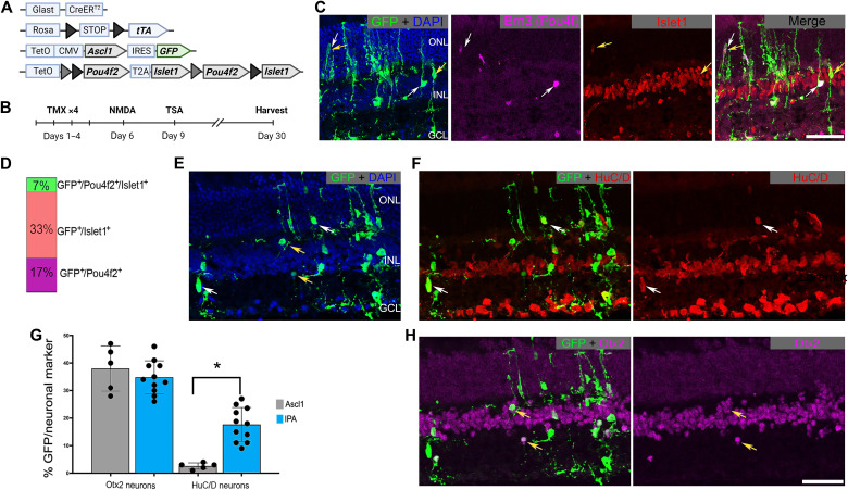
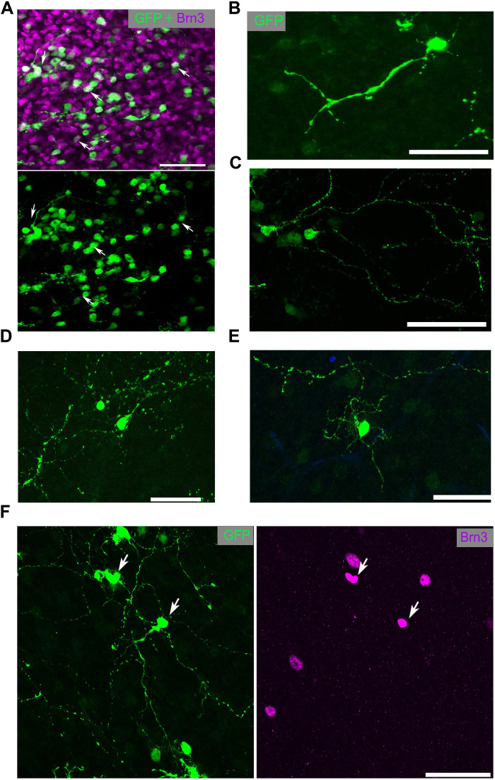
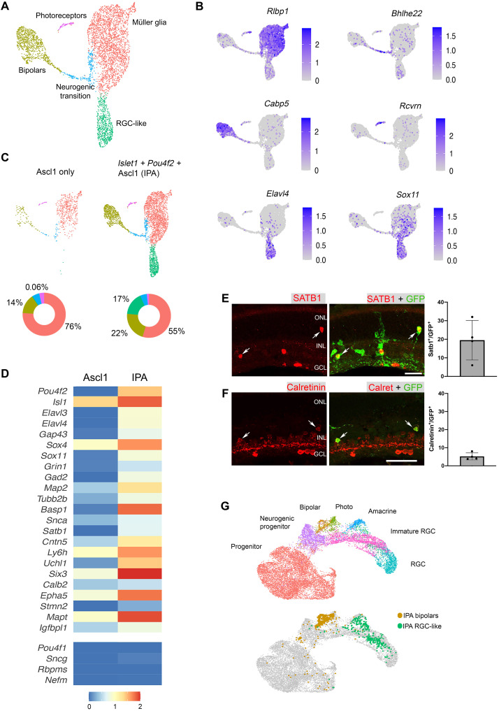
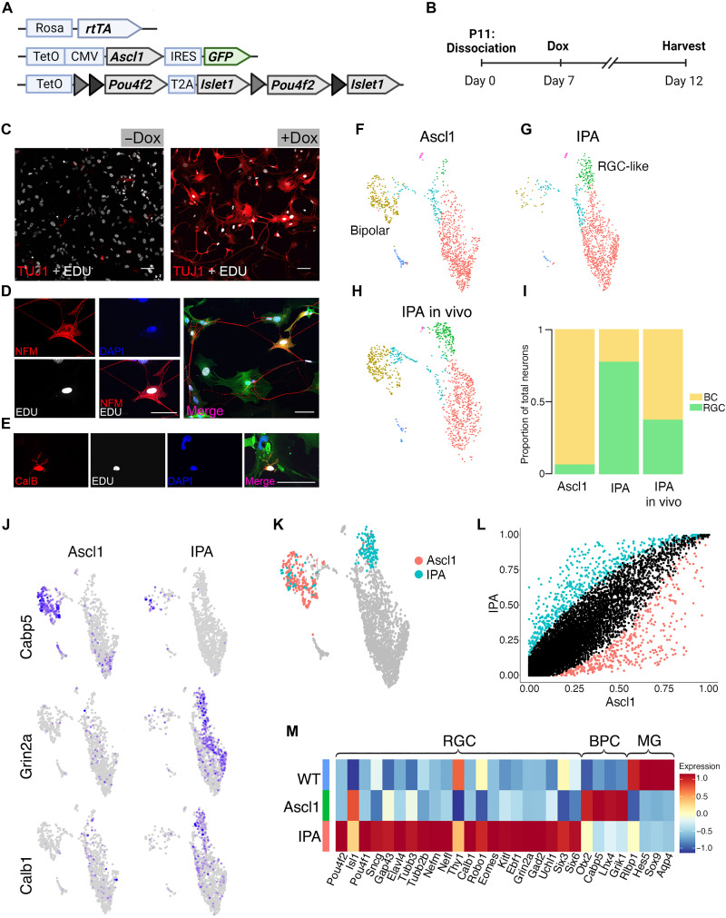
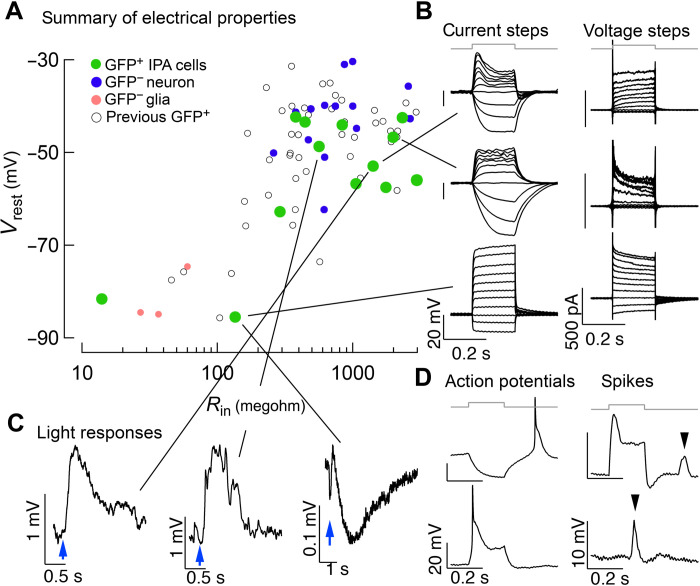
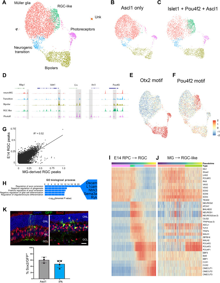
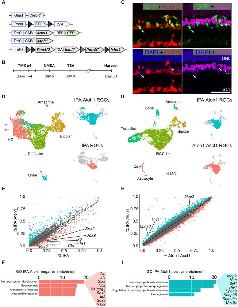
Many neurodegenerative diseases cause degeneration of specific types of neurons. For example, glaucoma leads to death of retinal ganglion cells, leaving other neurons intact. Neurons are not regenerated in the adult mammalian central nervous system. However, in nonmammalian vertebrates, glial cells spontaneously reprogram into neural progenitors and replace neurons after injury. We have recently developed strategies to stimulate regeneration of functional neurons in the adult mouse retina by overexpressing the proneural factor Ascl1 in Müller glia. Here, we test additional transcription factors (TFs) for their ability to direct regeneration to particular types of retinal neurons. We engineered mice to express different combinations of TFs in Müller glia, including Ascl1, Pou4f2, Islet1, and Atoh1. Using immunohistochemistry, single-cell RNA sequencing, single-cell assay for transposase-accessible chromatin sequencing, and electrophysiology, we find that retinal ganglion-like cells can be regenerated in the damaged adult mouse retina in vivo with targeted overexpression of developmental retinal ganglion cell TFs.


Figures

Fig. 1.
Fig. 1.. Pou4f2 and/or Islet1 stimulate regeneration of RGC-like neurons.
(A) Schematic depicting the transgenic constructs used to induce Ascl1 and Pou4f2/Islet1 specifically in MG. Pou4f2/Islet1 is surrounded by mutually exclusive floxed sites, leading to expression of Pou4f2, Islet1, or both in the presence of active Cre. (B) Experimental paradigm to induce retinal regeneration in adult mice. Tamoxifen (TMX). (C) Representative sections of the retina after intravitreal NMDA damage, showing transgenic expression of Pou4f2/Brn3 (purple) and/or Islet1 (red) in GFP+ lineage–traced MG. DAPI, 4′,6-diamidino-2-phenylindole. (D) Quantification of the percent of transgene-expressing MG that express Pou4f2, Islet1, or both. (E and F) Representative sections showing MG-derived neurons after the regeneration paradigm expressing HuC/D (red). (G) Quantification of the percent of GFP+ MG-derived neurons that express either HuC/D or Otx2. (H) Examples of MG-derived neurons expressing Otx2 (purple). Significance of difference was determined using an unpaired t test (asterisk = p < 0.0001); dots represent individual animals. Scale bars, 50 μm. ONL, outer nuclear layer; INL, inner nuclear layer; GCL, ganglion cell layer. Mouse schematic was made with Biorender.com.
Fig. 2.
Fig. 2.. IPA-stimulated MG-derived neurons display complex neuronal morphology.
(A) Retinal whole mounts stained for GFP (MG-derived cells; green) and Brn3 (purple). (B to E) Examples of the morphology of GFP+ MG-derived cells. (F) MG-derived (GFP+) cell with complex neurites colabeled with Brn3 (purple). Scale bars, 50 μm.
Fig. 3.
Fig. 3.. scRNA-seq analysis of Pou4f2/Islet1-stimulated neurons reveals molecular characteristics of RGCs.
(A) UMAP plot for FACS-sorted MG-derived cells after the IPA regeneration paradigm combined with a previous scRNA-seq dataset where Ascl1 only was used. (B) Feature plots highlight the major clusters of MG (Rlbp1), neurogenic transition (Bhlhe22), bipolars (Cabp5), photoreceptors (Rcvrn), and RGC-like cells (Elavl4 and Sox11). (C) The distribution of cells from each treatment projected onto a split UMAP plot. Donut plots represent the percent each cluster comprises of the dataset. (D) Heatmap comparing scRNA-seq datasets of Ascl1 only versus IPA treatment. Selected genes are depicted that are associated with RGCs. (E) Retinal sections stained for MG-derived cells (GFP) with Satb1 (red), and quantifications show the percent of GFP+ cells that are Satb1+. (F) Retinal sections stained for MG-derived cells (GFP) with Calretinin (red), and quantifications show the percent of GFP+ cells that are Calretinin+. (G) UMAP of IPA-derived neurons integrated with scRNA-seq of E14 mouse retina from Clark et al. (21), revealing that IPA neurons cluster similarly to immature RGCs. Scale bars, 50 μm.
Fig. 4.
Fig. 4.. Islet1 and Pou4f2 coinduction stimulates RGC-like neurons from MG in vitro.
(A) Schematic of transgenic construct to induce IPA in all primary MG in vitro by doxycycline. (B) Paradigm for inducing Ascl1-mediated neurogenesis in vitro. (C to E) Representative images of EdU+ MG-derived neurons expressing neuronal markers.) (C) EdU+ (white) MG-derived cell expressing Tuj1 (red). (D) MG-derived neuron expressing EdU (white), Neurofilament M (NFM; red), and the GFP transgene reporter (GFP). ( (E) MG-derived neuron expressing Calbindin (red) colabeling with EdU (white), DAPI, and GFP. (F to H) UMAP plots of cultured MG reprogrammed with Ascl1 (F) and IPA (G), integrated with cells from in vivo IPA regeneration model (H) as reference. (I) Stacked bar plot showing composition of neuronal clusters in each sample. BC, bipolar cell. (J) Feature plots highlighting differentially expressed genes in neuronal clusters of either reprogramming strategy. (K) Highlighted cells of the Ascl1 and IPA datasets used for downstream DGE analysis in (L). (M) Heatmap of genes differentially expressed in either the Ascl1 or IPA condition. WT, untreated cultured MG included for baseline values. Statistics for differential gene analysis: Wilcoxon Mann-Whitney test for significance (P < 0.05). Scale bars, 50 μm. Mouse schematic was made with Biorender.com.
Fig. 5.
Fig. 5.. Physiological profiling of IPA-induced neurons.
(A) Summary of electrical properties of cells in this study compared to endogenous neurons, endogenous glia, and MG-derived neurons from previous regeneration protocols (4, 5, 13). Resting potential and input resistance were estimated from current clamp recordings. (B) Examples of responses to current (left) and voltage (right) steps for three cells. (C) Three examples of cells that responded to light stimulus. (D) Examples of cells that displayed action potentials or similar events. The two left panels are responses to hyperpolarizing and depolarizing current steps from a cell that generated apparent Na+ spikes. The right two panels are responses from a cell that generated smaller discrete events, likely Ca2+ spikes.
Fig. 6.
Fig. 6.. scATAC reveals MG remodel chromatin to an RGC-like state in response to IPA treatment.
(A) Combined UMAP of GFP+ sorted MG and their progeny from the in vivo regeneration paradigm with Ascl1-only (B) or IPA treatment (C). (D) Coverage plots for known marker genes used to identify clusters. (E and F) chromVAR scores of Otx2 and Pou4f2 to highlight differential accessibility of their respective motifs. (G) Scatterplot comparing accessible motifs in E14 RGCs versus IPA-induced RGCs. (H) Top “GO biological process” results for peaks specific to E14 RGCs compared to IPA-derived RGC-like neurons. (I) Heatmap of the chromVAR activity scores of the top variable motifs for TFs found on the pseudotime lineage of E14 progenitor cells to RGCs. (J) Heatmap of the same chromVAR activity scores of the E14 motifs plotted on pseudotime from MG to RGC-like neuron after IPA treatment. (K) Retinal sections showing GFP+ MG-derived cells costained with the MG nuclei marker Sox2 (red) and quantification of GFP+ cells expressing Sox2. Scale bars, 50 μm.
Fig. 7.
Fig. 7.. The addition of Atoh1 to the IPA paradigm facilitates transition from a progenitor state to a differentiated neuron.
(A) Schematic of transgenic construct to express IPA with Atoh1 in MG. (B) Regeneration paradigm for inducing IPA:Atoh1 expression in MG in the damaged retina. (C) Representative immunofluorescence images of regenerated neurons from IPA:Atoh1 mice demonstrating MG-derived neurons (GFP+) are HuC/D+ (red) and not Otx2+ (purple). (D) Integrated UMAP of FACS-sorted MG-derived cells after regeneration paradigm with either IPA:Atoh1 or IPA-only overexpression. Highlighted in either blue or red are the RGC-like cells from each dataset that were subsetted for further comparative analysis. (E) Scatterplot highlighting differentially expressed genes between the RGC-like cells of the IPA:Atoh1 (blue) and IPA-only (red) regeneration paradigms. (F) GO analysis revealed that neurodevelopmental terms containing many retinal progenitor genes were down-regulated in the IPA:Atoh1 dataset versus IPA only. (G) Integrated UMAP of IPA:Atoh1 data as described above with previously generated Ascl1:Atoh1 dataset (13). Blue or red highlighting denotes RGC-like cells from each dataset compared in further analysis. (H) Scatterplot highlighting differentially expressed genes between the RGC-like cells of the IPA:Atoh1 and Ascl1:Atoh1 regeneration paradigms. (I) Bar plot of GO terms relating to neurite outgrowth enriched in the IPA:Atoh1 data. Known neuron projection genes listed are up-regulated with IPA:Atoh1 versus Ascl1:Atoh1. Mouse schematic was made with Biorender.com.

References

    1. L. Todd, T. A. Reh,Comparative biology of vertebrate retinal regeneration: Restoration of vision through cellular reprogramming. Cold Spring Harb. Perspect. Biol. 14,a040816 (2022). - PMC - PubMed
    1. A. Hamon, J. E. Roger, X. J. Yang, M. Perron,Müller glial cell-dependent regeneration of the neural retina: An overview across vertebrate model systems. Dev. Dyn. 245,727–738 (2016). - PMC - PubMed
    1. A. Bringmann, I. Iandiev, T. Pannicke, A. Wurm, M. Hollborn, P. Wiedemann, N. N. Osborne, A. Reichenbach,Cellular signaling and factors involved in Müller cell gliosis: Neuroprotective and detrimental effects. Prog. Retin. Eye Res. 28,423–451 (2009). - PubMed
    1. N. L. Jorstad, M. S. Wilken, W. N. Grimes, S. G. Wohl, L. S. VandenBosch, T. Yoshimatsu, R. O. Wong, F. Rieke, T. A. Reh,Stimulation of functional neuronal regeneration from Müller glia in adult mice. Nature 548,103–107 (2017). - PMC - PubMed
    1. N. L. Jorstad, M. S. Wilken, L. Todd, C. Finkbeiner, P. Nakamura, N. Radulovich, M. J. Hooper, A. Chitsazan, B. A. Wilkerson, F. Rieke, T. A. Reh,STAT signaling modifies Ascl1 chromatin binding and limits neural regeneration from Muller glia in adult mouse retina. Cell Rep. 30,2195–2208.e5 (2020). - PMC - PubMed

Publication types

Substances