. 2022 Nov 21:30:e20220316.
doi: 10.1590/1678-7757-2022-0316. eCollection 2022.

Macrophage M1 polarization mediated via the IL-6/STAT3 pathway contributes to apical periodontitis induced by Porphyromonas gingivalis

Affiliations

Macrophage M1 polarization mediated via the IL-6/STAT3 pathway contributes to apical periodontitis induced by Porphyromonas gingivalis

Xuan Chen et al. J Appl Oral Sci. .

Abstract

Objective: To investigate the involvement of IL-6/STAT3 signaling pathway activation in macrophage polarization and bone destruction related to apical periodontitis (AP) stimulated by Porphyromonas gingivalis.

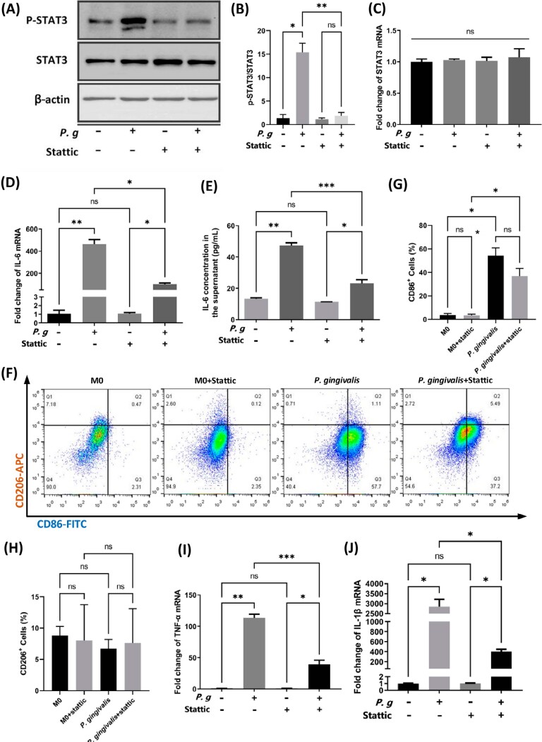
Methodology: Macrophage polarization, IL-6/STAT3 expression, and the presence of P. gingivalis were detected in human AP tissues via RT-qPCR, western blotting, and immunohistochemistry staining. Murine bone marrow derived macrophages were isolated and cultured with P. gingivalis W83 in vitro, and levels of macrophage IL-6 expression, STAT3 phosphorylation, and macrophage polarization with or without the selective STAT3 phosphorylation inhibitor Stattic (5 μM) were detected via ELISA, western blotting, RT-qPCR, and flow cytometry, respectively. P. gingivalis-induced murine AP models were constructed, and bone destruction and macrophage polarization in the apical region were evaluated. Transwell co-culture systems were used to investigate the effects of macrophages infected with P. gingivalis on osteogenesis and osteoclastogenesis.

Results: P. gingivalis was detected in human AP tissues that highly expressed IL-6/STAT3, and the M1 subtype of macrophages was more abundant in these tissues. P. gingivalis infection induced IL-6 expression, STAT3 phosphorylation, and M1 polarization of macrophages, while 5 μM of Stattic partially abolished these activation effects. Systemic STAT3 blockade via oral administration of Stattic at a dose of 25 mg kg-1 alleviated murine periapical bone resorption and apical infiltration of M1 macrophages induced by P. gingivalis infection in vivo. Furthermore, macrophages infected with P. gingivalis promoted bone destruction via secretion of IL-6, TNF-α, and RANKL, which hinder pre-osteoblast expression of Runx2 and accelerate pre-osteoclast expression of NFAT2.

Conclusions: The activation of IL-6/STAT3 signaling pathway is involved in mediating macrophages M1 polarization in the P. gingivalis induced apical inflammatory context and may also be intimately involved in the bone loss caused by P. gingivalis infection, directing the M1 macrophage infiltration during the progression of AP.


Conflict of interest statement

Conflict of interest

The authors declare no conflicts of interest.

Figures

Figure 1
Figure 1. Polarized macrophage distributions in human apical periodontitis tissues. (A), representative TSA-based immunofluorescence staining images. (B), statistical results of the MFI analysis. CD68, general marker for macrophages; iNOS, specific marker for M1 macrophages; CD206, specific marker for M2 macrophages; DAPI, nuclear dye. In the merge picture, open white arrows indicate typical M2 macrophages (CD68+CD206+), blue arrows indicate typical M1 macrophages (CD68+iNOS+). MFI, mean fluorescence density; A.U., arbitrary unit. Scale bars, 20 μm. **p<0.01.
Figure 2
Figure 2. IL-6/STAT3 expression and P. gingivalis presence in human apical periodontitis tissues. (A), representative western blot images for p-STAT3, STAT3, and β-actin. (B), grayscale analysis of p-STAT3 expression (relative to STAT3). (C), gene expression level of IL-6 in healthy pulps and apical tissues. (D), P. gingivalis detection in apical tissues via RT-qPCR using universal P. gingivalis primers, showing that P. gingivalis were present in samples #1 and #2. Healthy, healthy human dental pulps; AP, apical periodontitis lesion tissues. Sample numbers are indicated beside the dots. *p<0.05; ***p<0.001
Figure 3
Figure 3. P. gingivalis infection induced IL-6 expression, STAT3 phosphorylation, and macrophage M1 polarization in BMDMs. (A-C) IL-6 transcription (A), IL-6 secretion (B), and Stat3 transcription (C) in response to P. gingivalis stimulation for 12 h. (D) and (E), p-STAT3 expression changes upon P. gingivalis activation. (F), representative flow cytometry results of unstimulated BMDMs (M0) and P. gingivalis-stimulated BMDMs. (G) and (H), statistical results for CD86+ (G) and CD206+ (H) macrophages in flow cytometry. (I) and (J), RT-qPCR results for expression levels of other M1 markers in response to P. gingivalis infection. M0, unstimulated BMDMs; P. gingivalis, BMDMs stimulated by P. gingivalis for 12 h. *p<0.05; **p<0.01; ***p<0.001; ****p<0.0001; ns, not significant
Figure 4
Figure 4. STAT3 blocking attenuated P. gingivalis-induced IL-6 production, STAT3 phosphorylation, and M1 type polarization in murine primary macrophages. (A), representative image demonstrating the phosphorylation status of STAT3 induced by P. gingivalis and suppressed by Stattic. (B), grayscale analysis of p-STAT3 expression (relative to STAT3). (C) and (D), relative mRNA expression levels of STAT3 (C) and IL-6 (D) after P. gingivalis infection for 12 h in the presence or absence of 5 μM Stattic. (E), levels of IL-6 protein secreted in the culture supernatant of BMDMs in response to P. gingivalis infection with or without 5 μM Stattic. (F), representative flow cytometry results exhibiting the influence of Stattic on macrophage polarization status after P. gingivalis stimulation for 12 h. (G) and (H), statistical results for CD86+ (G) and CD206+ (H) macrophages in flow cytometry. M0, unstimulated BMDMs; P. g and P. gingivalis, BMDMs stimulated by P. gingivalis for 12 h. *p<0.05; **p<0.01; ***p<0.001; ns, not significant
Figure 5
Figure 5. P. gingivalis infection caused radiographically detectable apical lesions in vivo, and systemic blockade of STAT3 alleviated bone resorption in P. gingivalis-derived murine apical periodontitis. (A), representative 3-D reconstruction images of the infected mandibles. Areas in red indicate the soft tissues surrounding the root apexes; scale bars, 100 μm. (B), analysis of the bone volume fractions (BV/TV). (C), representative H&E staining results for the apical regions. Scale bars, 100 μm. Con, control group; PA, P. gingivalis-induced apical periodontitis group; Stattic, Stattic (supplemented with 0.5% carboxymethylcellulose) treatment of the AP group; CMC, 0.5% carboxymethylcellulose treatment of the AP group. BV, bone volume; TV, tissue volume. ***p<0.001, ****p<0.0001; ns, not significant. Red arrows in the H&E staining pictures indicate pulpal accesses; black arrow in the AP group indicates lateral root canal. PDL, periodontal ligament; DP, dental pulp; RE, root end; AB, alveolar bone; PL, periapical lesion
Figure 6
Figure 6. IHC staining of the apical regions of the mandibular first molars of the murine models. (A), representative IHC staining images of the Con, PA, Stattic, and CMC groups. Scale bars, 50 μm (×200) and 20 μm (×400), respectively. (B), statistical results for positive cell counting of the F4/80+, iNOS+, and CD206+ macrophages. Different markers on the bars indicate statistical differences. (C) and (D), percentages of the CD206+ (C) and iNOS+ (D) cells (relative to F4/80+ cells). (E) ratios of iNOS+ cells to CD206+ cells (M1/M2 ratios). Con, control group; PA, P. gingivalis-induced apical periodontitis group; Stattic, Stattic (supplemented with 0.5% carboxymethylcellulose) treatment of the AP group; CMC, 0.5% carboxymethylcellulose treatment of the AP group. *p<0.05; **p<0.01; ***p<0.001; ****p<0.0001; ns, not significant
Figure 7
Figure 7. Effects of P. gingivalis-infected macrophages on protein expression levels in MC3T3-E1 and RAW264.7. (A), schematic diagram of the co-culture system. (B), representative bands and grayscale analysis for Runx2 and Osterix protein expression levels in MC3T3-E1 cells. (C), representative bands and grayscale analysis for NFAT2 protein expression levels in RAW264.7 cells. (D) and (E), TNF-α (D) and RANKL (E) production in BMDMs after P. gingivalis infection for 12 h with or without 5 μM Stattic. M0, unstimulated BMDMs; MP.g, BMDMs stimulated with P. gingivalis for 12 h; MP.g+ST, BMDMs stimulated with P. gingivalis in the presence of 5 μM Stattic for 12 h. obi, osteoblastic-induced medium; hi-glu DMEM, complete high-glucose DMEM medium; oci, osteoclastic-induced medium, composed of hi-glu DMEM supplemented with 50 ng mL-1 RANKL. *p<0.05; **p<0.01; ***p<0.001; ****p<0.0001; ns, not significant
Figure 8
Figure 8. Schematic illustration. P. gingivalis infection polarizes macrophages toward the M1 phenotype via activating the IL-6/STAT3 pathway. The M1 macrophages accelerate apical inflammation and bone destruction via secretion of IL-6, TNF-α, and RANKL, which are pro-inflammatory mediators that can impede osteogenesis and promote osteoclastogenesis

References

    1. Howait M, Albassam A, Yamada C, Sasaki H, Bahammam L, Azuma MM, et al. Elevated expression of macrophage migration inhibitory factor promotes inflammatory bone resorption induced in a mouse model of periradicular periodontitis. J Immunol. 2019;202(7):2035–2043. doi: 10.4049/jimmunol.1801161. - DOI - PMC - PubMed
    1. Nair PN. Pathogenesis of apical periodontitis and the causes of endodontic failures. Crit Rev Oral Biol Med. 2004;15(6):348–381. doi: 10.1177/154411130401500604. - DOI - PubMed
    1. Marinho AC, Martinho FC, Leite FR, Nascimento GG, Gomes BP. Proinflammatory activity of primarily infected endodontic content against macrophages after different phases of the root canal therapy. J Endod. 2015;41(6):817–823. doi: 10.1016/j.joen.2015.01.017. - DOI - PubMed
    1. Chen SY, Chiang CF, Chiu KC, Cheng CW, Huang SM, Chen PH, et al. Macrophage phenotypes and Gas6/Axl signaling in apical lesions. J Dent Sci. 2019;14(3):281–287. doi: 10.1016/j.jds.2018.12.002. - DOI - PMC - PubMed
    1. Lamont RJ, Koo H, Hajishengallis G. The oral microbiota: dynamic communities and host interactions. Nat Rev Microbiol. 2018;16(12):745–759. doi: 10.1038/s41579-018-0089-x. - DOI - PMC - PubMed