. 2022 Nov 22;41(8):111675.
doi: 10.1016/j.celrep.2022.111675.

Dynamic quality control machinery that operates across compartmental borders mediates the degradation of mammalian nuclear membrane proteins

Affiliations

Dynamic quality control machinery that operates across compartmental borders mediates the degradation of mammalian nuclear membrane proteins

Pei-Ling Tsai et al. Cell Rep. .

Abstract

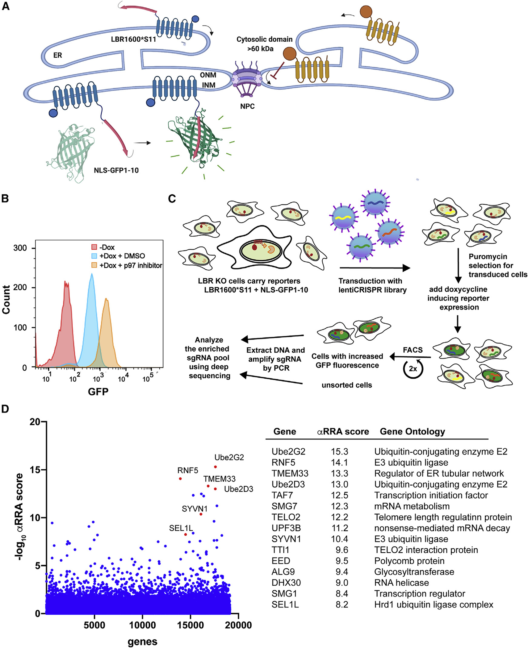
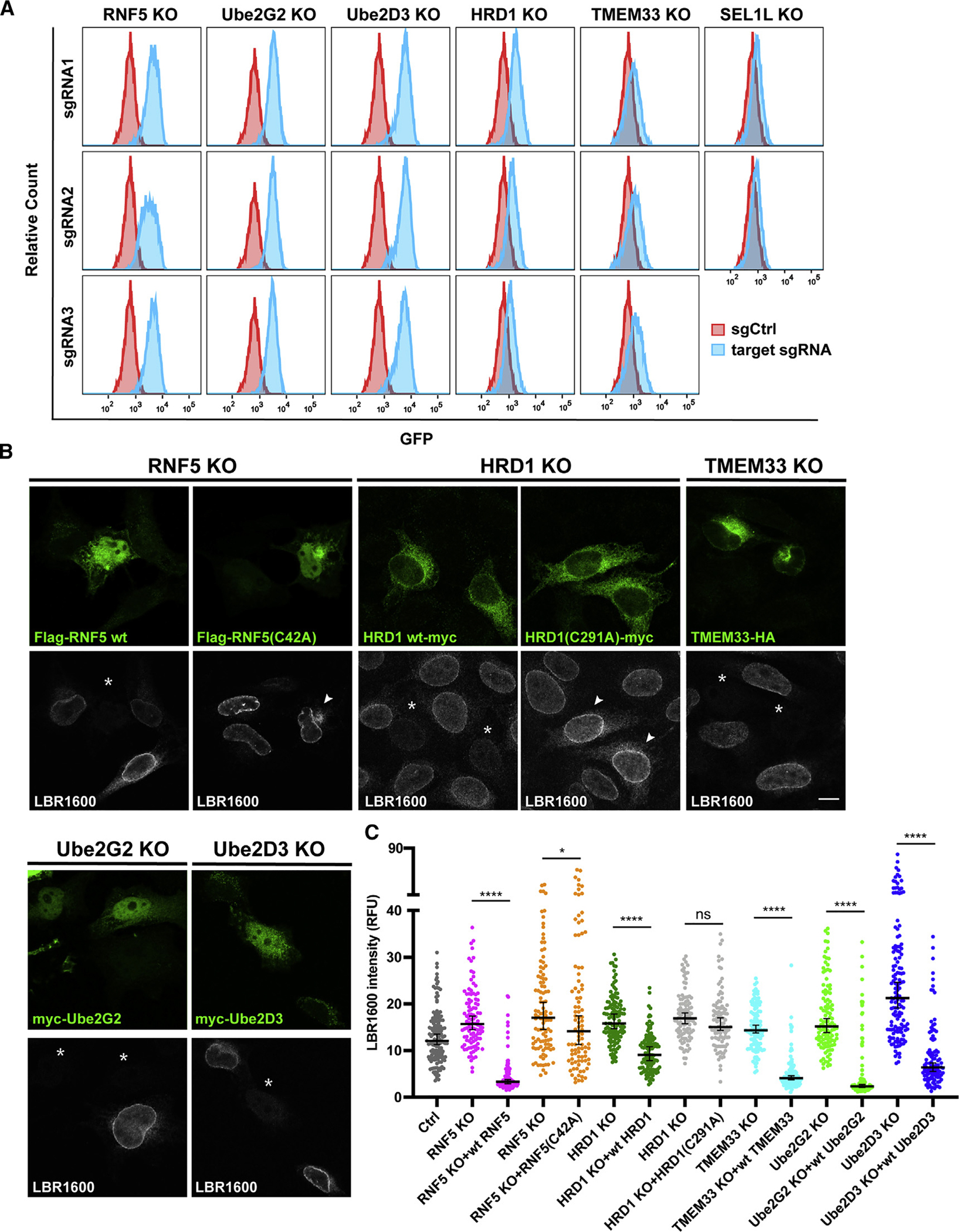
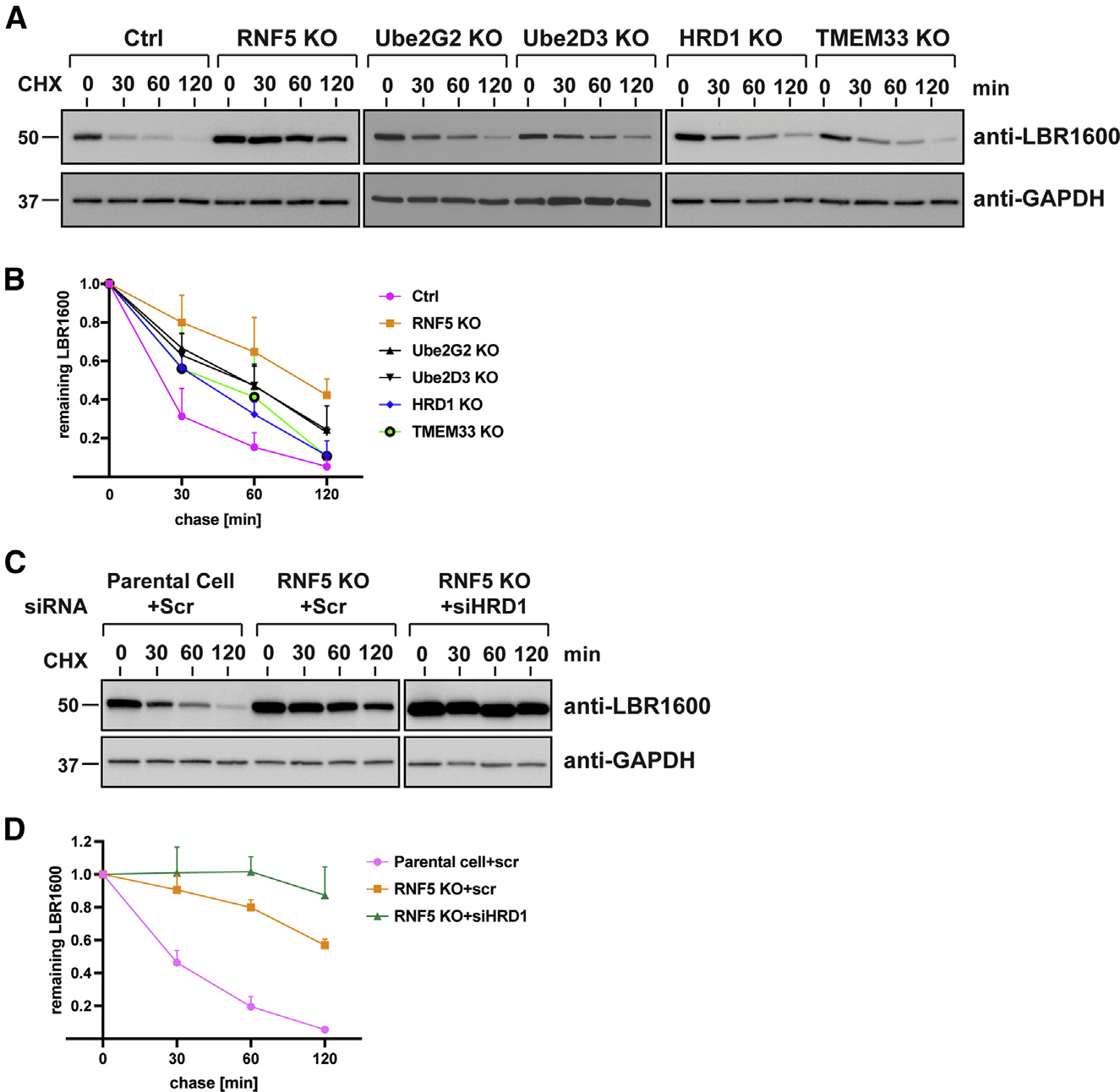
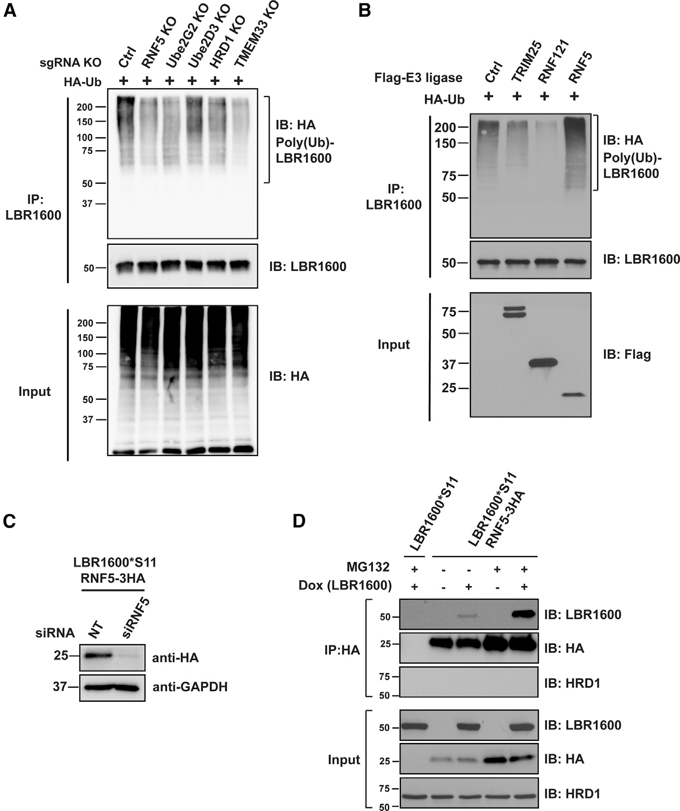
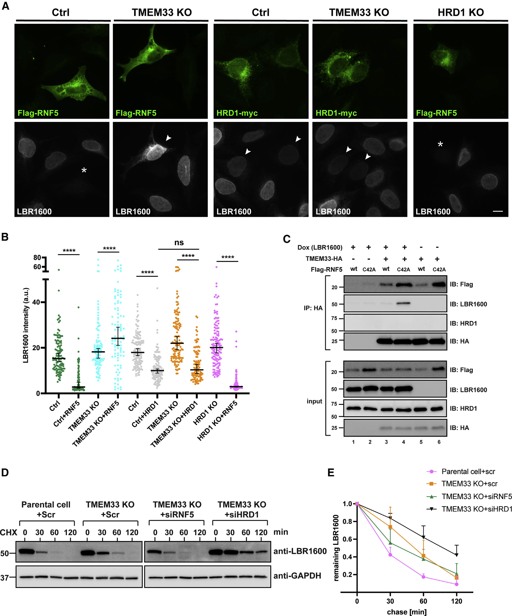
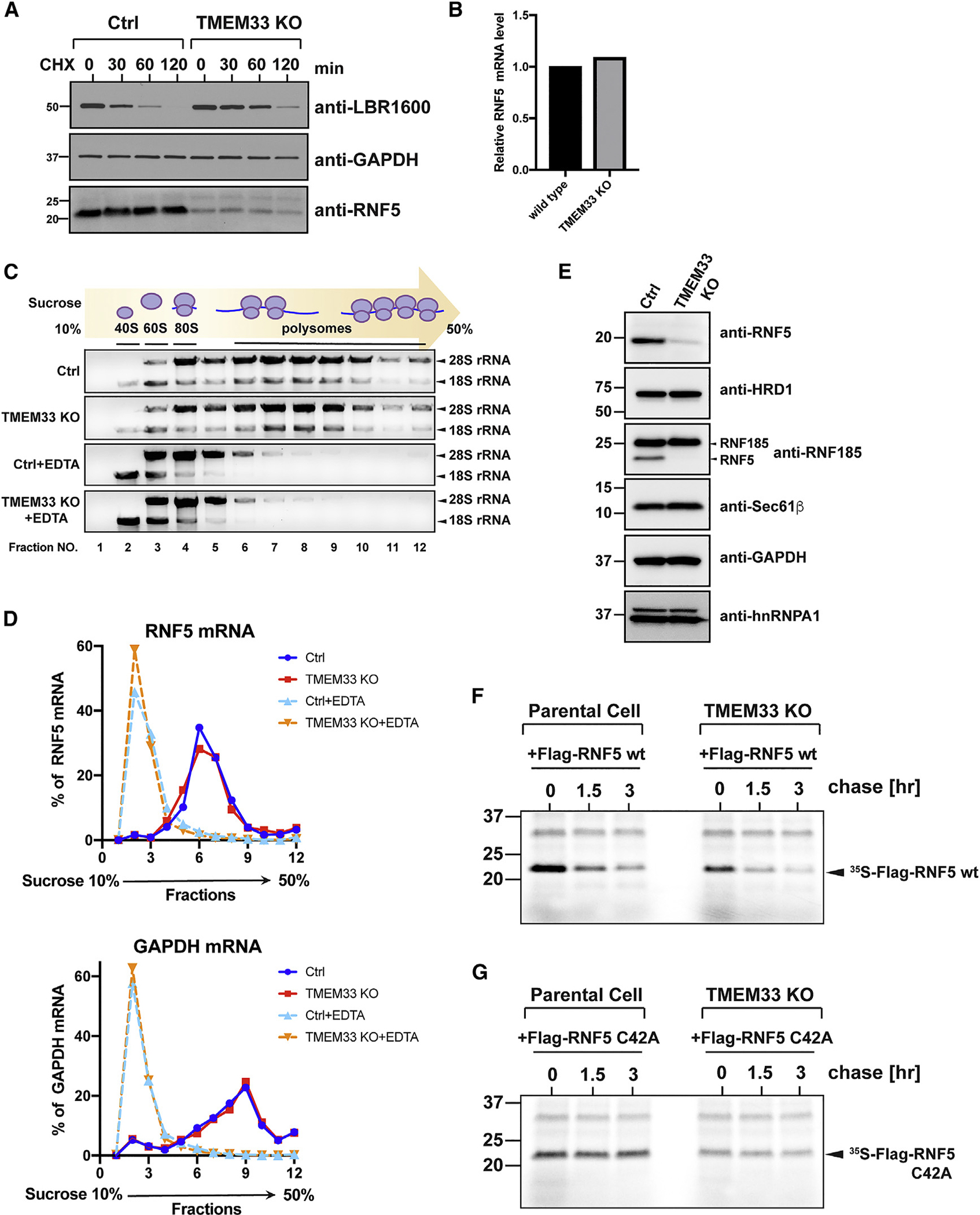
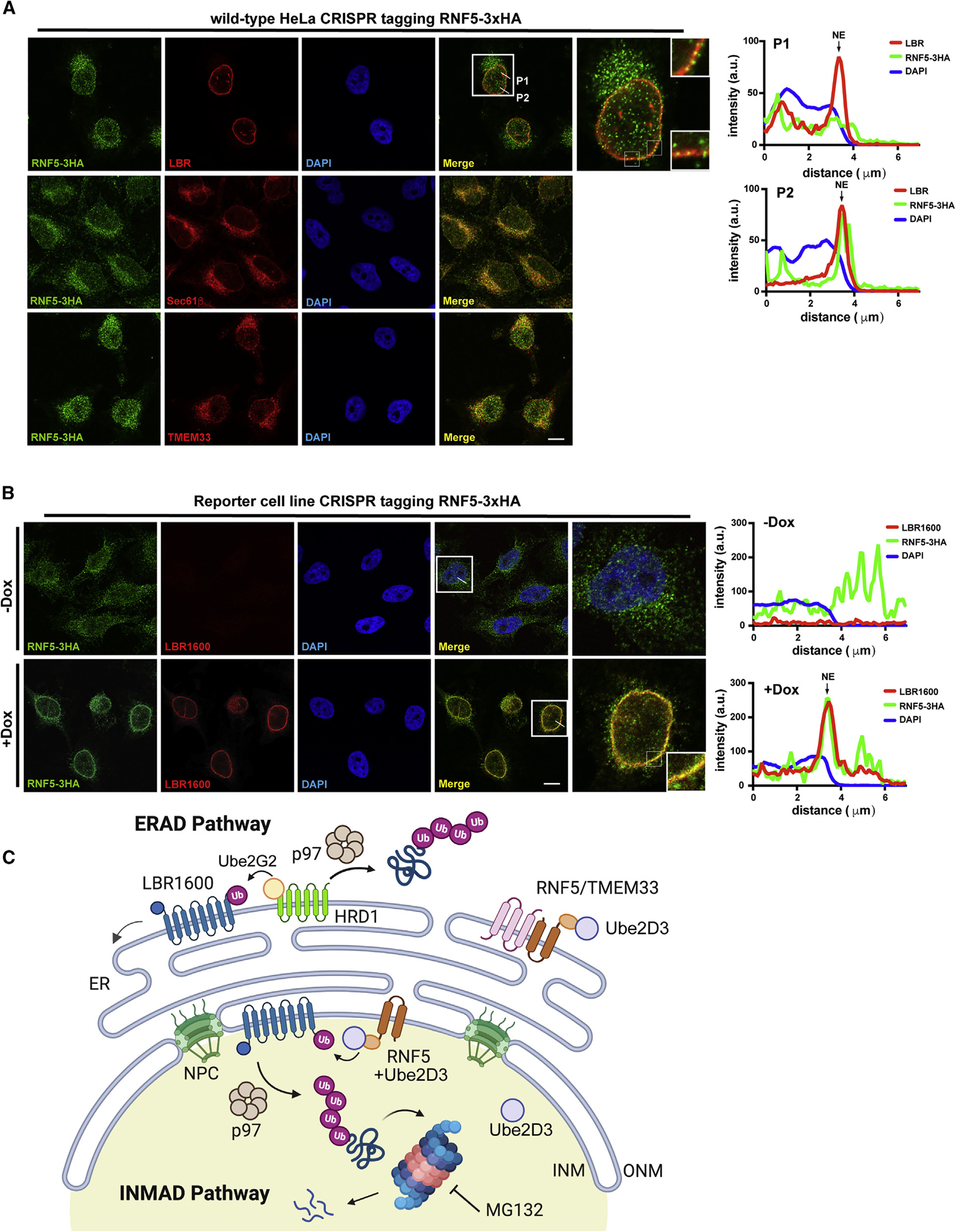
Many human diseases are caused by mutations in nuclear envelope (NE) proteins. How protein homeostasis and disease etiology are interconnected at the NE is poorly understood. Specifically, the identity of local ubiquitin ligases that facilitate ubiquitin-proteasome-dependent NE protein turnover is presently unknown. Here, we employ a short-lived, Lamin B receptor disease variant as a model substrate in a genetic screen to uncover key elements of NE protein turnover. We identify the ubiquitin-conjugating enzymes (E2s) Ube2G2 and Ube2D3, the membrane-resident ubiquitin ligases (E3s) RNF5 and HRD1, and the poorly understood protein TMEM33. RNF5, but not HRD1, requires TMEM33 both for efficient biosynthesis and function. Once synthesized, RNF5 responds dynamically to increased substrate levels at the NE by departing from the endoplasmic reticulum, where HRD1 remains confined. Thus, mammalian protein quality control machinery partitions between distinct cellular compartments to address locally changing substrate loads, establishing a robust cellular quality control system.

Keywords: CP: Molecular biology; CRISPR screen; ERAD; RNF5; TMEM33; nuclear envelopathies; proteasome; protein quality control; protein turnover; proteostasis; ubiquitin ligase.


Conflict of interest statement

Declaration of interests The authors declare no competing interests.

Figures

Figure 1.
Figure 1.. A genome-wide screen identifies genes required for INM protein turnover
(A) ER membrane proteins with large cytosolic domains cannot diffuse freely from the ER to the INM. Thus, a split-GFP system was employed to construct the LBR-based reporter for the screen. The C terminus of LBR1600* was fused to the last β strand of GFP that complements NLS-GFP1-10 lacking this C-terminal β strand, restoring GFP fluorescence in the nucleus. INM, inner nuclear membrane; ONM, outer nuclear membrane. GFP structure is adapted from PDB (2B3P). (B) The reporter cell line was treated with doxycycline for 24 h, followed by 5 μM p97 inhibitor CB-5083 or DMSO for 3 h, and subjected to flow cytometry. (C) Flowchart of the CRISPR screen. The reporter cell line was transduced with a lentiviral Brunello library, and cells with the highest GFP were enriched by two rounds of sorting at day 8 and day 13 post transduction. (D) Candidate genes identified in the CRISPR screen. Gene significance was analyzed by comparing the gene enrichment in high-GFP cells with the unsorted population using the MAGeCK algorithm. The x axis corresponds to targeted genes in alphabetical order; y axis, significant enrichment of each gene represented as −Log10 robust rank aggregation (α-RRA). Gene ontology and the α-RRA score of the top-scoring 15 genes are listed in the table. See also Figure S1.
Figure 2.
Figure 2.. Target validation of candidates involved in LBR1600*S11 turnover
(A) Three independent sgRNAs utilized in the original Brunello library were used to generate KO cells for the indicated genes. KO cells were induced to express the LBR1600*S11 and NLS-GFP1-10 overnight and analyzed using flow cytometry. Parental cells transduced with non-targeting sgRNA were used as control. (B) Representative confocal images of KO cells transfected with plasmids encoding corresponding WT proteins or ligase-inactive mutants to assess whether degradation of LBR1600*S11 can be rescued. Asterisks mark transfected cells devoid of LBR1600*S11; arrowheads indicate transfected cells having similar LBR1600*S11 levels compared with non-transfected cells. Scale bar, 10 μm. (C) Quantification of the rescuing ability determined by LBR1600*S11 fluorescence intensity. In each sample, about 100 transfected and non-transfected cells were quantified from three independent coverslips. Error bars indicate median with 95% confidence interval. p value was determined by t test. ****p < 0.0001; *p < 0.05; ns, not significant.
Figure 3.
Figure 3.. Degradation of LBR1600* depends on the E3 ligases RNF5 and HRD1, TMEM33, and the E2 ubiquitin-conjugating enzymes, Ube2G2 and Ube2D3
(A) LBR1600*S11 turnover was assessed in respective KO cells using a CHX-chase assay. Lysates from indicated time points were harvested and analyzed using immunoblotting. (B) Quantification of CHX assay in (A) via densitometry. The plot shows the mean of three independent experiments ± SD. (C) RNF5 KO cells were transfected with siRNAs for depleting HRD1 for 48 h, and CHX assays were performed as described in (A). (D) Quantification of CHX assay in (C). The plot shows the mean of three independent experiments ±SD.
Figure 4.
Figure 4.. RNF5, HRD1, TMEM33, Ube2G2, and Ube2D3 contribute to ubiquitylation of LBR1600*
(A) Reporter cell lines with the indicated KO condition were induced to express LBR1600*S11 and transfected with HA-ubiquitin, incubated for 24 h, and subjected to immunoprecipitation and immunoblotting using the indicated antibodies. (B) FLAG-tagged E3 ligases and HA-ubiquitin were co-transfected into cells expressing LBR1600*S11, followed by immunoprecipitation and immunoblotting as in (A). (C) The authentic RNF5 locus of the reporter cell line was tagged with HA, and validated by RNF5 depletion using siRNA and immunoblotting with anti-HA. (D) Endogenous RNF5 interacts with LBR1600*S11. The reporter cell line with RNF5-HA expressed from the authentic locus was treated with or without doxycycline and MG132, followed by immunoprecipitation with anti-HA antibody and immunoblotting with the indicated antibodies. See also Figure S2.
Figure 5.
Figure 5.. Epistasis analysis of RNF5, TMEM33, and HRD1
(A) FLAG-RNF5 or HRD1-myc were transiently expressed in WT, TMEM33 KO, or HRD1 KO cells expressing LBR1600*S11, followed by immunofluorescence microscopy using the indicated antibodies. Asterisks denote transfected cells devoid of LBR1600*S11; arrowheads indicate transfected cells with similar or moderate decreases in LBR1600*S11 levels compared with non-transfected cells. Scale bar, 10 μm. (B) Quantification of RNF5 effect on LBR1600*S11 degradation in TMEM33 KO and HRD1 KO cells. Images were quantified as in Figure 2; about 100 cells with moderate expression of FLAG-RNF5 were quantified in each sample. n = 100, N = 2. The plot shows median with 95% confidence interval. Data were analyzed using a t test. ****p <0.0001; ns, not significant. (C) Cells expressing LBR1600*S11 were co-transfected with plasmids encoding TMEM33-HA and FLAG-RNF5 WT or FLAG-RNF5 C42A, lysed and subjected to immunoprecipitation with anti-HA beads, followed by immunoblotting using the indicated antibodies. (D) Cycloheximide chase experiments were performed for the control or TMEM33 KO cells with the indicated siRNAs depleting RNF5 or HRD1. (E) Quantification of the CHX assay in (D). The plot shows the mean of three independent experiments ±SD.
Figure 6.
Figure 6.. TMEM33 is required for RNF5 biosynthesis
(A) CHX-chase assay was performed to monitor the stability of the indicated proteins in reporter and TMEM33 KO cells. (B) Relative mRNA levels of RNF5 were determined in control and TMEM33 KO cells using qPCR. (C) Homogenates obtained from reporter and TMEM33 KO cells were treated with or without 30 mM EDTA and subjected to centrifugation over a 10%–50% sucrose gradient. After ultracentrifugation, each gradient was fractionated, and RNAs were extracted and subjected to agarose gel electrophoresis. (D) RNA isolates from (C) were subjected to RT-qPCR. The distribution of mRNA along the gradient is represented as percentage of total mRNA. (E) Detergent extracts from reporter and TMEM33 KO cells were resolved by SDS-PAGE, followed by immunoblotting with indicated antibodies. Note that the anti-RNF185 antibody cross-reacts with RNF5 due to sequence similarity between those E3s. (F) The reporter cells and TMEM33 KO cells were transfected with FLAG-RNF5 WT, metabolically labeled with 35S-Cys/Met for 5 min, and chased in the presence of excess unlabeled Cys/Met. FLAG-RNF5 was retrieved from lysates harvested at the indicated time points via immunoprecipitation, resolved by SDS-PAGE, and visualized by autoradiography. (G) The reporter cells and TMEM33 KO cells transfected with FLAG-RNF5 C42A were subjected to pulse-chase analysis as described in (F).
Figure 7.
Figure 7.. RNF5 relocalizes to the NE upon LBR1600*S11 induction
(A) RNF5 of WT HeLa cells was tagged with 3xHA at the endogenous locus and co-stained with antibodies against HA and LBR (as INM marker) or Sec61β or TMEM33 (as ER markers). The white box marks the representative cell enlarged in the last panel. White lines mark the position of the line-scan profile of the fluorescence intensity of RNF5-3xHA (green), LBR (red), and DNA (blue) across the NE. Scale bar, 10 μm. (B) Reporter cell line with endogenously HA-tagged RNF5 was incubated with or without doxycycline for 24 h before immunofluorescence. Cells were co-stained with antibodies against HA and LBR1600*S11. White boxes denoted the enlarged area in the last panel and the white lines correspond to the line-scan profile as described in (A). Scale bar, 10 μm. Note that the anti-LBR antibody can recognize both WT LBR and LBR1600*S11; however, since this cell line is an LBR KO genetic background, LBR signal is not observed in the absence of doxycycline. (C) Proposed model of INM protein turnover. LBR1600* is synthesized at the ER and traffics along the ER membrane to the outer nuclear membrane (ONM) and inner nuclear membrane (INM), where it associates with the nuclear lamina and chromatin. A subset of misfolded LBR1600* is recognized by HRD1/Ube2G2 or RNF5-mediated ERAD and retrotranslocated into cytosol for degradation, whereas LBR1600* escaping ERAD is ubiquitylated by INM-resident RNF5/Ube2D3 and degraded in the nucleus through the action of p97 and the proteasome. See also Figures S3–S5 and S7.

References

    1. Bahmanyar S, and Schlieker C (2020). Lipid and protein dynamics that shape nuclear envelope identity. Mol. Biol. Cell 31, 1315–1323. 10.1091/mbc.E18-10-0636. - DOI - PMC - PubMed
    1. Pawar S, and Kutay U (2021). The diverse cellular functions of inner nuclear membrane proteins. Cold Spring Harb. Perspect. Biol. 13, a040477. 10.1101/cshperspect.a040477. - DOI - PMC - PubMed
    1. Gerace L, and Tapia O (2018). Messages from the voices within: regulation of signaling by proteins of the nuclear lamina. Curr. Opin. Cell Biol. 52, 14–21. 10.1016/j.ceb.2017.12.009. - DOI - PMC - PubMed
    1. Shin JY, and Worman HJ (2022). Molecular pathology of laminopathies. Annu. Rev. Pathol. 17, 159–180. 10.1146/annurevpathol-042220-034240. - DOI - PMC - PubMed
    1. Wu X, and Rapoport TA (2018). Mechanistic insights into ER-associated protein degradation. Curr. Opin. Cell Biol. 53, 22–28. 10.1016/j.ceb.2018.04.004. - DOI - PMC - PubMed

Publication types