. 2023 Jun 13;119(6):1403-1415.
doi: 10.1093/cvr/cvac174.

Disrupting circadian control of peripheral myogenic reactivity mitigates cardiac injury following myocardial infarction

Affiliations

Disrupting circadian control of peripheral myogenic reactivity mitigates cardiac injury following myocardial infarction

Jeffrey T Kroetsch et al. Cardiovasc Res. .

Abstract

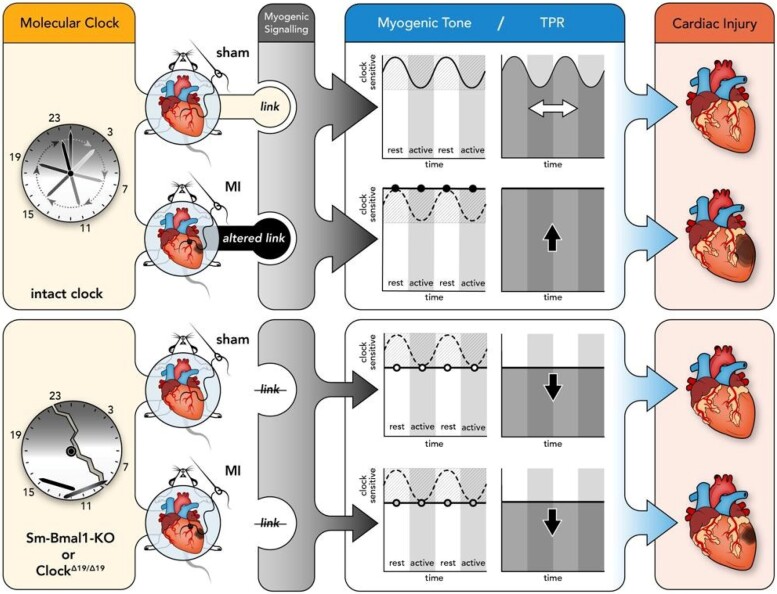
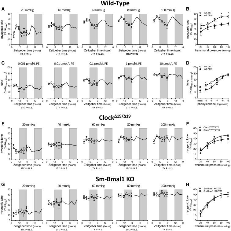
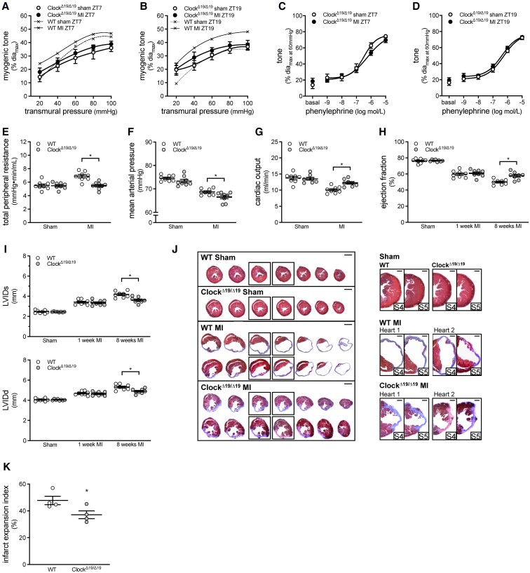
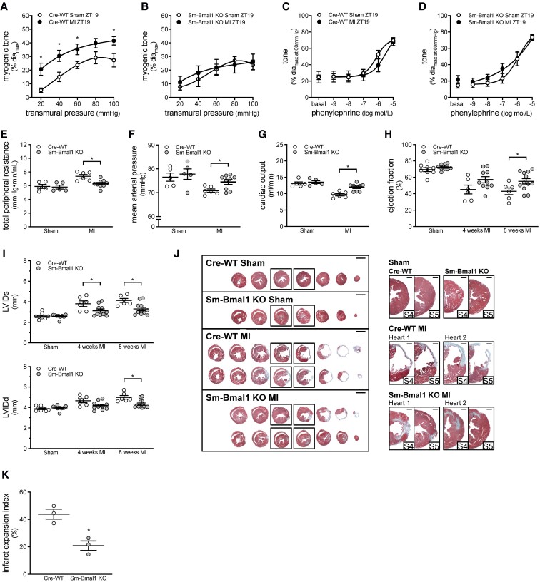
Aims: Circadian rhythms orchestrate important functions in the cardiovascular system: the contribution of microvascular rhythms to cardiovascular disease progression/severity is unknown. This study hypothesized that (i) myogenic reactivity in skeletal muscle resistance arteries is rhythmic and (ii) disrupting this rhythmicity would alter cardiac injury post-myocardial infarction (MI).

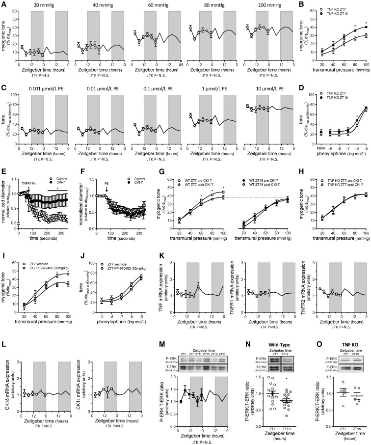
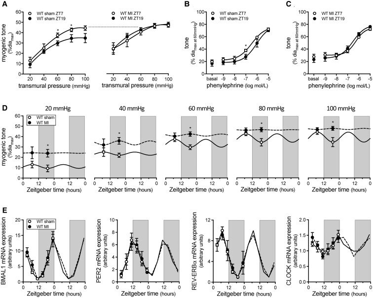
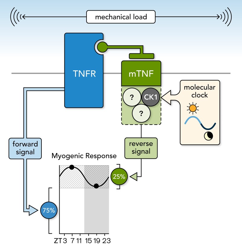
Methods and results: Cremaster skeletal muscle resistance arteries were isolated and assessed using standard pressure myography. Circadian rhythmicity was globally disrupted with the ClockΔ19/Δ19 mutation or discretely through smooth muscle cell-specific Bmal1 deletion (Sm-Bmal1 KO). Cardiac structure and function were determined by echocardiographic, hemodynamic and histological assessments. Myogenic reactivity in cremaster muscle resistance arteries is rhythmic. This rhythm is putatively mediated by the circadian modulation of a mechanosensitive signalosome incorporating tumour necrosis factor and casein kinase 1. Following left anterior descending coronary artery ligation, myogenic responsiveness is locked at the circadian maximum, although circadian molecular clock gene expression cycles normally. Disrupting the molecular clock abolishes myogenic rhythmicity: myogenic tone is suspended at the circadian minimum and is no longer augmented by MI. The reduced myogenic tone in ClockΔ19/Δ19 mice and Sm-Bmal1 KO mice associates with reduced total peripheral resistance (TPR), improved cardiac function and reduced infarct expansion post-MI.

Conclusions: Augmented microvascular constriction aggravates cardiac injury post-MI. Following MI, skeletal muscle resistance artery myogenic reactivity increases specifically within the rest phase, when TPR would normally decline. Disrupting the circadian clock interrupts the MI-induced augmentation in myogenic reactivity: therapeutics targeting the molecular clock, therefore, may be useful for improving MI outcomes.

Keywords: Casein kinase 1; Systemic hemodynamics; Total peripheral resistance; Tumour necrosis factor; Vascular smooth muscle cells.


Conflict of interest statement

Conflict of interest: J.T.K. and D.L. have consulted for Qanatpharma AG (Stans, Switzerland) and Aphaia Pharma AG (Zug, Switzerland) within the last 36 months. J.H. is an employee of Qanatpharma AG. S.S.B. is a founder and executive board member of Qanatpharma AG and Aphaia Pharma AG. Aphaia Pharma AG had no financial or intellectual involvement in this article. Qanatpharma AG played no role in study design, data collection/analysis, decision to publish, or the preparation of the publication. Aside from J.T.K., D.L., J.H., and SSB, none of the other authors have relationships to industry.

Figures

Graphical Abstract
Graphical Abstract
Figure 1
Figure 1
Myogenic responsiveness displays circadian rhythmicity. (A) Myogenic tone in wild-type (WT) cremaster arteries plotted over Zeitgeber time (n = 4–6) and (B) at ZT7 and ZT19 (n = 4–6). (C) Phenylephrine (PE)-stimulated vasoconstriction in WT cremaster arteries plotted over Zeitgeber time (n = 4–6) and (D) at ZT7 and ZT19 (n = 4–6). (E) Myogenic tone in ClockΔ19/Δ19 mutant cremaster arteries plotted over Zeitgeber time (n = 5–7) and (F) at ZT7 and ZT19 (n = 5–6). (G) Myogenic tone in Sm-Bmal1 KO cremaster arteries plotted over Zeitgeber time (n = 5–6) and (H) at ZT7 and ZT19 (n = 5–6). Data in Panels A, C, E, and G are double-plotted for visualization purposes and are statistically analysed for a circadian rhythm with JTK_Cycle. Panels B, D, F, and H are statistically analysed with t-tests or Mann–Whitney tests (unpaired single comparisons), with * denoting P < 0.05 at the respective transmural pressure or phenylephrine concentration.
Figure 2
Figure 2
Rhythmic myogenic reactivity depends on TNF reverse signalling. (A) Myogenic tone in tumour necrosis factor knockout (TNF KO) cremaster arteries plotted over Zeitgeber time (n = 5–6) and (B) at ZT7 and ZT19 (n = 5–6). (C) Phenylephrine (PE)-stimulated vasoconstriction in TNF KO cremaster arteries plotted over Zeitgeber time (n = 5–6) and (D) at ZT7 and ZT19 (n = 5–6). (E–G) Effect of in vitro CK1 inhibition (10 nmol/L CKI-7, 30 min) on (E) sTNFR1-Fc stimulated vasoconstriction (n = 4–5), (F) phenylephrine-stimulated vasoconstriction (n = 5) and (G) myogenic tone at ZT7 (n = 6) and ZT19 (n = 5) in wild-type cremaster arteries. (H) Effect of in vitro CK1 inhibition on myogenic tone in TNF KO cremaster arteries at ZT7 (n = 5). (I–J) Effect of in vivo CK1 inhibition (30 mg/kg PF670462 i.p.; 24 h) on cremaster artery (I) myogenic tone and (J) phenylephrine-stimulated vasoconstriction (both n = 5; both at ZT7). (K) TNF, TNFR1, and TNFR2 mRNA expression (normalized to HMBS mRNA expression) in cremaster arteries plotted over Zeitgeber time (n = 5–7 for each point). (L) CK1 delta and CK1 epsilon mRNA expression (normalized to HMBS mRNA expression) in cremaster arteries plotted over Zeitgeber time (n = 2 for each point). (M) pressure-stimulated (40–100 mmHg) ERK1/2 phosphorylation levels in wild-type cremaster arteries plotted over Zeitgeber time (n = 5). (N and O) ERK1/2 phosphorylation levels at ZT7 and ZT19 in (N) wild-type (n = 15) and (O) TNF KO cremaster arteries (n = 5). Data in Panels A, C, K, L, and M are double-plotted for visualization purposes and are statistically analysed for a circadian rhythm with JTK_Cycle. Panels B, D, E, F, I, and J are statistically analysed as unpaired single comparisons using t-tests or Mann–Whitney tests, with * denoting P < 0.05. Panels G, H, N, and O are statistically analysed as paired single comparisons using paired t-tests or Wilcoxon tests, with * denoting P < 0.05.
Figure 3
Figure 3
Myogenic rhythmicity is altered following myocardial infarction. (A) Myogenic tone in wild-type (WT) cremaster arteries isolated at ZT7 and ZT19 from sham-operated mice (n = 6–9) and mice with a myocardial infarction (MI; n = 7). Phenylephrine-stimulated vasoconstriction in cremaster arteries isolated at ZT7 and ZT19 from (B) sham-operated mice (n = 6–9) and (C) MI mice (n = 7). (D) Myogenic tone in sham-operated and MI mice plotted over Zeitgeber time (n = 6–9). (E) Bmal1, Per2, Rev-Erbα and Clock mRNA expression (normalized to HMBS mRNA expression) in cremaster arteries from sham and MI mice plotted over Zeitgeber time (n = 3–4). Panels A-D are statistically analysed as unpaired single comparisons using t-tests or Mann–Whitney tests, with * denoting P < 0.05. Data in Panels D and E are double-plotted for visualization purposes; data in Panel E are statistically analysed for a circadian rhythm with JTK_Cycle.
Figure 4
Figure 4
Global ClockΔ19/Δ19 mutation attenuates myogenic responsiveness and improves cardiac function following myocardial infarction. Myogenic tone at (A) ZT7 (n = 7–8) and (B) ZT19 (n = 7–8) in ClockΔ19/Δ19 cremaster arteries isolated from sham-operated mice and mice with a myocardial infarction (MI). Traces from sham and MI wild-type (WT) mice are reproduced from Figure 3A for qualitative comparison purposes. Phenylephrine-stimulated vasoconstriction at (C) ZT7 (n = 7–8) and (D) ZT19 (n = 7–8) in ClockΔ19/Δ19 cremaster arteries isolated from sham-operated and MI mice. Pressure–volume hemodynamic assessments of (E) total peripheral resistance, (F) mean arterial pressure, and (G) cardiac output in WT and ClockΔ19/Δ19 mutant mice with sham or MI procedure (n = 8). Echocardiographic assessments of (H) ejection fraction and (I) systolic and diastolic left ventricular internal diameter (LVIDs and LVIDd) in WT and ClockΔ19/Δ19 mutant mice with sham or MI procedure (n = 8). (J) Cardiac histology was processed at 8 weeks post-MI. Shown are representative histological images of cardiac infarcts in WT and ClockΔ19/Δ19 mutant mice with sham or MI procedure; serial sections are shown on the left (bar = 2 mm) and close-ups of the infarct region in serial sections 4–5 are shown on the right (bar = 500 µm). (K) Quantification of infarct expansion from the histological images (n = 4). All data are statistically analysed as unpaired single comparisons using t-tests or Mann–Whitney tests, with * denoting P < 0.05.
Figure 5
Figure 5
Smooth muscle Bmal1 deletion attenuates myogenic responsiveness and improves cardiac function following myocardial infarction. Myogenic tone at ZT19 in (A) Cre-WT (n = 7) and (B) Sm-Bmal1 KO (n = 7) cremaster arteries isolated from sham-operated mice and mice with a myocardial infarction (MI). Phenylephrine-stimulated vasoconstriction at ZT19 in (C) Cre-WT (n = 7) and (D) Sm-Bmal1 KO (n = 7) cremaster arteries isolated from sham-operated and MI mice. Pressure–volume hemodynamic assessments of (E) total peripheral resistance, (F) mean arterial pressure and (G) cardiac output in Cre-WT and Sm-Bmal1 KO mice with sham or MI procedure (n = 5–9). Echocardiographic assessments of (H) ejection fraction and (I) systolic and diastolic left ventricular internal diameter (LVIDs and LVIDd) in Cre-WT and Sm-Bmal1 KO mice with sham or MI procedure (n = 6–12). (J) Cardiac histology was processed at 8 weeks post-MI. Shown are representative histological images of cardiac infarcts in Cre-WT and Sm-Bmal1 KO mice with sham or MI procedure; serial sections are shown on the left (bar = 2 mm) and close-ups of the infarct region in serial sections 4–5 are shown on the right (bar = 500 µm). (K) Quantification of infarct expansion from the histological images (n = 3). All data are statistically analysed as unpaired single comparisons using t-tests or Mann–Whitney tests, with * denoting P < 0.05.
Figure 6
Figure 6
Proposed mechanotransduction mechanism mediating myogenic reactivity in skeletal muscle resistance arteries. Shown is the proposed molecular mechanism regulating myogenic responsiveness in skeletal muscle resistance arteries. Membrane-bound tumour necrosis factor (mTNF) and a tumour necrosis factor receptor (TNFR) form a mechanosensitive pair. Upon mechanical stimulation, both mTNF and TNFR generate signals; the signal generated by TNFR regulates a larger proportion of myogenic tone than mTNF (∼75% vs. 25%) and is not rhythmic. The mTNF signalosome incorporates CK1 as a pivotal element for the propagation of the mTNF-dependent reverse signal; the mTNF signalosome also incorporates a yet undefined element that is under circadian control. Accordingly, the mTNF reverse signal and the ∼25% of the myogenic response that it controls display circadian rhythmicity.

References

    1. Drexler H. Changes in the peripheral circulation in heart failure. Curr Opin Cardiol 1995;10:268–273. - PubMed
    1. Freis ED, Schnaper HW, Johnson RL, Schreiner GE. Hemodynamic alterations in acute myocardial infarction. I. Cardiac output, mean arterial pressure, total peripheral resistance, central and total blood volumes, venous pressure and average circulation time. J Clin Invest 1952;31:131–140. - PMC - PubMed
    1. Pfeffer MA, Braunwald E. Ventricular remodeling after myocardial infarction. Experimental observations and clinical implications. Circulation 1990;81:1161–1172. - PubMed
    1. Eaton LW, Weiss JL, Bulkley BH, Garrison JB, Weisfeldt ML. Regional cardiac dilatation after acute myocardial infarction: recognition by two-dimensional echocardiography. N Engl J Med 1979;300:57–62. - PubMed
    1. Kroetsch JT, Levy AS, Zhang H, Aschar-Sobbi R, Lidington D, Offermanns S, Nedospasov SA, Backx PH, Heximer SP, Bolz SS. Constitutive smooth muscle tumour necrosis factor regulates microvascular myogenic responsiveness and systemic blood pressure. Nat Commun 2017;8:14805. - PMC - PubMed

Publication types

Substances