. 2022 Dec 8;126(48):10185-10193.
doi: 10.1021/acs.jpcb.2c06557. Epub 2022 Nov 23.

Resonant Inelastic Soft X-ray Scattering and X-ray Emission Spectroscopy of Solid Proline and Proline Solutions

Affiliations

Resonant Inelastic Soft X-ray Scattering and X-ray Emission Spectroscopy of Solid Proline and Proline Solutions

Frank Meyer et al. J Phys Chem B. .

Abstract

The amino group of proline is part of a pyrrolidine ring, which makes it unique among the proteinogenic amino acids. To unravel its full electronic structure, proline in solid state and aqueous solution is investigated using X-ray emission spectroscopy and resonant inelastic soft X-ray scattering. By controlling the pH value of the solution, proline is studied in its cationic, zwitterionic, and anionic configurations. The spectra are analyzed within a "building-block principle" by comparing with suitable reference molecules, i.e., acetic acid, cysteine, and pyrrolidine, as well as with spectral calculations based on density functional theory. We find that the electronic structure of the carboxyl group of proline is very similar to that of other amino acids as well as acetic acid. In contrast, the electronic structure of the amino group is significantly different and strongly influenced by the ring structure of proline.


Conflict of interest statement

The authors declare no competing financial interest.

Figures

Figure 1
Figure 1
(a) Chemical structure of proline and the used reference systems (from left to right): cysteine, pyrrolidine, and acetic acid. Proline and cysteine are shown in the zwitterionic state and pyrrolidine in the cationic state. (b) All possible charge configurations of proline, together with the characteristic pKa values for the respective transitions; the isoelectric point of proline is 6.30.
Figure 2
Figure 2
C (a), N (b), and O (c) K-edge RIXS maps of an evaporated proline film. The X-ray emission intensity is color-coded (see the scale bar above the maps) and plotted as a function of emission (abscissa) and excitation (ordinate) energies. Blue represents zero and white indicates the strongest intensity.
Figure 3
Figure 3
O K RIXS maps of proline in aqueous solutions at three different pH values (0.8, 6.8, and 13.0), representing the cationic, zwitterionic, and anionic configurations. The absorption onset of water is visible in the top part of the maps. The X-ray emission intensity is color-coded, as given by the scale bar above the maps.
Figure 4
Figure 4
O K RIXS spectra of proline (black curves) in aqueous solution at various pH values (0.8, 6.8, and 13.0), representing its cationic, zwitterionic, and anionic configurations. For comparison, the spectra of solid proline (black, top) and acetic acid (red curves) at pH 0.2 and 12.8 (taken from ref (17)) are also shown. The acetic acid spectra were aligned with the proline spectra (shifted by +0.3 eV in emission energy). The low pH spectra represent a neutral carboxyl group, while the neutral and high-pH solutions correspond to a deprotonated carboxyl group (see Figure 1b). The spectra of proline were extracted from the RIXS maps in Figure 3.
Figure 5
Figure 5
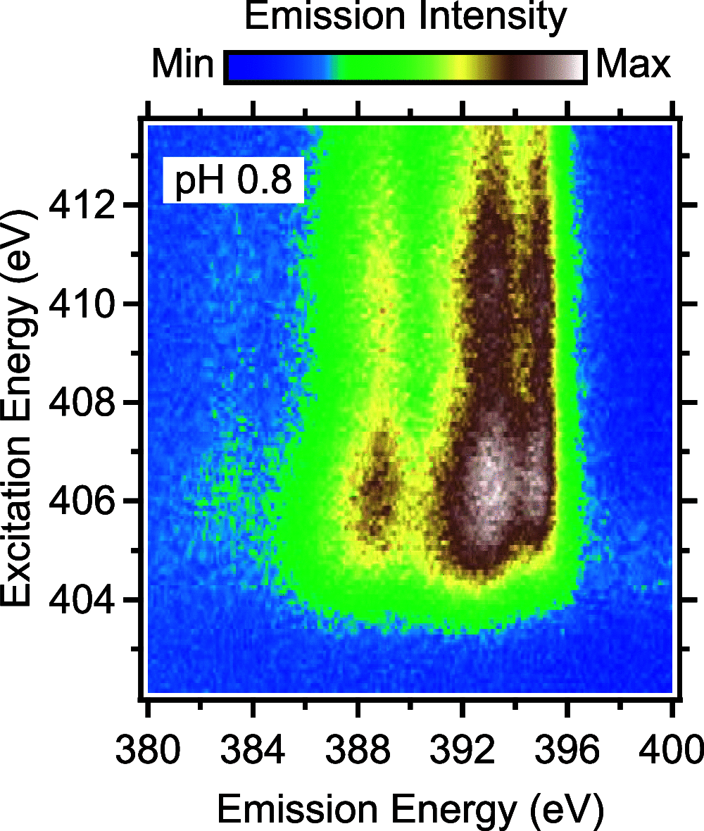
N K RIXS map of proline in aqueous solution at pH 0.8 representing the cationic configuration. The X-ray emission intensity is color-coded, as given by the scale bar above the map.
Figure 6
Figure 6
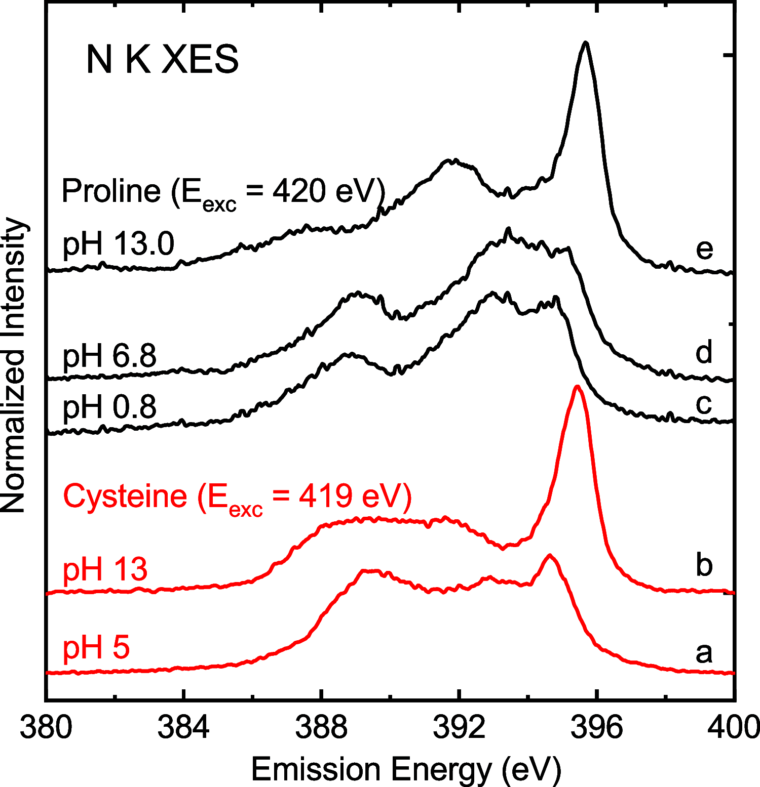
Nonresonant (Eexc = 419 eV) N K XES spectra of proline in aqueous solution at various pH values (0.8, 6.8, and 13.0) representing the cationic (c), zwitterionic (d), and anionic (e) configurations. For comparison, nonresonant (Eexc = 420 eV) N K XES spectra of cysteine in zwitterionic (a) and anionic (b) configurations are shown (first published in ref (17)).
Figure 7
Figure 7
N K-edge RIXS maps of pyrrolidine in aqueous solution at pH 1.4 and 13.6, representing the cationic and neutral configurations, respectively. The X-ray emission intensity is color-coded, as shown in the scale bar above the maps.
Figure 8
Figure 8
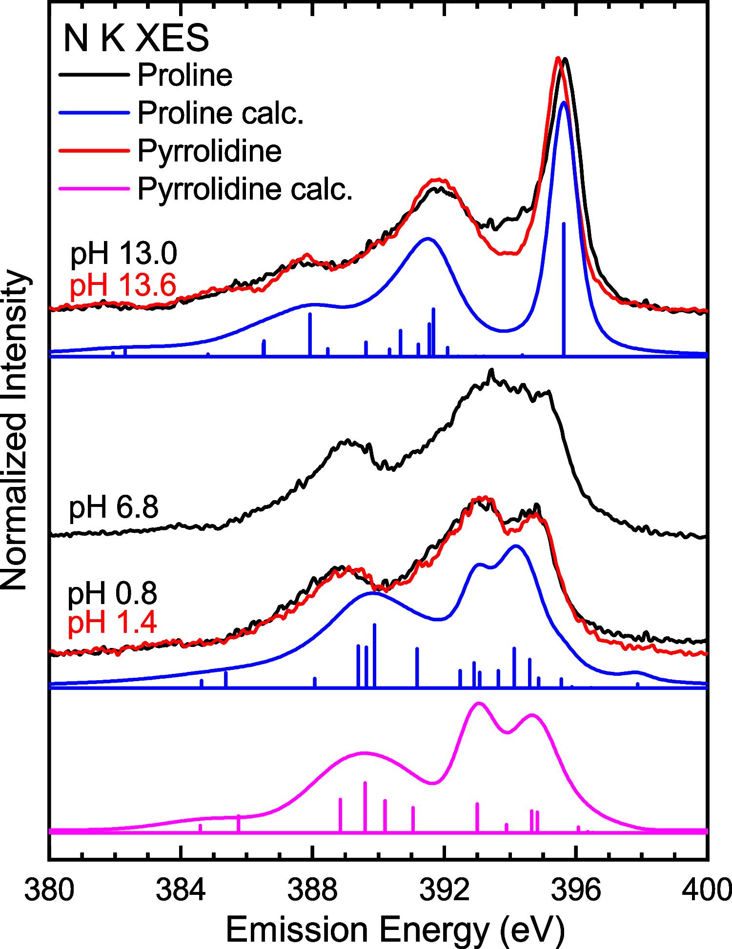
Nonresonant N K-edge XES spectra of proline (black curves, Eexc = 419 eV) and pyrrolidine (red curves, Eexc = 424 eV) in aqueous solution at pH values corresponding to the cationic (pH 0.8 and 1.4, resp.), zwitterionic (pH 6.8), and anionic (pH 13.0 and 13.6, resp.) configurations. The spectra of pyrrolidine were extracted from the RIXS maps in Figure 7. The experimental data are complemented by the calculated X-ray emission energy positions and intensities of isolated cationic and neutral proline (blue bars). The calculation for an isolated protonated pyrrolidine ion is shown in magenta. The blue and magenta curves were derived by applying a Gaussian broadening for which the FWHM was increased at lower emission energies.

References

    1. Wagner I.; Musso H. New Naturally Occurring Amino Acids. Angew. Chem., Int. Ed. 1983, 22, 816–828. 10.1002/anie.198308161. - DOI
    1. Zubavichus Y.; Fuchs O.; Weinhardt L.; Heske C.; Umbach E.; Denlinger J. D.; Grunze M. Soft X-Ray-Induced Decomposition of Amino Acids: An XPS, Mass Spectrometry, and NEXAFS Study. Radiat. Res. 2004, 161, 346–358. 10.1667/RR3114.1. - DOI - PubMed
    1. Cavalleri O.; Gonella G.; Terreni S.; Vignolo M.; Floreano L.; Morgante A.; Canepa M.; Rolandi R. High Resolution X-ray Photoelectron Spectroscopy of L-Cysteine Self-Assembled Films. Phys. Chem. Chem. Phys. 2004, 6, 4042–4046. 10.1039/b405516k. - DOI
    1. Plekan O.; Feyer V.; Richter R.; Coreno M.; de Simone M.; Prince K. C.; Carravetta V. Investigation of the Amino Acids Glycine, Proline, and Methionine by Photoemission Spectroscopy. J. Phys. Chem. A 2007, 111, 10998–11005. 10.1021/jp075384v. - DOI - PubMed
    1. Kamada M.; Sugiyama H.; Takahashi K.; Azuma J.; Kitajima S.; Ogawa K.; Sumimoto M.; Hori K.; Fujimoto H. Photoelectron Spectroscopic Study of Electronic Structures of L-Cysteine. J. Phys. Soc. Jpn. 2010, 79, 03470910.1143/JPSJ.79.034709. - DOI

Publication types