. 2022 Nov 22;13(1):7166.
doi: 10.1038/s41467-022-34878-2.

Functionally integrating nanoparticles alleviate deep vein thrombosis in pregnancy and rescue intrauterine growth restriction

Affiliations

Functionally integrating nanoparticles alleviate deep vein thrombosis in pregnancy and rescue intrauterine growth restriction

Juan Cheng et al. Nat Commun. .

Abstract

There is still unmet demand for effective, safe, and patient-friendly anti-thrombotics to treat deep vein thrombosis (DVT) during pregnancy. Here we first engineer a bioactive amphiphile (TLH) by simultaneously conjugating Tempol and linoleic acid onto low molecular weight heparin (LMWH), which can assemble into multifunctional nanoparticles (TLH NP). In pregnant rats with DVT, TLH NP can target and dissolve thrombi, recanalize vessel occlusion, and eradicate the recurrence of thromboembolism, thereby reversing DVT-mediated intrauterine growth restriction and delayed development of fetuses. Mechanistically, therapeutic effects of TLH NP are realized by inhibiting platelet aggregation, facilitating thrombolysis, reducing local inflammation, attenuating oxidative stress, promoting endothelial repair, and increasing bioavailability. By decorating with a fibrin-binding peptide, targeting efficiency and therapeutic benefits of TLH NP are considerably improved. Importantly, LMWH nanotherapies show no toxicities to the mother and fetus at the dose 10-time higher than the examined therapeutic dosage.


Conflict of interest statement

H.B.Q., J.C., S.Q.Z., and X.Y.J. are inventors in a pending patent filed by the China National Intellectual Property Administration (No. 202210798192.X, July 06 2022) related to TLH NP and CTLH NP, but the rights belong to the Women and Children’s Hospital of Chongqing Medical University. All other authors declare that they have no competing interests.

Figures

Fig. 1
Fig. 1. Schematic illustration of engineering of a multifunctional anti-thrombotic nanotherapy for targeted treatment of deep vein thrombosis (DVT) during pregnancy.
a Development of a nanotherapy with multiple bioactivities by self-assembly of a multifunctional low molecular weight heparin (LMWH) derivative (i.e., CTLH). Covalent conjugation of LMWH with bioactive moieties of linoleic acid (LA) and Tempol affords an amphiphile with multiple pharmacological activities, which is further functionalized with a thrombus-targeting peptide CREKA to obtain CTLH that can assemble into a multifunctional nanotherapy CTLH NP. b A sketch showing targeted treatment of DVT in pregnant rats by CTLH NP.
Fig. 2
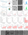
Fig. 2. Design, construction, and characterization of multifunctional anti-thrombotic nanotherapies based on LMWH-derived materials.
a, b Schematic illustration of nanotherapies self-assembled by linoleic acid (LA)-conjugated LMWH (LH) (a) or Tempol/LA-conjugated LMWH (TLH) (b). ch Typical TEM images (c, d), SEM images (e, f), and size distribution profiles (g, h) of LH NP (c, e, g) and TLH NP (d, f, h). i Changes in the mean diameter of LH NP and TLH NP after incubation with PBS or serum for predetermined time periods. jm Typical excitation fluorescence spectra (j, l) and the corresponding plots of the intensity ratio I338/I333 as a function of Log C (k, m) for pyrene in the presence of increasing concentrations (g/L) of LH (j, k) or TLH (l, m). nq Dose-dependent elimination of superoxide anion (n), DPPH radical (o), H2O2 (p), and hypochlorite (q) by TLH NP. r The anti-FXa activity of LMWH, LH NP, and TLH NP. sw Effects of different formulations on PT (s), APTT (t), TT (u), TAT (v), and BCI (w). Of note, BCI increases with increased free hemoglobin in solutions. A higher BCI means stronger anti-thrombotic activity for the examined formulations. x SEM images showing thrombin-stimulated platelets after separate treatment with PBS, Tempol, LMWH, LH NP, or TLH NP. Platelets treated with PBS alone served as the normal control. Data in ch, x are representative of six independent samples. Scale bar, 3 μm. Data in i, nw are mean ± s.d. (n = 6 independent samples). Statistical significance was assessed by one-way ANOVA with post hoc LSD tests. *p < 0.05, **p < 0.01, ***p < 0.001. Source data are provided as a Source Data file.
Fig. 3
Fig. 3. Targeting effects of LH NP and TLH NP in pregnant rats with stenosis-induced DVT.
a A schematic diagram showing the establishment of a stenosis-induced DVT model in pregnant rats. b, c Ex vivo fluorescence images (b) and quantitative data (c) show the accumulation of Cy5-LH NP or Cy5-TLH NP in normal veins or veins with stenosis-induced thrombosis at 4 h after i.v. injection in pregnant rats at G10. df Immunofluorescence analysis of the co-localization of Cy5-LH NP or Cy5-TLH NP with vWF-positive endothelial cells (d), MPO-positive neutrophils (e), and CD61-positive platelets (f) in cryosections of left iliac veins of DVT rats. Pregnant rats (at G10) with stenosis-induced DVT were administered with Cy5-LH NP or Cy5-TLH NP via i.v. injection. At 4 h after administration, rats were euthanized and left iliac veins were isolated for analysis. Scale bars, 40 μm. g Ex vivo imaging of placentas and fetuses at 4 h after i.v. injection of Cy5-LH NP or Cy5-TLH NP in normal or DVT pregnant rats (at G15). h Microscopic observation of placental sections. Upper panel, bright field images; lower panel, fluorescence images. Scale bars, 2 mm. In all cases, pregnant rats with thrombosis were treated with the same volume of saline in the saline group. Data in b, dh are representative of six independent samples. Data in c are mean ± s.d. (n = 6 independent samples). Statistical significance was assessed by one-way ANOVA with post hoc LSD tests. ***p < 0.001. Source data are provided as a Source Data file.
Fig. 4
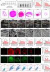
Fig. 4. Therapeutic effects of LH NP and TLH NP in pregnant rats with stenosis-induced DVT.
a Schematic illustration of the treatment regimens. b Representative digital photos of left iliac veins with thrombi isolated from pregnant rats after treatment with different formulations. Scale bar, 1 mm. c, d The normalized weight (c) and length (d) of thrombi in left iliac veins after different treatments. e H&E-stained histopathological sections of left iliac veins with thrombi. Scale bar, 400 μm. f Ultrasound images of left iliac veins. The yellow dashed lines indicate the vascular endothelium, while the red dashed lines indicate thrombi. Scale bar, 1 mm. g SEM observation of the endothelial surface of left iliac veins with thrombi and after different treatments. Scale bar, 10 μm. hj Relative mRNA levels of TNF-α, IL-6, NF-κB, PAI-1, TAFI, and vWF in left iliac veins. k, l Fluorescence images of DHE-stained cryosections (k) and quantitative analysis of relative ROS levels (l) in left iliac veins. Scale bar, 400 μm. m, n Immunofluorescence images showing MPO-positive neutrophils in cryosections (m) and quantified MPO levels (n) in left iliac veins. Scale bar, 400 μm. o, p Representative flow cytometric profiles (o) and quantified levels (p) of CD11b+/RP-1+ neutrophils in left iliac veins. In all these studies, left iliac veins of pregnant rats at G10 were ligated to induce DVT. At 6 h after the formation of stenosis-induced thrombi, diseased rats were daily administered with saline (the model group), free Tempol (8 mg/kg, i.v.), free LMWH (5 mg/kg, s.c.), LH NP (at 5 mg/kg of LMWH, i.v.), or TLH NP (at 5 mg/kg of LMWH, i.v.) for 5 days. In the normal group, healthy pregnant rats with sham operation were treated with saline. Data in b, eg, k, m, o are representative of six independent samples. Data in cdh–jlnp are mean ± s.d. (n = 6 independent samples). Statistical significance was assessed by one-way ANOVA with post hoc LSD tests. *p < 0.05, **p < 0.01, ***p < 0.001. Source data are provided as a Source Data file.
Fig. 5
Fig. 5. Inhibition of DVT-induced embryonic developmental disorders and early fetal growth delay in pregnant rats by LMWH-derived nanotherapies.
a, b Digital photos show gross morphological appearance of representative rat fetuses (a) and placentas (b) from normal or DVT pregnant rats at G17. One fetus or placenta was randomly selected from each rat of different groups (n = 6 independent animals). Scale bar, 1 cm. c, d Fetal (c) and placental (d) weights of different groups. Both fetuses and placentas were from 6 pregnant rats in each group (independent animals). e H&E-stained histological sections of isolated fetuses at G17. Scale bar, 4 mm. f Whole slide and high-magnification images of H&E-stained placental sections. Scale bars, 4 mm (upper) and 500 μm (lower). De, decidua; Jz, junctional zone; Lz, labyrinth zone. g Whole-mount skeletal staining of fetuses via Alizarin red and Alcian blue in different groups at G17. Scale bar, 500 μm. In these studies, stenosis-induced DVT in pregnant rats was established at G10. After thrombus formation, pregnant rats in the model group were treated with saline alone, while other groups were separately administered with free Tempol (8 mg/kg, i.v.), LMWH (5 mg/kg, s.c.), LH NP (at 5 mg/kg of LMWH, i.v.), or TLH NP (at 5 mg/kg of LMWH, i.v.) for 5 days. In the normal group, healthy pregnant rats with the sham operation were treated with saline. All fetuses and placentas were excised from uteruses at G17 for analyses. Data in box plots c, d show the mean value and extend from 25 to 75%, while the whiskers extend from the minimal to maximal values, which are based on all fetuses and placentas from 6 pregnant rats in each group. Statistical significance was assessed by one-way ANOVA with post hoc LSD tests. *p < 0.05, **p < 0.01, ***p < 0.001. Data in eg are representative of six independent samples. Source data are provided as a Source Data file.
Fig. 6
Fig. 6. Design, construction, characterization, and in vitro anti-coagulant/anti-thrombotic effects of an active targeting nanotherapy CTLH NP.
a Schematic illustration of engineering of CTLH NP. bd TEM (b) and SEM (c) images as well as the size distribution (d) of CTLH NP. e, f Typical digital photographs (e) and quantified BCI values (f) showing anti-coagulant effects of TLH NP and CTLH NP. Blood samples were incubated with different formulations and then coagulation was induced by CaCl2 for 5 min. After washing with deionized water, the absorbance of hemoglobin was determined to calculate BCI. g SEM observation of platelet aggregation. After pretreatment with PBS, TLH NP, or CTLH NP for 0.5 h, platelet aggregation was induced by thrombin. Platelets treated with PBS alone served as a negative control. Scale bar, 3 μm. hi Representative digital photos (h) and quantified dissolution degrees (i) of thrombi at various time points after treatment with PBS, TLH NP, CTLH NP, or URK. j, k Confocal microscopy images (j) and quantitative analysis (k) indicate binding interactions between coagulated platelets and Cy5-TLH NP or Cy5-CTLH NP. Coagulation was induced by thrombin, and platelets were stained with FITC-labeled anti-CD61 antibody (green). Scale bars, 10 μm. Data in be, g, h, j are representative of six independent samples. Data in f, i, k are mean ± s.d. (n = 6 independent samples). Statistical significance was assessed by one-way ANOVA with post hoc LSD tests (f) and unpaired t-test (k). **p < 0.01, ***p < 0.001. Source data are provided as a Source Data file.
Fig. 7
Fig. 7. In vivo targeting effects of CTLH NP in pregnant rats with stenosis-induced DVT.
a, b Ex vivo fluorescence images (a) and quantitative data (b) showing the accumulation of Cy5-TLH NP and Cy5-CTLH NP in normal veins and veins with thrombi at 4 h after injection in pregnant rats at G10. For the saline group, DVT rats were treated with saline. c Typical SEM images indicate the presence of CTLH NP on the endothelial surface of the left iliac vein with thrombosis. The control group was treated with saline. CTLH NP is illustrated in purple. Scale bars, 1 μm. d Two-photon microscopy observation of the co-localization of Cy3-CTLH NP with FITC-labeled fibrin in the left iliac vein with stenosis-induced DVT at 4 h after i.v. injection of Cy3-CTLH NP. Scale bar, 50 μm. eg Immunofluorescence analysis of the co-localization of Cy5-TLH NP or Cy5-CTLH NP with vWF-positive endothelial cells (e), MPO-positive neutrophils (f), and CD61-positive platelets (g) in cryosections of left iliac veins. Pregnant rats (at G10) with DVT were administered with Cy5-TLH NP or Cy5-CTLH NP by i.v. injection. At 4 h after administration, rats were euthanized and left iliac veins were isolated for analysis. Scale bars, 40 μm. h Ex vivo imaging of placentas and fetuses at 4 h after i.v. injection of Cy5-TLH NP or Cy5-CTLH NP in normal or DVT pregnant rats (at G15). i Microscopic observation of placental sections. Upper panel, bright field images; lower panel, fluorescence images. Scale bars, 2 mm. In the saline group, DVT rats were treated with saline. Data in b are mean ± s.d. (n = 6 independent samples). Statistical significance was assessed by one-way ANOVA with post hoc LSD tests. ***p < 0.001. Data in a, ci are representative of six independent samples. Source data are provided as a Source Data file.
Fig. 8
Fig. 8. Therapeutic effects of the active targeting nanotherapy CTLH NP in pregnant rats with stenosis-induced DVT.
a Representative digital photos showing thrombi in left iliac veins isolated from DVT pregnant rats after treatment with different formulations. Scale bar, 1 mm. b, c The normalized weight (b) and length (c) of thrombi in left iliac veins after different treatments. d H&E-stained sections of left iliac veins. Scale bar, 400 μm. e Ultrasound imaging of left iliac veins. The yellow dashed lines indicate the vascular endothelium, while the red dashed lines indicate thrombi. Scale bar, 1 mm. f SEM observation of the luminal surface of left iliac veins after different treatments. Scale bar, 10 μm. gl Relative mRNA levels TAFI, PAI-1, TNF-α, IL-6, NF-κB, and vWF in left iliac veins of different groups. m, n Fluorescence images of DHE-stained cryosections (m) and quantitative analysis of relative ROS levels (n) in left iliac veins. Scale bar, 400 μm. o, p Immunofluorescence analysis of MPO-positive neutrophils in cryosections (o) and quantification of MPO levels (p) in left iliac veins. Scale bar, 400 μm. In all these studies, left iliac veins of pregnant rats at G10 were ligated to induce DVT. At 6 h after the formation of thrombosis, DVT rats were daily administered with saline (the model group), TLH NP (5 mg/kg of LMWH, i.v.), or CTLH NP (5 mg/kg of LMWH, i.v.) for 5 days. In the normal group, healthy pregnant rats with sham operation were treated with saline. Data in a, df, m, o are representative of six independent samples. Data in b, c, gl, n, p are mean ± s.d. (n = 6 independent samples). Statistical significance was assessed by one-way ANOVA with post hoc LSD tests. *p < 0.05, **p < 0.01, ***p < 0.001. Source data are provided as a Source Data file.
Fig. 9
Fig. 9. Inhibition of DVT-induced embryonic developmental disorders and early fetal growth delay in pregnant rats by the active targeting nanotherapy CTLH NP.
a Digital photos of representative rat fetuses and placentas from normal or DVT pregnant rats at G17. One fetus or placenta was randomly selected from each rat of different groups (n = 6 independent animals). Scale bar, 1 cm. b Fetal and placental weights of different groups. Both fetuses and placentas were from 6 pregnant rats in each group (independent animals). c H&E-stained histological sections of isolated fetuses at G17. Scale bar, 4 mm. d Whole slide and high-magnification images of H&E-stained placental sections. Scale bars, 2 mm (upper) and 500 μm (lower). De, decidua; Jz, junctional zone; Lz, labyrinth zone. e Whole-mount skeletal staining of fetuses via Alizarin red and Alcian blue. Scale bar, 200 μm. In this study, stenosis-induced DVT in pregnant rats was established at G10. After thrombus formation, pregnant rats in the model group were treated with saline, while other two groups were separately administered with TLH NP and CTLH NP at 5 mg/kg of LMWH by daily i.v. injection for 5 days. For the normal group, healthy pregnant rats with the sham operation were treated with saline. All fetuses and placentas were excised from uteruses at G17 for analyses. Data in box plots b show the mean value and extend from 25 to 75%, while the whiskers extend from the minimal to maximal values, which are based on all fetuses and placentas from six pregnant rats in each group. Statistical significance was assessed by one-way ANOVA with post hoc LSD tests. **p < 0.01, ***p < 0.001. Data in ce are representative of six independent samples. Source data are provided as a Source Data file.

Similar articles

Cited by

References

    1. Heit JA. Epidemiology of venous thromboembolism. Nat. Rev. Cardiol. 2015;12:464–474. doi: 10.1038/nrcardio.2015.83. - DOI - PMC - PubMed
    1. Greer IA. Pregnancy complicated by venous thrombosis. N. Engl. J. Med. 2015;373:540–547. doi: 10.1056/NEJMcp1407434. - DOI - PubMed
    1. Devis P, Knuttinen MG. Deep venous thrombosis in pregnancy: incidence, pathogenesis and endovascular management. Cardiovasc. Diagn. Ther. 2017;7:S309–S319. doi: 10.21037/cdt.2017.10.08. - DOI - PMC - PubMed
    1. Meng K, Hu X, Peng X, Zhang Z. Incidence of venous thromboembolism during pregnancy and the puerperium: a systematic review and meta-analysis. J. Matern. Fetal Neonatal Med. 2015;28:245–253. doi: 10.3109/14767058.2014.913130. - DOI - PubMed
    1. Greer IA, Thomson AJ. Management of venous thromboembolism in pregnancy. Best. Pract. Res. Clin. Obstet. Gynaecol. 2001;15:583–603. doi: 10.1053/beog.2001.0202. - DOI - PubMed

Publication types

Substances