Review
. 2023 May;24(5):276-294.
doi: 10.1038/s41576-022-00550-0. Epub 2022 Nov 23.

The nexus between RNA-binding proteins and their effectors

Affiliations
Review

The nexus between RNA-binding proteins and their effectors

Shiyang He et al. Nat Rev Genet. 2023 May.

Abstract

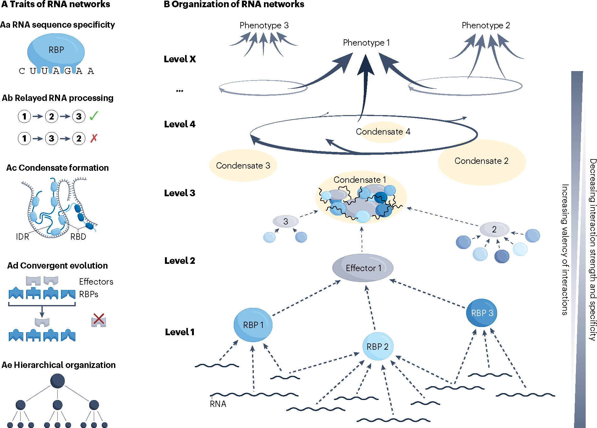
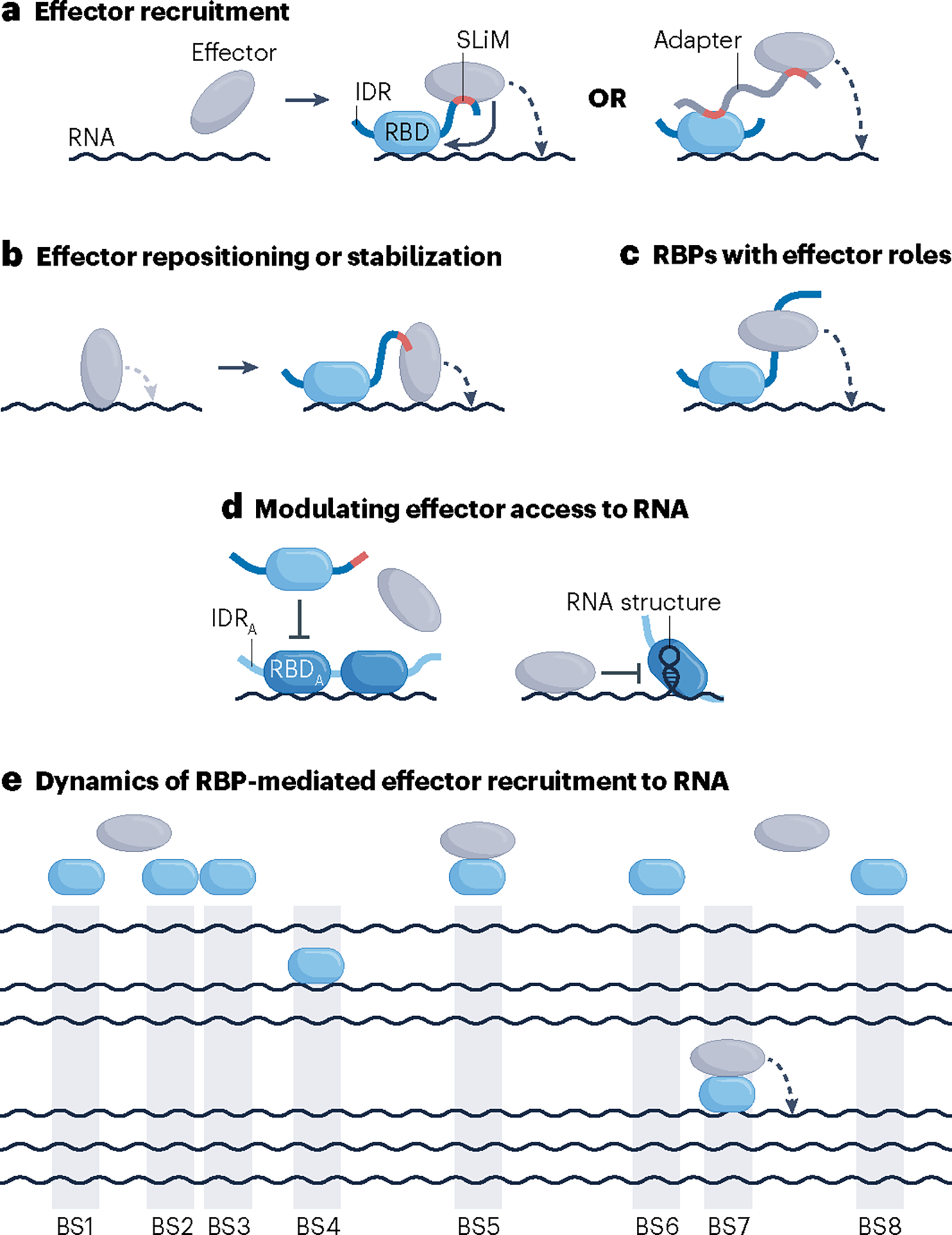
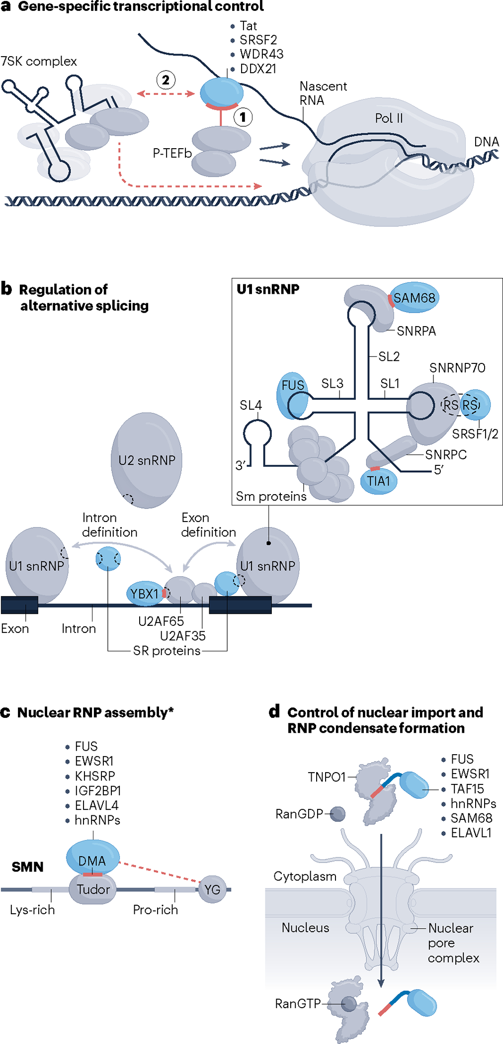
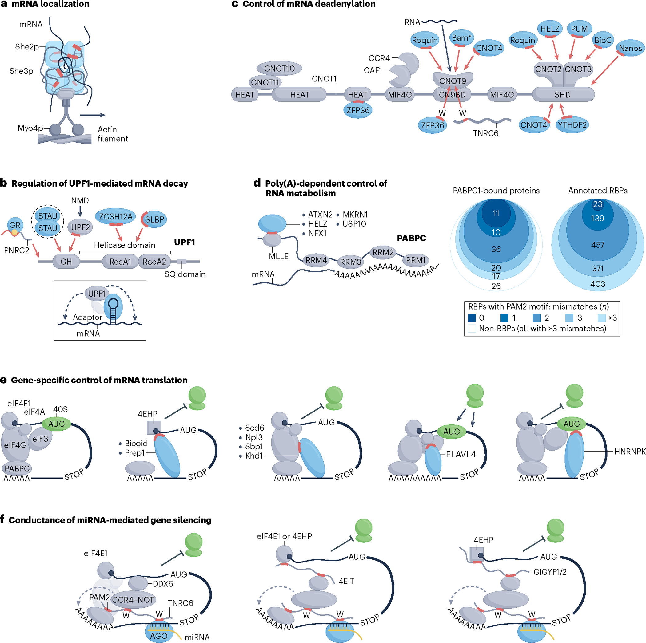
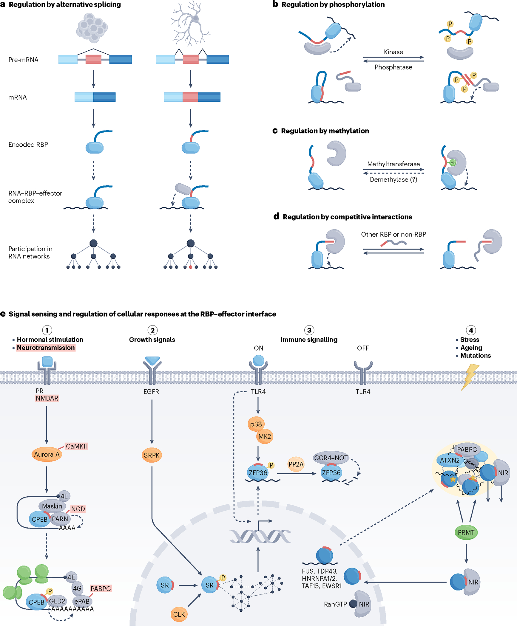
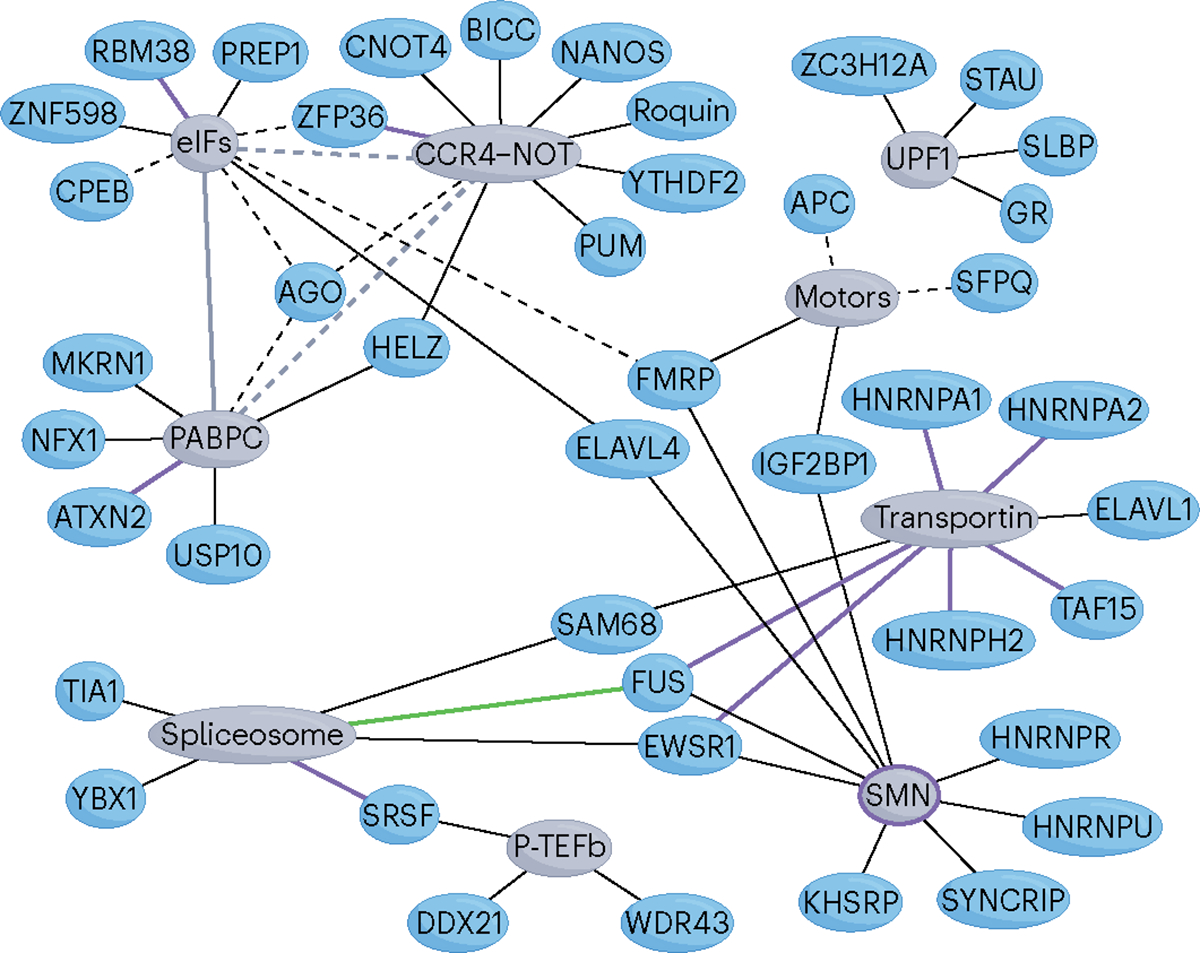
RNA-binding proteins (RBPs) regulate essentially every event in the lifetime of an RNA molecule, from its production to its destruction. Whereas much has been learned about RNA sequence specificity and general functions of individual RBPs, the ways in which numerous RBPs instruct a much smaller number of effector molecules, that is, the core engines of RNA processing, as to where, when and how to act remain largely speculative. Here, we survey the known modes of communication between RBPs and their effectors with a particular focus on converging RBP-effector interactions and their roles in reducing the complexity of RNA networks. We discern the emerging unifying principles and discuss their utility in our understanding of RBP function, regulation of biological processes and contribution to human disease.


Conflict of interest statement

Competing interests

The authors declare no competing interests.

Figures

Fig. 1 |
Fig. 1 |. Traits and organization of RNA networks associated with the management of regulatory complexity.
A, Traits of RNA networks. Aa, The recognition of specific RNA sequences and/or structures allows trans-acting factors, including RNA-binding proteins (RBPs) and small RNAs, such as microRNAs (miRNAs), to act upon some but not other transcripts. This trait also allows for synchronized processing of multiple, often functionally related transcripts. Recognition of specific RNA features defines the most elementary regulatory level of RNA networks that determines which transcripts will be controlled by which trans-acting factors. Different RNA specificities of different RBPs considerably simplify the challenge of coordinated regulation, in addition to allowing for network adaptation through rewiring of RNA targets,. Ab, Relayed RNA processing refers to the correct sequence of post-transcriptional processing events. For instance, a typical pipeline would ensure that RNA splicing occurs prior to RNA export and localization to a distal intracellular region, followed by localized translation and that RNA degradation occurs last. Any other sequence of events could be detrimental to cell homeostasis. General molecular and cellular organization, e.g. association of the splicing machinery with RNA polymerase II (Pol II) and separation of nuclear from cytoplasmic components, respectively, as well as more specific molecular interactions contribute to the correct relay of RNA processing events,. Correct and rapid relay of RNA processing events secures directness and energy-efficient regulation of RNA processing. Ac, The formation of ribonucleoprotein (RNP) condensates increases local concentration of RBP–RNA modules along with their effector complexes. RNP condensation is driven primarily by intrinsically disordered regions (IDRs) of different RBP molecules, which multimerize through numerous weak, non-specific interactions, and is aided by transient secondary structures formed within IDRs as well as contributions from the associated RNA. RNP condensation can stabilize the association of individual RBPs with their recognition motifs on RNA and contribute to correct RNA folding that may be required for processing,. In addition, increased local concentration of effector proteins and RNA can increase the rate of biochemical reactions, or assist in storage or transport of these molecules. Upon extensive RNP condensation, a physicochemical phenomenon of liquid–liquid phase separation occurs through which various types of RNA granules, including stress granules, P-bodies, splicing speckles, neuronal granules, and others, are generated. Such granules, also referred to as ‘membraneless organelles’, exist in liquid-like and occasionally solid-like physical states that exhibit distinct physiological roles,. Mechanisms that govern the formation and dissolution of RNP condensates include membrane surfaces, molecular chaperones, including nuclear import receptors, RNA helicases, and post-translational modifications of condensate components,. Ad, Convergent molecular evolution has an important role at different levels of RNA processing and contributes to the hierarchical structure of RNA networks. At the RBP–RNA level, convergence has been observed in RNA-targeting specificities of RBPs and in the evolutionary adaptation of RNA molecules to a particular mode of post-transcriptional processing, including alternative splicing and mRNA decay,,. Convergent evolution also occurs at the level of RBPs interacting with their effector molecules. Short linear motifs (SLiMs), which are typically located in IDRs of RBPs or adaptor proteins, are specifically recognized by cognate domains of effectors and frequently evolve in a convergent manner,. These examples point to a broad potential of convergent evolution to unify the fates of different transcripts by a common regulatory step. Ae, Hierarchical organization is commonly observed in biological networks and is thought to evolve due to the high cost associated with creation and maintenance of network connections. B, Hierarchically wired networks, including RNA networks, not only exhibit fewer connections, but also adapt faster to the environment and show higher overall performance compared to non-hierarchical networks. RNA networks show several hierarchical regulatory levels, with control at higher levels having broader effects on RNA processing. RBD, RNA-binding domain.
Fig. 2 |
Fig. 2 |. Modes and dynamics of RBP-dependent effector engagement with RNA.
a, RNA-binding proteins (RBPs) (blue shapes) can recruit effectors to target RNA via direct or indirect protein–protein interactions (PPIs) that typically entail a short linear motif (SLiM; red), which typically resides in an intrinsically disordered region (IDR) of the RBP or an adaptor protein, and a structured domain located in the effector. Indirect interactions can involve additional proteins or can be mediated by non-coding RNAs (not shown). Upon recruitment, the effector can exert activity in cis, that is, on the RBP-bound RNA (dashed curved arrow) and occasionally also on the recruiting RBP (solid curved arrow), or in trans, that is, on other molecules (not shown). b, Instead of serving a recruiting role, some RBP–effector interactions may facilitate repositioning or stabilization of a pre-bound effector to modulate its activity. c,d, Certain activities of RBPs do not entail contacts with effector molecules, either because RBPs themselves operate as effectors, as is the case for RBPs with enzymatic activities,– (panel c), or because they operate by modulating effector access to RNA, such as heterogeneous nuclear ribonucleoproteins (hnRNPs) in regulation of splicing (panel d). e, RBP-mediated recruitment of an effector to RNA is transient and occurs infrequently. Shown is a hypothetical steady-state scenario in which copies of an RBP (blue ovals), the number of which matches the number of RBP-binding sites (BS1–BS8) on RNA (black wavy lines), compete for a limiting number of available effector molecules (grey ovals). Only an RNA–RBP–effector assembly can process RNA in cis (dashed arrow). IDRA, IDR of antagonizing RBP; RBD, RNA-binding domain; RBDA, RBD of antagonizing RBP.
Fig. 3 |
Fig. 3 |. Converging RBP–effector interactions regulating (peri-)nuclear RNA processing.
a, RNA-binding protein (RBP)-dependent release of paused RNA polymerase II (Pol II) by positive transcription elongation factor b (P-TEFb). RBPs can stimulate relocation of P-TEFb from a local 7SK complex to the vicinity of Pol II either by recruiting P-TEFb via direct protein–protein interactions (PPIs) while bound to nascent RNA (step 1) or indirectly by associating with or disassociating from the 7SK complex (step 2). b, RBPs directly interact with spliceosomal components, including U1 small nuclear ribonucleoprotein (snRNP), U2 snRNP and U2AF subunits, to promote the early stages of spliceosome assembly. Illustrated is an overview of all interactions (left) along with a zoomed-in view of the U1 snRNP (right). Grey arrows denote intron or exon definition interactions, several of which are mediated by RS domains (dashed sections of shapes) of SR proteins, such as SRSF1 and SRSF2, and components of the spliceosome. RBPs other than SR proteins, including YBX1, SAM68 and TIA1, use short stretches of their intrinsically disordered regions (IDRs) (red dashes) to contact the indicated spliceosomal proteins. FUS recognizes the stem–loop (SL) region 3 (SL3) of the U1 small nuclear RNA (snRNA). Sm proteins are seven core spliceosomal proteins that make up a stable ring-like structure. c, Dimethylated arginines (DMAs) in the RGG/RG-rich regions of the indicated RBPs are recognized by the aromatic cage within the Tudor domain of survival motor neuron protein (SMN). Dashed red line denotes additional IDR-mediated interactions of some of the listed RBPs with the YG box domain (YG). The asterisk indicates that the same RBP–SMN interactions might also participate in processes other than RNP assembly in the nucleus. For clarity, RBP-bound RNA is not drawn. d, RBPs that use a proline-tyrosine-rich nuclear localization sequence (PY-NLS) short linear motif (SLiM) (red dash) to interact with transportin 1 (TNPO1) for their nuclear import. The disaggregase activity of TNPO1 is not indicated (Fig. 4e). Drawings of multiple RBPs binding to the same effector molecule in individual panels solely illustrate that different RBPs can bind to a particular effector; they do not imply simultaneous interactions of multiple RBPs with different segments of the same effector or competition between different RBPs for binding to a particular region of an effector molecule.
Fig. 4 |
Fig. 4 |. Converging RBP–effector interactions regulating cytoplasmic RNA processing.
a, Myo4p motor protein-mediated transport of She2p/She3p RNA-binding protein (RBP)-bound mRNA along an actin filament, a process required for asymmetrical mRNA localization in budding yeast. Red segments indicate intrinsically disordered regions (IDRs) of She3p in contact with She2p and mRNA. b, Interactions of RBPs with the RNA helicase UPF1 implicated in regulation of mRNA decay. Red highlights represent IDR segments that form key protein–protein interactions (PPIs) required for activation of specific RBP-mediated mRNA decay pathways. Yellow dot denotes glucocorticoid, the ligand required for efficient association of the glucocorticoid receptor (GR) with the PNRC2 adaptor. Dashed oval indicates dimerization of Staufen (STAU) proteins. Red arrows point to approximate (domain-resolution) sites of contact on UPF1. Drawing in the inset illustrates a generalized mode of RBP-stimulated UPF1-mediated mRNA decay. Dashed arrows denote indirect stimulation in cis of exo- or endo-ribonucleolytic cleavage or translational repression. c, Schematic of the CCR4–NOT complex with indicated protein subunits and domains of the largest, CNOT1 subunit. Blue shapes indicate regulatory RBPs that bind to CCR4–NOT whereas red and black arrows point to sites of contact of RBPs and RNA, respectively. Red dash denotes short linear motif (SLiM)-containing IDR segments and letters W indicate tryptophan residues that form PPIs with CCR4–NOT. The asterisk indicates that the RNA-binding capacity of Bam is currently uncertain. The grey wavy line represents TNRC6 proteins that can serve as adaptors to connect RBPs with CCR4–NOT. RBP-bound mRNA has been omitted for clarity. d, RBPs that use a PAM2 or a PAM2-like SLiM (red dash) to interact with the MLLE domain of cytoplasmic poly(A)-binding protein (PABPC) bound to a poly(A) RNA sequence (left). Numbers of human PABPC1-bound proteins or annotated human RBPs that contain a PAM2 or a PAM2-like SLiM shown in proportional Venn diagrams. Total pools of proteins in each group were defined previously, and are listed in Supplementary Table 3. The two PAM2 motifs, LIG_PAM2_1 and LIG_PAM2_2, annotated in the ELM database were considered as canonical (no mismatching residues; darkest shade of blue). Proteins that harbour motifs with one, two or three residues that deviate from either of the canonical PAM2 motifs are indicated in progressively lighter shades of blue (right). Note that the PAM2-like SLiM of makorin 1 (MKRN1) contains three residues that mismatch LIG_PAM2_1. None of the 26 PABPC1-bound non-RBPs harbours any PAM2 or PAM2-like motifs (that is, those with zero to three mismatches). e, Direct RBP–effector interactions that regulate translation initiation. Red segments of mRNA-bound RBPs denote IDRs that interact with different initiation factors or the 40S ribosomal subunit, as indicated. f, Cooperation of RBP–adaptor–effector conduits in miRNA–AGO-mediated gene silencing. Several co-associated molecules and processes have been omitted for clarity. Blunt or sharp arrows towards or away from the ribosomes (green shapes) indicate repressive or activating net effect on translation, respectively. Drawings of multiple RBPs binding to the same effector molecule in panels bd solely illustrate that different RBPs can bind to a particular effector; they do not imply simultaneous interactions of multiple RBPs with different segments of the same effector or competition between different RBPs for binding to a particular region of an effector molecule. NMD, nonsense-mediated decay; RRM, RNA recognition motif.
Fig. 5 |
Fig. 5 |. Physiological regulation at the interface of RBPs and their effectors.
a–d, Modes of pre-translational (panel a) and post-translational (panels b–d) regulation of RNA-binding protein (RBP)–effector interactions involved in the control of biological processes. a, Tissue-specific alternative splicing, illustrated here as skipping or inclusion of the small linear motif (SLiM)-encoding alternative exon (red) in stem cells (left) or neurons (right), respectively, can facilitate rewiring of RNA networks. b, Phosphorylation, which is implemented by kinases and removed by phosphatases, most often disrupts RBP–effector interactions and occurs in RBP regions flanking the SLiM segment (red) that contacts the effector surface (top). Interactions of SR proteins with components of the spliceosome present a particular case in which phosphorylation of the RS domains (red) of both interacting partners stimulates contact establishment (bottom). c, Monomethylation or dimethylation of arginine residues of RBPs can stimulate RBP–effector interactions, typically through recognition of a methylated arginine by the Tudor domain, as illustrated, or can weaken the affinity of RBPs for effectors, as in the case of the RBP–transportin 1 (TNPO1) interactions (not shown). It is unclear whether demethylation of RBPs also occurs in vivo (denoted by a question mark), although arginine demethylating activity has been ascribed to a handful of enzymes. d, Competition between an RBP and other RBPs or non-RBPs for a common binding site on an effector can exert a direct regulatory effect on RNA processing. Competition of RanGTP with RNA-free RBPs for binding to nuclear import receptors can be considered as having an indirect effect on RNA processing (Fig. 3d). Curved dashed arrows depict effector activity on RNA in cis. e, The interface of RBPs and their effectors serves as a sensor of intracellular and extracellular signals as well as a regulator of cellular responses to signalling. Illustrated are signal transduction pathways (pathways 1–4) that trigger responses through distinct modes of post-transcriptional RNA processing. Hormonal stimulation of oocytes triggers their maturation, in part, via phosphorylation-dependent reconfiguration of cytoplasmic polyadenylation element binding protein (CPEB)–effector interactions. This turns CPEB from a repressor to an activator of polyadenylation-induced translation, a process that is crucial for germ-cell development. A highly similar pathway leading to CPEB activation is triggered upon synaptic stimulation of neurons and plays a key part in synaptic plasticity (terms in red, where indicated, are specific to the neuronal pathway). Green shapes indicate translocating ribosomes (pathway 1). Activities of SR proteins are modulated by external and internal signals via phosphorylation by SR protein kinases (SRPKs) and CDC-like kinases (CLKs), respectively, with the capacity to trigger a systemic response through changes in numerous alternative splicing events (pathway 2). Regulation of RBP–effector interactions via phosphorylation has a central role in securing a timely response to immune signalling, as well as its resolution. Upon stimulation with lipopolysaccharide (LPS), phosphorylation prevents association of ZFP36 with CCR4–NOT to help stabilize the induced and ZFP36-bound pro-inflammatory mRNAs. This response is rapidly reversed once the signalling subsides via protein phosphatase 2A (PP2A)-mediated dephosphorylation of ZFP36 and the ensuing recruitment of CCR4–NOT, which deadenylates the ZFP36-bound transcripts, which are then rapidly degraded (pathway 3). A series of largely nuclear RBPs (dark blue circles) with prion-like domains operate as splicing factors but partially also shuttle to the cytoplasm where they take on additional roles. Upon cellular stress, these and other aggregation-prone RBPs, such as ataxin 2 (ATXN2), potentially with their bound RNA, relocate to ribonucleoprotein (RNP) condensates/stress granules where they are kept functionally inert. Effectors that moonlight as RBP chaperones, including nuclear import receptors (NIRs) and cytoplasmic poly(A)-binding protein (PABPC), assist by preventing irreversible aggregation of RBPs in part through their recognition via nuclear localization sequence (NLS) and PAM2 motifs (red), respectively. Condensation properties as well as NIR interactions and nuclear import of some RBPs are additionally regulated by methylation by protein arginine methyltransferases (PRMTs). Excessive stress, RBP mutations (yellow asterisks) and ageing can prolong the time that RBPs spend in a condensed state, increasing the risk of RBP aggregation and neuronal degeneration. NIRs can act as disaggregases, with an intrinsic capacity to dissolve certain types of aberrant RNP condensates (pathway 4). CaMKII, calcium/calmodulin-dependent protein kinase II; EGFR, epidermal growth factor receptor; ePAB, embryonic poly(A) binding protein; MK2, MAPK-activated protein kinase 2; NMDAR, N-methyl-d-aspartate receptor; PR, progesterone receptor; TLR4, Toll-like receptor 4.
Fig. 6 |
Fig. 6 |. A network view of the nexus.
Shown is a compilation of converging RNA-binding protein (RBP)–effector interactions that have been characterized at both the molecular and functional levels. Note that several RBPs (blue shapes) connect (black lines) to more than one effector (grey shapes) and that there also exist RBP-independent interactions between different effectors (grey lines). Solid and dashed lines denote direct and indirect (adaptor-mediated) protein–protein interactions (PPIs), respectively. The green line that connects FUS and the spliceosome represents an RBP–RNA interaction. Purple lines represent disease-associated or therapeutically targeted interactions (Table 2). The purple circle around survival motor neuron protein (SMN) indicates spinal muscular atrophy (SMA)-linked mutations of SMN that weaken its affinity for multiple RBPs. Only mammalian RBPs are shown. Motor proteins (motors; KIF11, KIF3C, KIF5A, KIF3AB) and translation initiation factors (eIFs; eIF4E, eIF4E2 (4EHP), eIF4G) have been grouped together and are shown as a single effector. Non-converging PPIs discussed in the text, TDP43–Kapβ2/a1 and MATR3–TREX, are omitted. CPEB, cytoplasmic polyadenylation element binding protein; MKRN1, makorin 1; PABPC, cytoplasmic poly(A)-binding protein; STAU, Staufen.

Similar articles

Cited by

References

    1. Licatalosi DD & Darnell RB RNA processing and its regulation: global insights into biological networks. Nat. Rev. Genet. 11, 75–87 (2010). - PMC - PubMed
    1. Chen M & Manley JL Mechanisms of alternative splicing regulation: insights from molecular and genomics approaches. Nat. Rev. Mol. Cell Biol. 10, 741–754 (2009). - PMC - PubMed
    1. Scotti MM & Swanson MS RNA mis-splicing in disease. Nat. Rev. Genet. 17, 19–32 (2016). - PMC - PubMed
    1. Jankowsky E & Harris ME Specificity and nonspecificity in RNA–protein interactions. Nat. Rev. Mol. Cell Biol. 16, 533–544 (2015). - PMC - PubMed
    1. Ramskold D, Wang ET, Burge CB & Sandberg R An abundance of ubiquitously expressed genes revealed by tissue transcriptome sequence data. PLoS Comput. Biol. 5, e1000598 (2009). - PMC - PubMed

Publication types