. 2022 Nov 23;13(11):987.
doi: 10.1038/s41419-022-05436-x.

LINC00924-induced fatty acid metabolic reprogramming facilitates gastric cancer peritoneal metastasis via hnRNPC-regulated alternative splicing of Mnk2

Affiliations

LINC00924-induced fatty acid metabolic reprogramming facilitates gastric cancer peritoneal metastasis via hnRNPC-regulated alternative splicing of Mnk2

Qiuming He et al. Cell Death Dis. .

Abstract

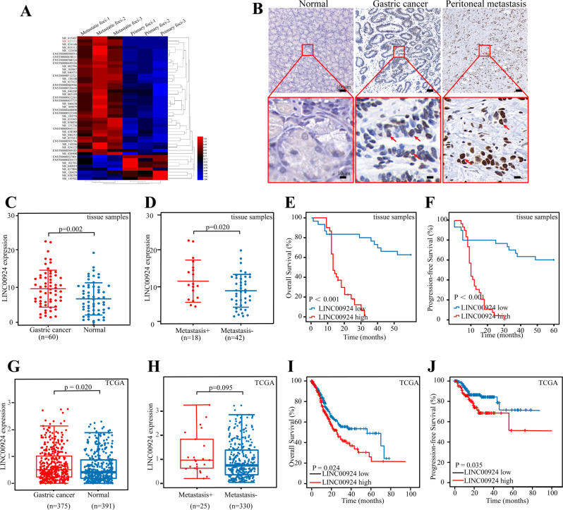
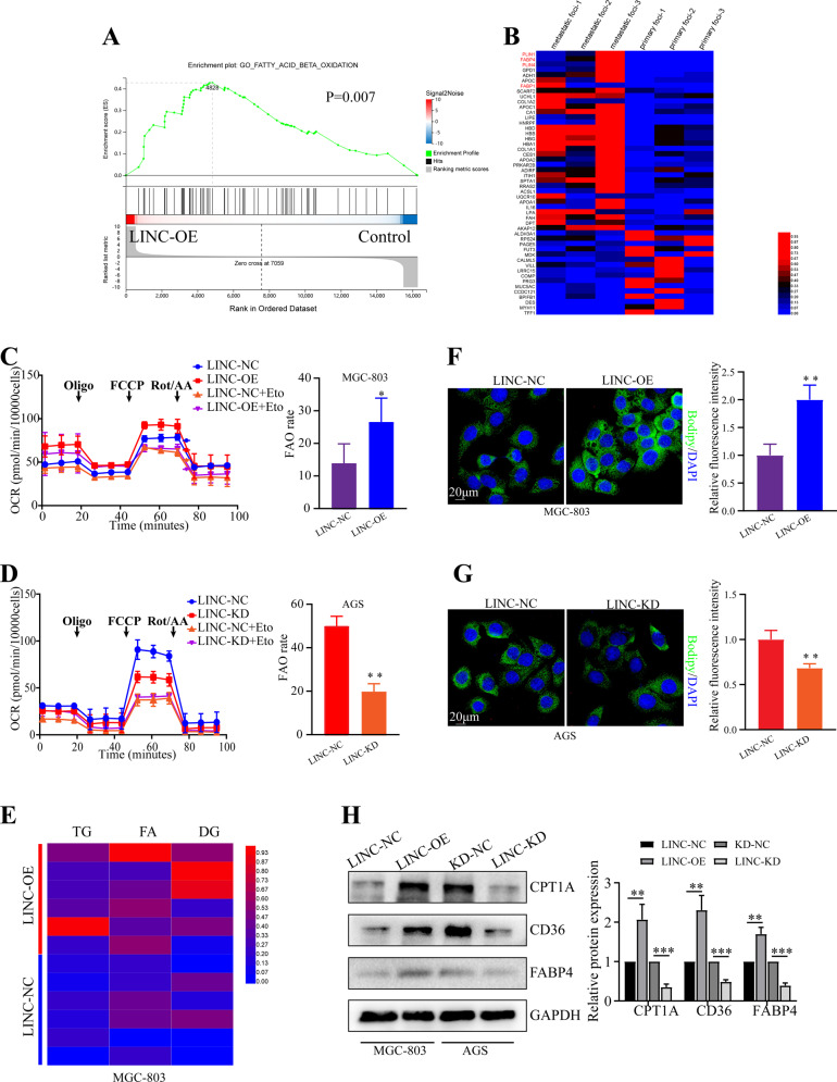
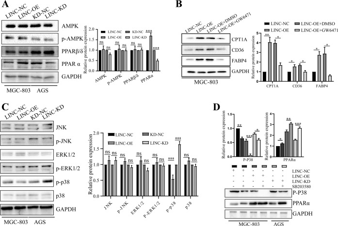
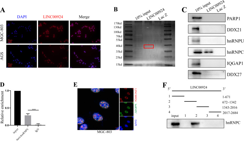
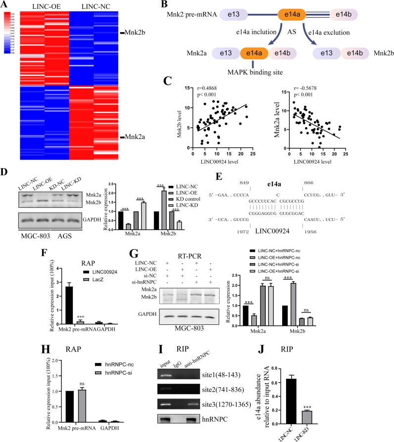
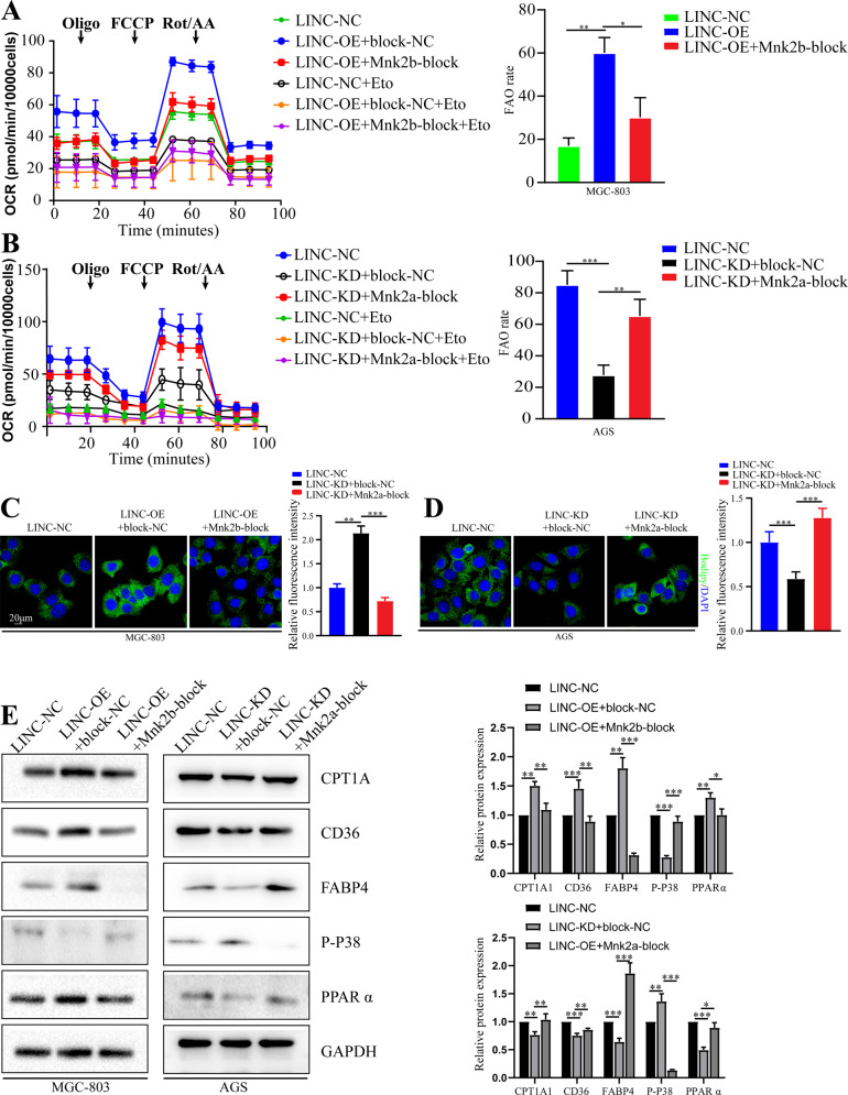
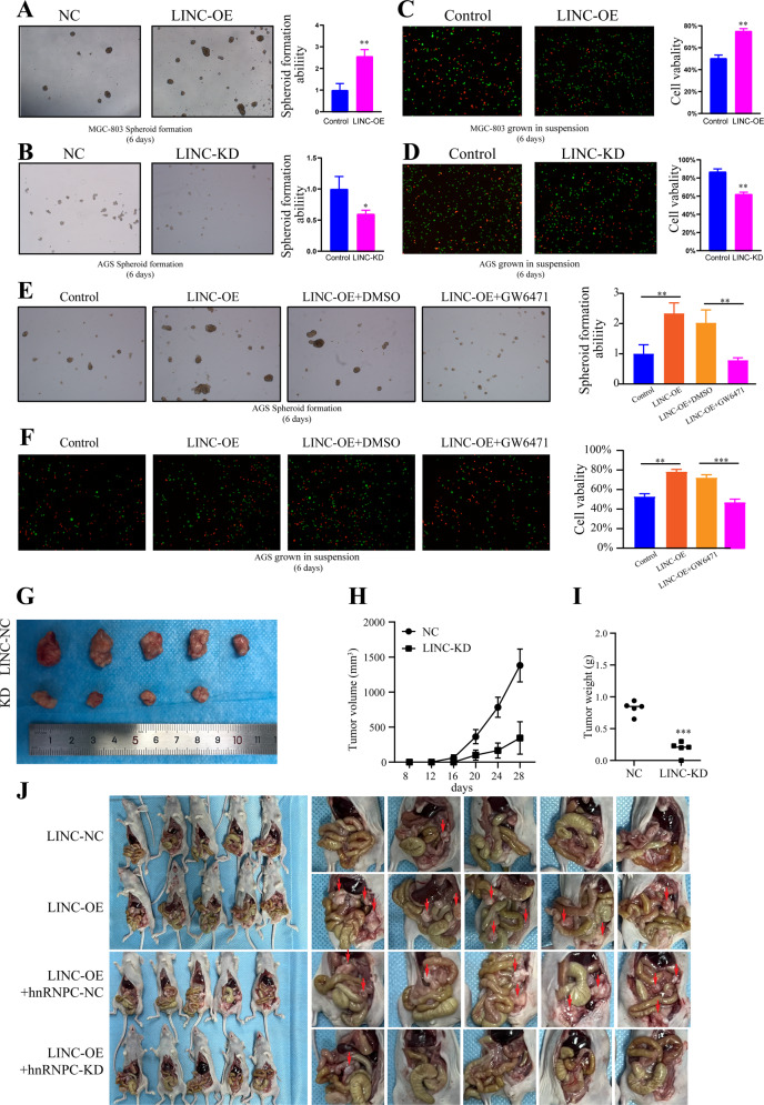
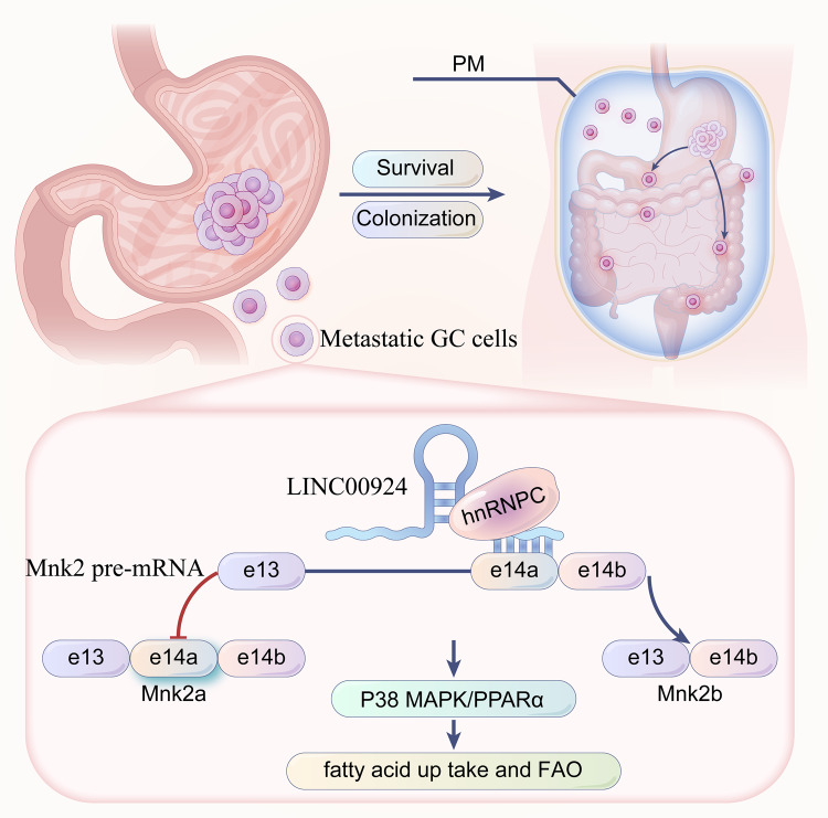
The molecular mechanism underlying gastric cancer (GC) peritoneal metastasis (PM) remains unclear. Here, we identified LINC00924 as a GC PM-related lncRNA through Microarray sequencing. LINC00924 was highly expressed in GC, and its high expression is associated with a broad range of PM. Via RNA sequencing, RNA pulldown assay, mass spectrometry, Seahorse, Lipidomics, spheroid formation and cell viability assays, we found that LINC00924 promoted fatty acid (FA) oxidation (FAO) and FA uptake, which was essential for matrix-detached GC cell survival and spheroid formation. Regarding the mechanism, LINC00924 regulated the alternative splicing (AS) of Mnk2 pre-mRNA by binding to hnRNPC. Specifically, LINC00924 enhanced the binding of hnRNPC to Mnk2 pre-mRNA at e14a, thus downregulating Mnk2a splicing and regulating the p38 MAPK/PPARα signaling pathway. Collectively, our results demonstrate that LINC00924 plays a role in promoting GC PM and could serve as a drug target.


Conflict of interest statement

The authors declare no competing interests.

Figures

Fig. 1
Fig. 1. Identification of GC PM related LINC00924.
A Differential expression of lncRNAs in 3 pairs of GC primary and peritoneal metastatic lesions identified by lncRNA microarrays. B Representative ISH staining for LINC00924 expression in normal tissue, GC primary lesions and peritoneal metastatic lesions. C qRT–PCR analysis of LINC00924 expression levels in GC and normal tissues. D qRT–PCR analysis of LINC00924 expression levels in metastasis-positive (metastasis + ) and metastasis-negative (metastasis -) GC patients. E-F: The associations of LINC00924 expression with five-year OS (E) and five-year DFS (F) were analyzed by Kaplan–Meier survival analysis. G: qRT–PCR analysis of LINC00924 expression levels in GC and normal tissues from TCGA. H: qRT–PCR analysis of LINC00924 expression levels in metastasis-positive (metastasis + ) and metastasis-negative (metastasis -) GC patients from TCGA. I, J: Kaplan–Meier survival analysis curve calculated from 375 GC patients from TCGA. *p < 0.05, **p < 0.01, ***p < 0.001.
Fig. 2
Fig. 2. LINC00924 expression promotes FAO and FA uptake.
A GSEA of the RNA-sequencing (RNA-seq) data from LINC00924-OE and MGC803-NC cells. B Differential expression of proteins in 3 pairs of GC primary lesions and peritoneal metastatic lesions determined by protein profile analysis. C, D Oxygen consumption rate (OCR) analysis in MGC803-NC/LINC-OE and AGS-NC/LINC-KD cell lines using palmitate as a metabolic substrate. MGC803 and AGS cells were treated with the CPT1 inhibitor (+ETO) or vehicle (−ETO). E Heatmap presenting differences in TG, FA, and DG from lipidomics between MGC803-NC and LINC-OE cell lines. F Lipid droplet staining in AGS-NC and LINC-OE cells. G Lipid droplet staining in MGC803-NC and LINC-KD cells. H WB analysis of CPT1A, CD36, and FABP4 in MGC803-NC and LINC-OE cells lines and in AGS-NC and LINC-KD cell lines. The protein levels were quantified with ImageJ.
Fig. 3
Fig. 3. p38/PPARα signaling pathway is central to LINC00924 mediated FAO and FA uptake.
A WB analysis of AMPK, p-AMPK, PPARα and PPARβ/δ in MGC803-NC and LINC-OE cell lines and in AGS-NC and LINC-KD cell lines. B WB analysis of CPT1A, CD36, and FABP4 in MGC803-NC, LINC-OE, and LINC-KD cells treated with GW6471 or DMSO. C WB analysis of JNK, p-JNK, ERK1/2, p-ERK1/2, p-p38, and p38 in MGC803-NC and LINC-OE cell lines and in AGS-NC and LINC-KD cell lines. D WB analysis of p-P38 and PPARα in MGC803-NC, LINC-OE, and LINC-KD cells treated or not treated with SB203580.
Fig. 4
Fig. 4. LINC00924 physically interacts with hnRNPC.
A FISH results showing the nuclear localization of LINC00924 (red) in MGC803 and AGS cells. The nuclei were stained with DAPI. B Silver staining of biotinylated LINC00924-interacting proteins. Red boxes indicate specific bands. C WB results used to confirm the mass spectrometry results. D RIP analysis conducted using an anti-hnRNPC antibody to validate the interaction between hnRNPC and LINC00924. E Confocal RNA-FISH (red) and immunofluorescence (IF) (green) images indicating the colocalization of LINC00924 and hnRNPC. F Results of the LINC00924 serial deletion assay performed to localize the interaction region.
Fig. 5
Fig. 5. LINC00924 regulates the P38/PPARα signaling pathway via hnRNPC regulated Mnk2 pre-mRNA AS.
A Heatmap presenting significantly differentially expressed transcripts between MGC803-NC and LINC00924-OE cell lines. B A schematic model of Mnk2 AS. MNK2 pre-mRNA can be alternatively spliced to yield two isoforms: Mnk2a and Mnk2b. Mnk2a contains e14a, a binding site for MAP kinases, while Mnk2b does not [30]. C The correlations of LINC00924 expression with Mnk2b (B) and Mnk2a (C) expression in GC tissues. D RT–PCR analysis of Mnk2a and Mnk2b in MGC803-NC and LINC-OE cell lines and in AGS-NC and LINC-KD cell lines. E Schematic illustration of base pairing between LINC00924 and the e14a of Mnk2. F qRT–PCR detection of e14a enrichment by LINC00924 using a RAP assay. G RT–PCR analysis of MGC803-NC and LINC-OE cell lines treated with si-hnRNPC or NC. H hnRNPC knockdown did not disrupt the RNA–RNA duplex formed by LINC00924 and e14a. I RIP analysis conducted using an anti-hnRNPC antibody to validate the interaction between hnRNPC and the e14a of Mnk2. J LINC00924 is essential for the formation of the RNA–RNA duplex by LINC00924 and e14a.
Fig. 6
Fig. 6. Mnk2 AS is essential for LINC00924 regulating lipid metabolic reprogramming.
A OCR analysis of MGC803-NC and LINC-OE cell lines treated with Mnk2b-block SSO or NC using palmitate as a metabolic substrate. B OCR analysis of AGS-NC and LINC-KD cell lines treated with Mnk2a-block SSO or NC using palmitate as a metabolic substrate. C Lipid droplet staining of AGS-NC and LINC-OE cells treated with Mnk2a-block SSO or NC. D Lipid droplet staining if MGC803-NC and LINC-KD cells treated with Mnk2a-block SSO or NC. E WB analysis of CPT1A, CD36, FABP4, p-P38, and PPARα in MGC803-NC and LINC-OE cells treated with Mnk2b-block SSO or NC and AGS-NC and LINC00924-KD cells treated with Mnk2a-block SSO or NC.
Fig. 7
Fig. 7. LINC00924 promotes matrix-detached GC cells survival and metastasis in vitro and in vivo.
A 3D spheroid formation assay of MGC803-NC and LINC-OE cells. Results were quantified as fold changes. B 3D spheroid formation assay of AGS-NC and LINC-KD cells. Results were quantified as fold changes. C Live and dead staining of MGC803-NC and LINC-OE cells after culture on 3D ultralow attachment plates for 6 days (with live cells stained in green and dead cells stained in red). Results were quantified as fold changes. D Live and dead staining of AGS-NC and LINC-KD cells after culture on 3D ultralow attachment plates for 6 days (with live cells stained in green and dead cells stained in red). Results were quantified as fold changes. E 3D spheroid formation assay of MGC803-NC and LINC-OE cells treated or not treated with GW6471. Results were quantified as fold changes. F Live and dead staining of AGS-NC and LINC-KD cells treated or not treated with GW6471 after culture on 3D ultralow attachment plates for 6 days (with live cells stained in green and dead cells stained in red). Results were quantified as fold changes. G The morphological characteristics of subcutaneous tumor xenografts in the AGS-NC and LINC-KD groups. H Subcutaneous tumor volume in the AGS-NC and LINC-KD groups. I Subcutaneous tumor weight in the AGS-NC and LINC-KD groups. J The morphological characteristics of peritoneal tumor xenografts in the LINC-NC, LINC-OE, LINC-OE + hnRNPC-NC and LINC-KD + hnRNPC-KD groups.
Fig. 8
Fig. 8
LINC00924-induced fatty acid metabolic reprogramming facilitates gastric cancer peritoneal metastasis via hnRNPC-regulated alternative splicing of Mnk2.

References

    1. Sung H, Ferlay J, Siegel RL, Laversanne M, Soerjomataram I, Jemal A, et al. Global Cancer Statistics 2020: GLOBOCAN Estimates of Incidence and Mortality Worldwide for 36 Cancers in 185 Countries. CA: A Cancer J Clin. 2021;71:209–49. - PubMed
    1. Fujitani K, Yang HK, Mizusawa J, Kim YW, Terashima M, Han SU, et al. Gastrectomy plus chemotherapy versus chemotherapy alone for advanced gastric cancer with a single non-curable factor (REGATTA): a phase 3, randomised controlled trial. Lancet Oncol. 2016;17:309–18.. doi: 10.1016/S1470-2045(15)00553-7. - DOI - PubMed
    1. Headley MB, Bins A, Nip A, Roberts EW, Looney MR, Gerard A, et al. Visualization of immediate immune responses to pioneer metastatic cells in the lung. Nature. 2016;531:513–7. doi: 10.1038/nature16985. - DOI - PMC - PubMed
    1. Kleinschmidt EG, Miller NLG, Ozmadenci D, Tancioni I, Osterman CD, Barrie AM, et al. Rgnef promotes ovarian tumor progression and confers protection from oxidative stress. Oncogene. 2019;38:6323–37.. doi: 10.1038/s41388-019-0881-8. - DOI - PMC - PubMed
    1. Nieman KM, Kenny HA, Penicka CV, Ladanyi A, Buell-Gutbrod R, Zillhardt MR, et al. Adipocytes promote ovarian cancer metastasis and provide energy for rapid tumor growth. Nat Med. 2011;17:1498–503. doi: 10.1038/nm.2492. - DOI - PMC - PubMed

Publication types

MeSH terms