. 2022 Nov 23;12(1):20173.
doi: 10.1038/s41598-022-20096-9.

Modular automated microfluidic cell culture platform reduces glycolytic stress in cerebral cortex organoids

Affiliations

Modular automated microfluidic cell culture platform reduces glycolytic stress in cerebral cortex organoids

Spencer T Seiler et al. Sci Rep. .

Abstract

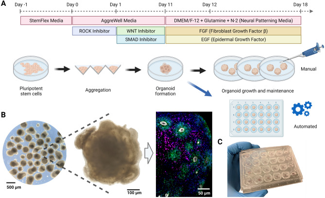
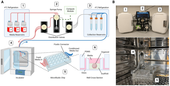
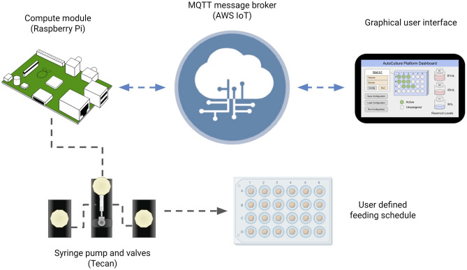
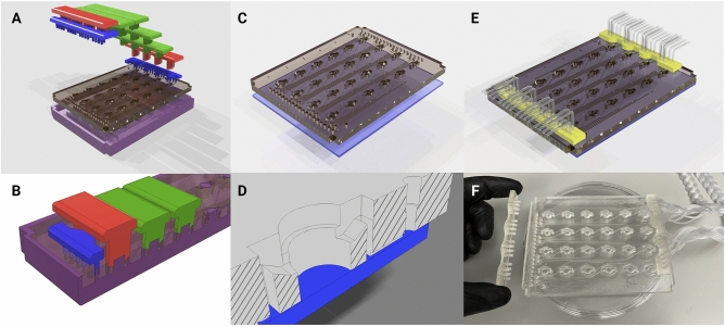
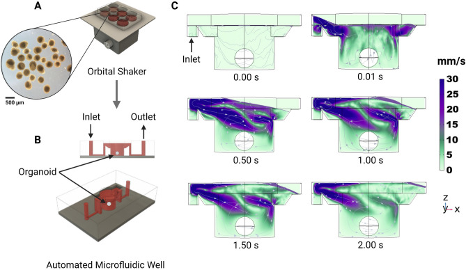
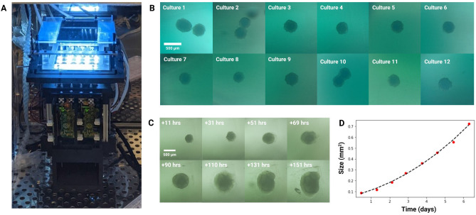
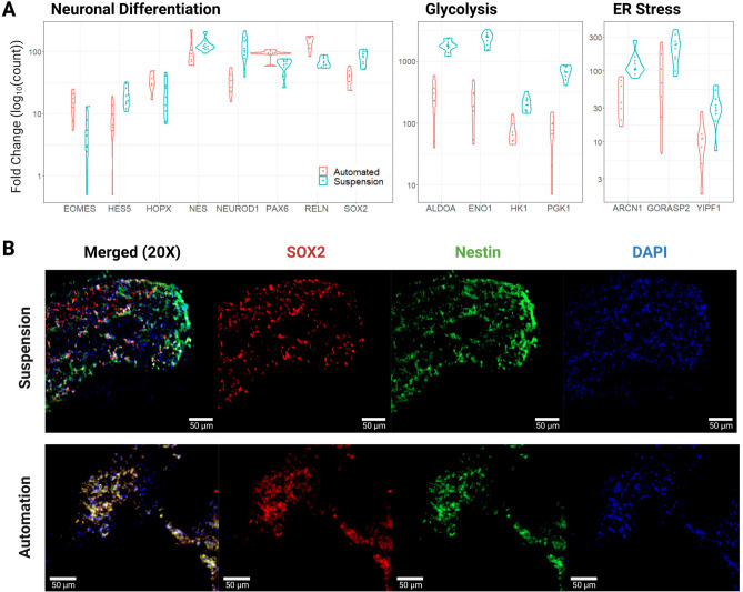
Organ-on-a-chip systems combine microfluidics, cell biology, and tissue engineering to culture 3D organ-specific in vitro models that recapitulate the biology and physiology of their in vivo counterparts. Here, we have developed a multiplex platform that automates the culture of individual organoids in isolated microenvironments at user-defined media flow rates. Programmable workflows allow the use of multiple reagent reservoirs that may be applied to direct differentiation, study temporal variables, and grow cultures long term. Novel techniques in polydimethylsiloxane (PDMS) chip fabrication are described here that enable features on the upper and lower planes of a single PDMS substrate. RNA sequencing (RNA-seq) analysis of automated cerebral cortex organoid cultures shows benefits in reducing glycolytic and endoplasmic reticulum stress compared to conventional in vitro cell cultures.


Conflict of interest statement

S.T.S and G.L.M. are founders of OrganOmics, a company that may be affected by the research reported in the enclosed paper. All other authors declare no competing interests.

Figures

Figure 1
Figure 1
Overview of the human cerebral organoid generation protocol. (A) Human pluripotent stem cells are expanded in traditional 2D culture, dissociated, aggregated into microwells, and matured into 3D organoid cultures using defined media conditions to promote cerebral cortex tissue differentiation. In this study, on day 12 post-aggregation, organoids were either kept in suspension and maintained manually (black arrow) or transferred to individual wells of a microfluidic chip and maintained in automation (blue arrow). (B) Images of cerebral organoid cultures. Bright-field images at low (left) and high (center) magnification under standard culture conditions show organoid morphology and heterogeneity. Immunofluorescence stains on week 5 for PAX6 (green, radial glia progenitor cells), CTIP2 (BCL11B) (magenta, excitatory projection neurons), ZO-1 (TJP1) (white, tight junction proteins on radial glia endfeet, apical surface of the neural tube), show characteristic ventricular zone-like rosette structures with radial glia surrounded by neurons. Nuclei stained with DAPI (blue). (C) Image of the PDMS microfluidic chip. The custom cell culture chip, modeled after a standard 24-well plate, houses organoids for automated experiments.
Figure 2
Figure 2
Design and implementation of the automated, microfluidic culture platform. (A) Illustration of the automated, microfluidic organoid culture platform, the Autoculture. (B) Front view images of the Autoculture. (1) Refrigerator with reagent reservoirs. (2) Syringe pump, distribution valves, and control interface. (3) Refrigerator with conditioned media collection reservoirs. These components reside on a lab bench directly above the cell culture incubator. (4) Microfluidic tubes enter through an incubator port and connect to the (5) microfluidic well plate chip inside the incubator. (6) Cross-sectional diagram of a single well containing an organoid culture.
Figure 3
Figure 3
IoT Cloud integration: a graphical user interface hosted on the internet relays messages to the Autoculture platform via MQTT to start, monitor and end experiments.
Figure 4
Figure 4
Fabrication of the PDMS microfluidic chip. (A) Graphical rendering of the interlocking mold pattern for the PDMS substrate in the microfluidic chip assembly. (B) Interlocking mounts (blue, red, and green) affix to the base mold (purple) and define microfluidic geometries upon the poured PDMS that are retained as the substrate cures. (C) The PDMS substrate is removed from the mold and bonded to glass. (D) A cross-sectional rendering of the chip. Fluid enters from microfluidic inlets on the surface and follows channels sealed by glass on the bottom to wells with open access from the top. (E) A 3D-printed fluidic interface plate (yellow) connects 24 fluidic microtube lines with the inlets/outlets of the microfluidic chip. (F) Microfluidic chip (center) with an example of the fluidic interface plate (left) and fully installed fluidic interface plate (right).
Figure 5
Figure 5
Computational fluid dynamics simulated flow in the individual tissue culture wells using the automated system. (A) Organoid cultures are expanded on well plates rotated on an orbital shaker. (B) After 12 days, organoids are transplanted into the automated microfluidic well. (C) One well of the proposed automated system with constant fluid injection over 2 s with velocity streamlines.
Figure 6
Figure 6
Longitudinal monitoring of organoid development. (A) The Autoculture microfluidic chip sits on a remote-controlled, IoT-enabled, 24-well automated imaging system. (B) Bright-field images of twelve individual 12-day-old cerebral cortex cultures at day 1 of automated feeding. (C) Longitudinal imaging of “Culture 4” during the experiment. (D) Projected area expansion of “Culture 4” during the experiment. This was obtained using a computer vision algorithm.
Figure 7
Figure 7
Transcription and immunofluorescent imaging results. (A) Pairwise comparisons of expression for select genes associated with neural differentiation, glycolysis, and ER stress. Results were statistically significant with an adjusted p value ≤ 0.05, except for HOPX and NES of neural differentiation. The “Automated” data represent 7 biological replicates and the “Suspension” data represent 4 biological replicates with 6 technical replicates (B) Immunofluorescent stains for SOX2, Nestin, and DAPI of Suspension and Automated organoid sections show congruent progenitor markers.

Similar articles

Cited by

References

    1. Gey GO, Coffman WD, Kubicek MT. Tissue culture studies of the proliferative capacity of cervical carcinoma and normal epithelium. Cancer Res. 1952;12:264–265.
    1. Scherer WF, Syverton JT, Gey GO. Studies on the propagation in vitro of poliomyelitis viruses. IV. Viral multiplication in a stable strain of human malignant epithelial cells (strain hela) derived from an epidermoid carcinoma of the cervix. Exp. Med. 1953;97:695–710. doi: 10.1084/jem.97.5.695. - DOI - PMC - PubMed
    1. Ringe J, Kaps C, Burmester G-R, Sittinger M. Stem cells for regenerative medicine: Advances in the engineering of tissues and organs. Naturwissenschaften. 2002;89:338–351. doi: 10.1007/s00114-002-0344-9. - DOI - PubMed
    1. Patel M, Yang S. Advances in reprogramming somatic cells to induced pluripotent stem cells. Stem Cell Rev. Rep. 2010;6:367–380. doi: 10.1007/s12015-010-9123-8. - DOI - PMC - PubMed
    1. Rossi G, Manfrin A, Lutolf MP. Progress and potential in organoid research. Nat. Rev. Genet. 2018;19:671–687. doi: 10.1038/s41576-018-0051-9. - DOI - PubMed

Publication types