

Functions of retinal astrocytes and Müller cells in mammalian myopia
- PMID: 36418970
- PMCID: PMC9686084
- DOI: 10.1186/s12886-022-02643-0
Functions of retinal astrocytes and Müller cells in mammalian myopia
Abstract
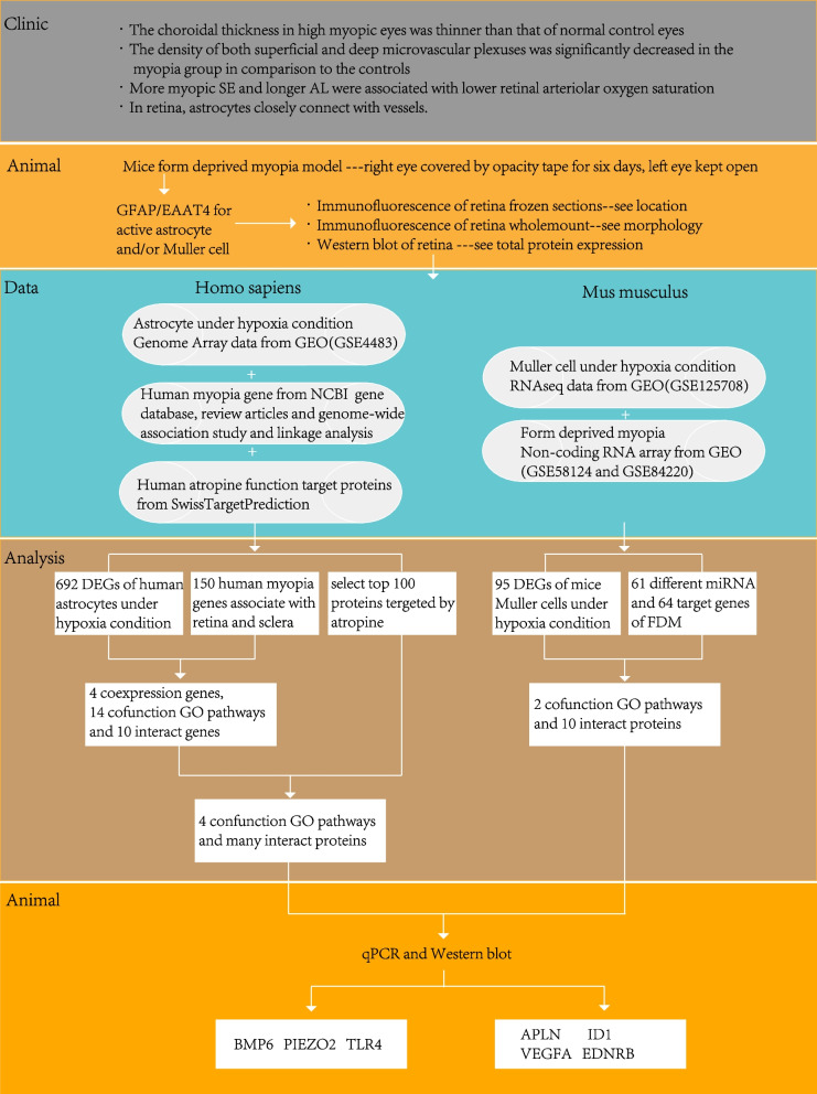
Background: Changes in the retina and choroid blood vessels are regularly observed in myopia. However, if the retinal glial cells, which directly contact blood vessels, play a role in mammalian myopia is unknown. We aimed to explore the potential role and mechanism of retinal glial cells in form deprived myopia.
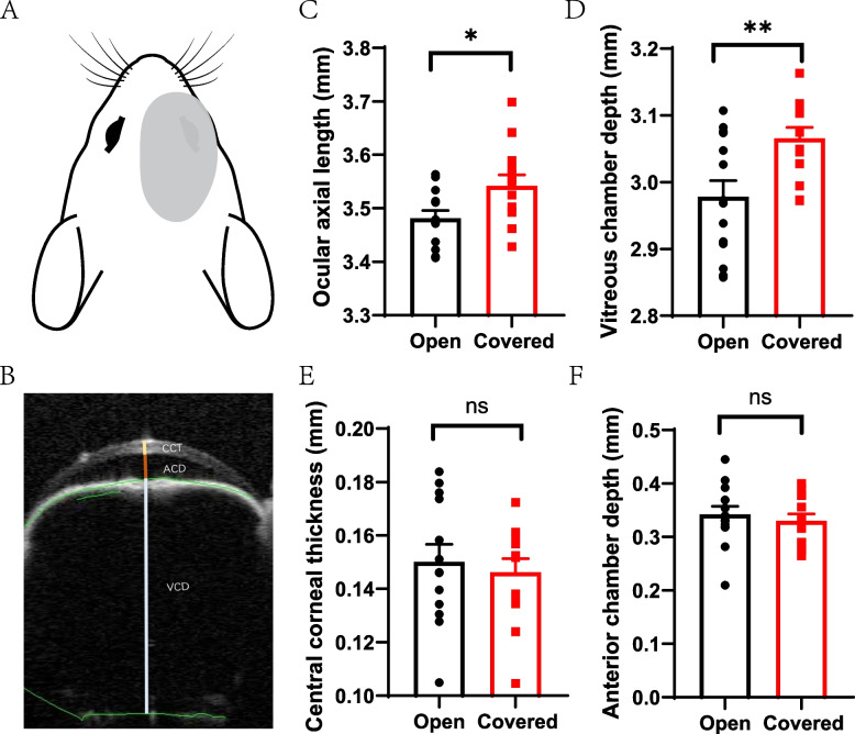
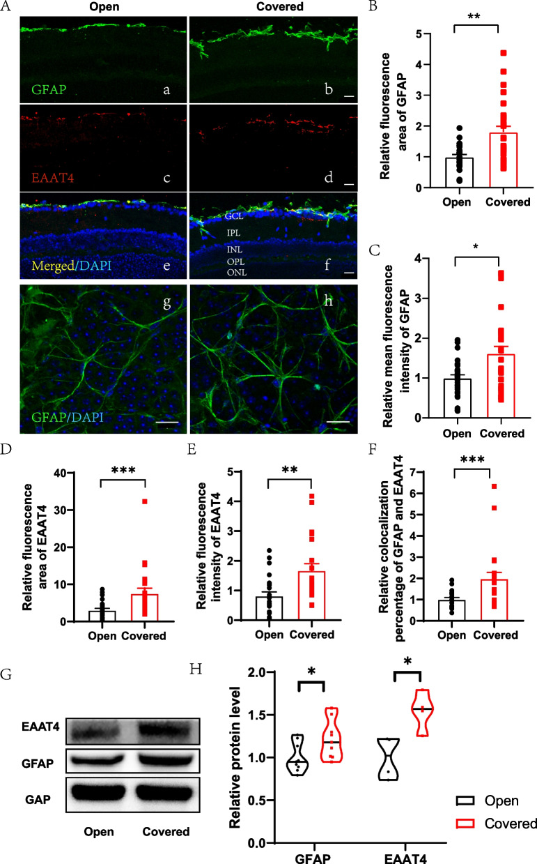
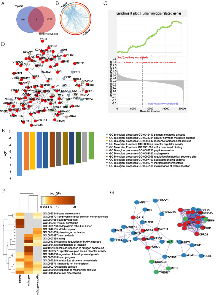
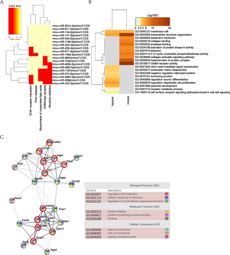
Methods: We adapted the mice form-deprivation myopia model by covering the right eye and left the left eye open for control, measured the ocular structure with anterior segment optical coherence tomography, evaluated changes in the morphology and distribution of retinal glial cells by fluorescence staining and western blotting; we also searched the online GEO databases to obtain relative gene lists and confirmed them in the form-deprivation myopia mouse retina at mRNA and protein level.
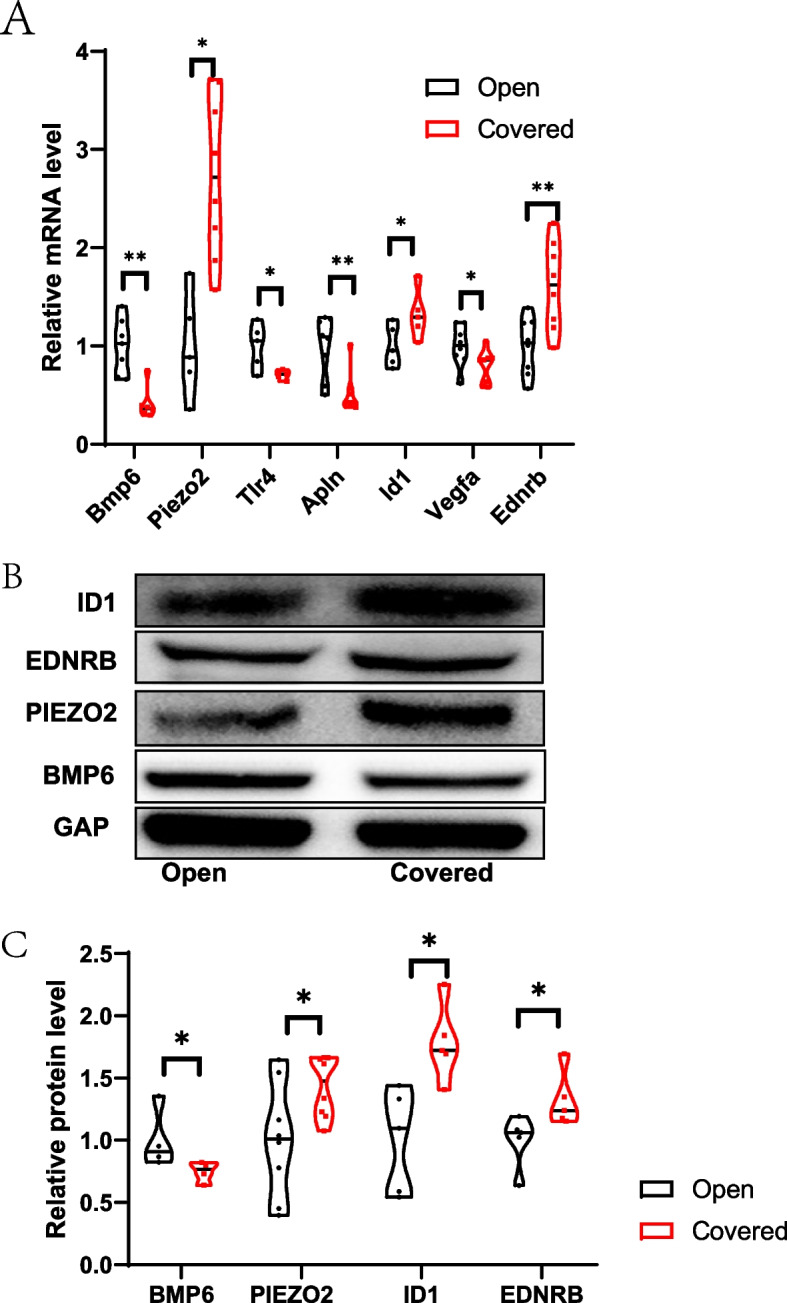
Results: Compared with the open eye, the ocular axial length (3.54 ± 0.006 mm v.s. 3.48 ± 0.004 mm, p = 0.027) and vitreous chamber depth (3.07 ± 0.005 mm v.s. 2.98 ± 0.006 mm, p = 0.007) in the covered eye became longer. Both glial fibrillary acidic protein and excitatory amino acid transporters 4 elevated. There were 12 common pathways in human myopia and anoxic astrocytes. The key proteins were also highly relevant to atropine target proteins. In mice, two common pathways were found in myopia and anoxic Müller cells. Seven main genes and four key proteins were significantly changed in the mice form-deprivation myopia retinas.
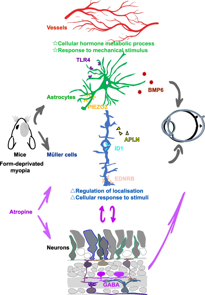
Conclusion: Retinal astrocytes and Müller cells were activated in myopia. They may response to stimuli and secretory acting factors, and might be a valid target for atropine.
Keywords: Astrocyte; Atropine; Gene set enrichment analysis; Myopia; Müller cell.
© 2022. The Author(s).
Conflict of interest statement
The authors declare that they have no competing interests.
Figures
Similar articles
-
Retinal glia in myopia: current understanding and future directions.Front Cell Dev Biol. 2024 Dec 20;12:1512988. doi: 10.3389/fcell.2024.1512988. eCollection 2024. Front Cell Dev Biol. 2024. PMID: 39759766 Free PMC article. Review.
-
Atropine Affects the Outer Retina During Inhibiting Form Deprivation Myopia in Guinea Pigs.Curr Eye Res. 2022 Apr;47(4):614-623. doi: 10.1080/02713683.2021.2009515. Epub 2022 Jan 12. Curr Eye Res. 2022. PMID: 35021941
-
Expression profiles of nestin and synemin in reactive astrocytes and Müller cells following retinal injury: a comparison with glial fibrillar acidic protein and vimentin.Mol Vis. 2010 Nov 27;16:2511-23. Mol Vis. 2010. PMID: 21139996 Free PMC article.
-
Glio-vascular modifications caused by Aquaporin-4 deletion in the mouse retina.Exp Eye Res. 2016 May;146:259-268. doi: 10.1016/j.exer.2016.03.019. Epub 2016 Mar 24. Exp Eye Res. 2016. PMID: 27018215
-
Glia-neuron interactions in the mammalian retina.Prog Retin Eye Res. 2016 Mar;51:1-40. doi: 10.1016/j.preteyeres.2015.06.003. Epub 2015 Jun 23. Prog Retin Eye Res. 2016. PMID: 26113209 Review.
Cited by
-
Characterization of Retinal VIP-Amacrine Cell Development During the Critical Period.Cell Mol Neurobiol. 2024 Feb 5;44(1):19. doi: 10.1007/s10571-024-01452-x. Cell Mol Neurobiol. 2024. PMID: 38315298 Free PMC article.
-
Proteomic analysis of CD29+ Müller cells reveals metabolic reprogramming in rabbit myopia model.Sci Rep. 2024 Oct 14;14(1):24072. doi: 10.1038/s41598-024-75637-1. Sci Rep. 2024. PMID: 39402218 Free PMC article.
-
Understanding amblyopia from the perspective of neurovascular units: changes in the retina and brain.Front Cell Dev Biol. 2025 Jun 27;13:1590009. doi: 10.3389/fcell.2025.1590009. eCollection 2025. Front Cell Dev Biol. 2025. PMID: 40655949 Free PMC article. Review.
-
Sustained Experimental Myopia Exacerbates the Effect of Eye Growth on Retinal Ganglion Cell Density and Function.Int J Mol Sci. 2025 Mar 20;26(6):2824. doi: 10.3390/ijms26062824. Int J Mol Sci. 2025. PMID: 40141465 Free PMC article.
-
Retinal glia in myopia: current understanding and future directions.Front Cell Dev Biol. 2024 Dec 20;12:1512988. doi: 10.3389/fcell.2024.1512988. eCollection 2024. Front Cell Dev Biol. 2024. PMID: 39759766 Free PMC article. Review.
References
-
- Wallman J, Winawer J. Homeostasis of eye growth and the question of myopia. Neuron. 2004;43(4):447–468. - PubMed
MeSH terms
Substances
Grants and funding
LinkOut - more resources
Full Text Sources
