. 2022 Jun 24:3:100059.
doi: 10.1016/j.fsirep.2022.100059. eCollection 2022 Dec.

Rapid and sensitive detection of pathogenic Elizabethkingia miricola in black spotted frog by RPA-LFD and fluorescent probe-based RPA

Affiliations

Rapid and sensitive detection of pathogenic Elizabethkingia miricola in black spotted frog by RPA-LFD and fluorescent probe-based RPA

Meihua Qiao et al. Fish Shellfish Immunol Rep. .

Abstract

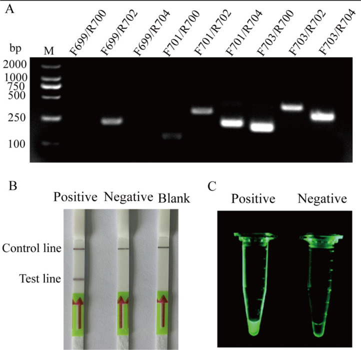
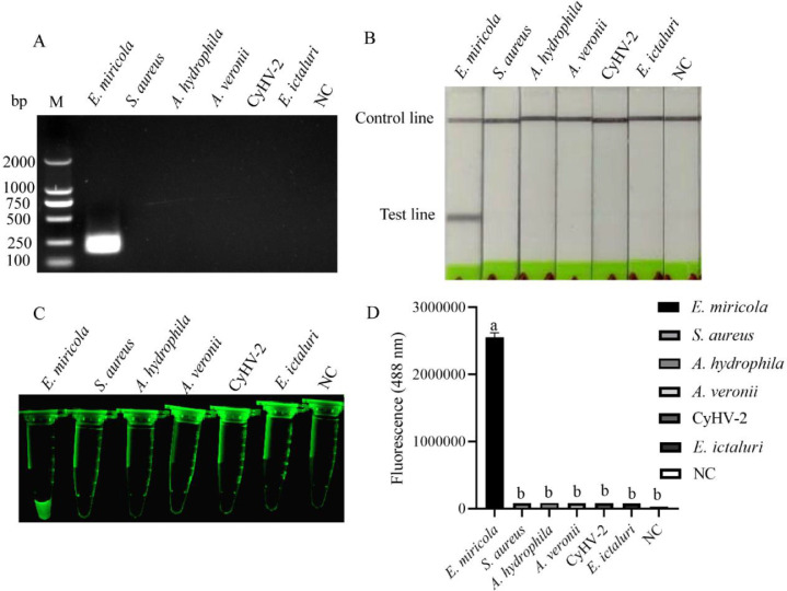
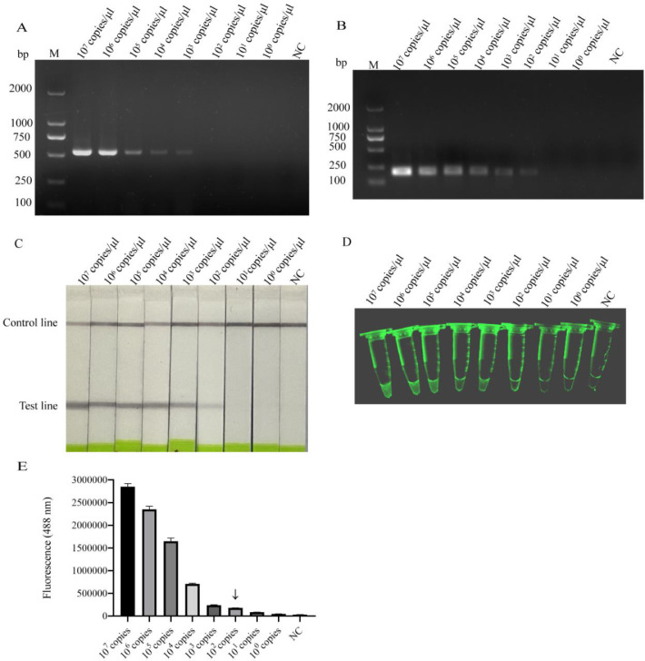
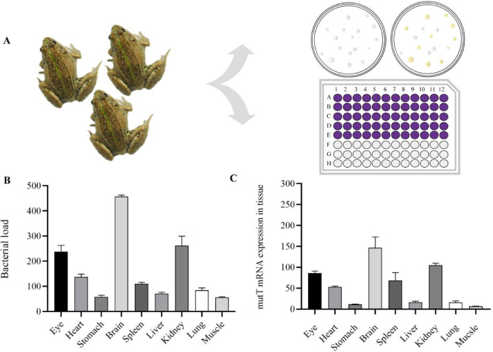
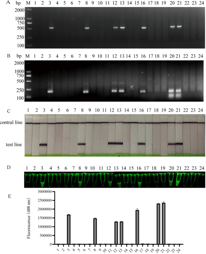
Elizabethkingia miricola is a highly infectious pathogen, which causes high mortality rate in frog farming. Therefore, it is urgent to develop a rapid and sensitive detection method. In this study, two rapid and specific methods including recombinase polymerase amplification combined with lateral flow dipstick (RPA-LFD) and fluorescent probe-based recombinase polymerase amplification (exo RPA) were established to effectively detect E. miricola, which can accomplish the examination at 38 °C within 30 min. The limiting sensitivity of RPA-LFD and exo RPA (102 copies/μL) was ten-fold higher than that in generic PCR assay. The specificities of the two methods were verified by detecting multiple DNA samples (E. miricola, Staphylococcus aureus, Aeromonas hydrophila, Aeromonas veronii, CyHV-2 and Edwardsiella ictaluri), and the result showed that the single band was displayed in E. miricola DNA only. By tissue bacterial load and qRT-PCR assays, brain is the most sensitive tissue. Random 24 black spotted frog brain samples from farms were tested by generic PCR, basic RPA, RPA-LFD and exo RPA assays, and the results showed that RPA-LFD and exo RPA methods were able to detect E. miricola accurately and rapidly. In summary, the methods of RPA-LFD and exo RPA were able to detect E. miricola conveniently, rapidly, accurately and sensitively. This study provides prospective methods to detect E. miricola infection in frog culture.

Keywords: Elizabethkingia miricola; Lateral flow dipstick; Pelophylax nigromaculatus; Recombinase polymerase amplification (RPA); exo RPA.


Conflict of interest statement

The authors declare that they have no known competing financial interests or personal relationships that could have appeared to influence the work reported in this paper.

Figures

Fig 1
Fig. 1
Screening the optimal primers and validating probes for the detection of E. miricola. 2% agarose gel electrophoresis of RPA products produced by nine sets of primer combinations was performed (A). Availability of mutT1 probe by RPA-LFD assay (B). Positive: in the reaction system, E. miricola DNA sample was used as template, and probe was mutT1 probe. Negative: in the reaction system, H2O was used as template and probe was mutT1 probe. Blank: in the reaction system, no template was used and probe is mutT1 probe. Availability of mutT2 probe by exo RPA assay (C). Positive: in the reaction system, E. miricola DNA sample was used as template, and probe was mutT2 probe. Negative: in the reaction system, H2O was used as template and probe was mutT2 probe.
Fig 2
Fig. 2
Specificity detection of RPA, RPA-LFD and exo RPA. The samples for RPA,RPA-LFD and exo RPA specificity assays were different pathogens. RPA products were exhibited by agarose gel electrophoresis (A), lateral flow dipstick (B), fluorescence imaging system (C). The fluorescence intensities of exo RPA products were detected by the enzyme-labeled instrument (D). “NC” indicates negative control. (n = 3). Different lowercase letters indicate significant differences between groups.
Fig 3
Fig. 3
Sensitivities of PCR, RPA, RPA-LFD and exo RPA assays. The pMD19-mutT standard plasmid was diluted to 10°–107 copies/μL, and then used to detect minimum detectable concentration by PCR (A), RPA (B), RPA-LFD (C) and exo RPA (D). The fluorescence intensities of exo RPA products were detected by the enzyme-labeled instrument (E), the symbol ↓ represents the limit of visible fluorescence. “NC” indicates negative control. (n = 3).
Fig 4
Fig. 4
Screening tissues with high bacterial load. Pattern diagram of bacterial load detection methods (A). Tissue bacterial load was detected by dilution coated plate method (B), and the expression of mutT in tissues was detected by qRT-PCR (C).
Fig 5
Fig. 5
Evaluation of the clinical performance of PCR, RPA, RPA-LFD and exo RPA. Random 24 frog brain DNA were tested by conventional PCR (A), RPA (B), RPA-LFD (C) and exo RPA (D). The fluorescence intensity of the 24 exo RPA products were detected with an enzyme-labeled instrument (E).

Similar articles

Cited by

References

    1. Kim K.K., Kim M.K., Lim J.H., Park H.Y., Lee S.T. Transfer of Chryseobacterium meningosepticum and Chryseobacterium miricola to Elizabethkingia gen. nov. as Elizabethkingia meningoseptica comb. nov. and Elizabethkingia miricola comb. nov. Int. J. Syst. Evol. Microbiol. 2005;55(3):1287–1293. - PubMed
    1. Trimpert J., Eichhorn I., Vladimirova D., Haake A., Schink A.K., Klopfleisch R., Lubke-Becker A. Elizabethkingia miricola infection in multiple anuran species. Transbound Emerg. Dis. 2020;68(2):931–940. - PubMed
    1. Elizabeth C., Isabel C., Carla F., Fernando C. Joint infection due to Elizabethkingia miricola. Span. J. Chemother. 2020;33(2):141–142. - PMC - PubMed
    1. Gupta P., Zaman K., Mohan B., Taneja N. Elizabethkingia miricola: a rare non-fermenter causing urinary tract infection. World J. Clin. Cases. 2017;5(5):187–190. - PMC - PubMed
    1. Hu R.X., Yuan J.F., Meng Y., Wang Z., Gu Z.M. Pathogenic Elizabethkingia miricola infection in cultured black-spotted frogs, China, 2016. Emerg. Infect. Dis. 2016;23(12):2055–2059. - PMC - PubMed

LinkOut - more resources