. 2022 Nov 7:12:1007305.
doi: 10.3389/fonc.2022.1007305. eCollection 2022.

Homotypic cell-in-cell structures as an adverse prognostic predictor of hepatocellular carcinoma

Affiliations

Homotypic cell-in-cell structures as an adverse prognostic predictor of hepatocellular carcinoma

Ruizhi Wang et al. Front Oncol. .

Abstract

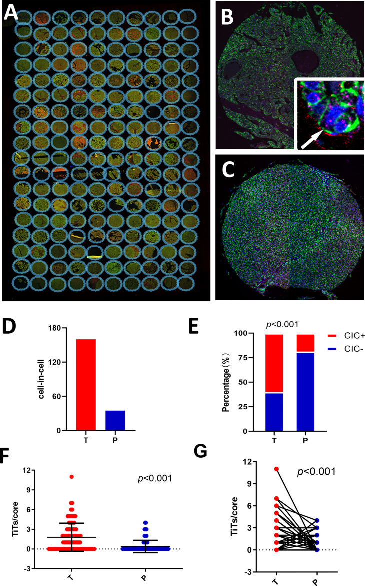
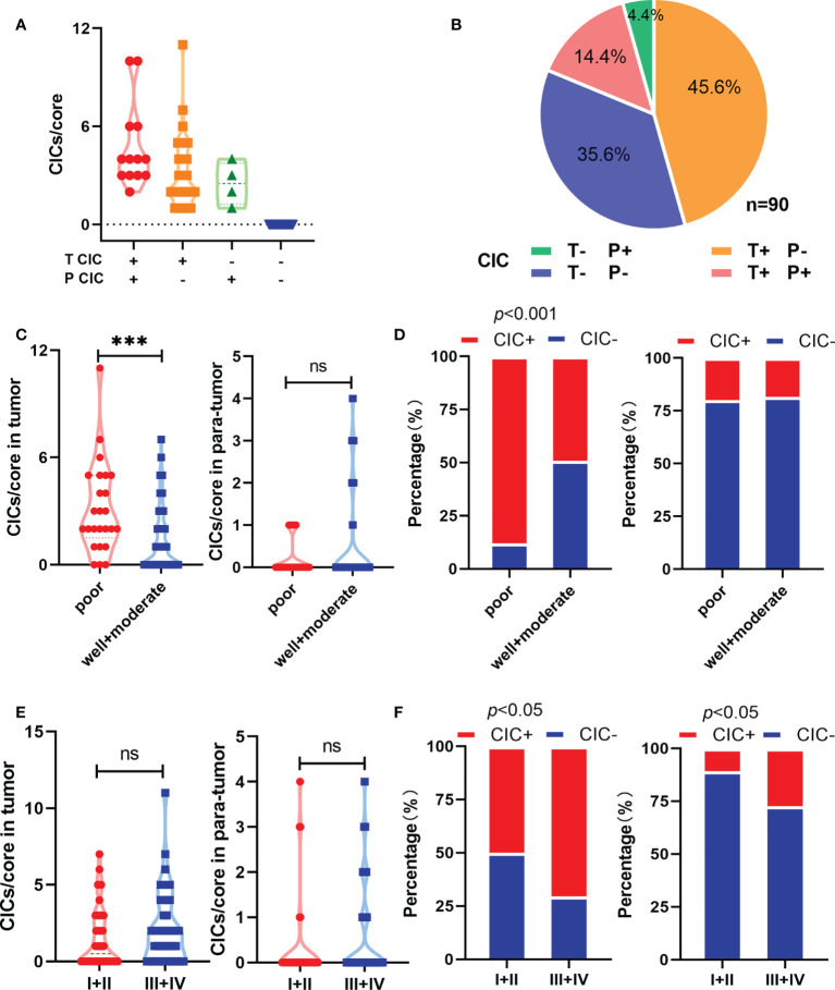
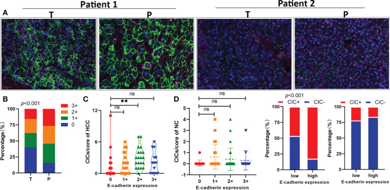
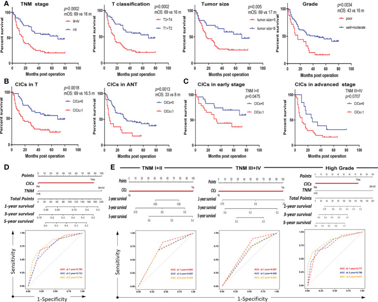
Hepatocellular carcinoma (HCC) is one of the most common malignant liver tumors. A homotypic cell-in-cell structure (hoCIC) refers to one or more cells internalized into the same type as their neighbors, which predominantly occurs in multiple tumors. The objective of this study was to investigate the prognostic value of hoCICs in HCC and its relationship with other clinicopathological features. By immunostaining analysis of a panel of HCC tissues, we found that hoCICs were prevalent in tumor tissues (54/90) but not in para-tumor tissues (17/90). The presence of hoCICs in tumor tissues was closely associated with E-cadherin expression. The presence of CICs was identified as significantly associated with poor survival rates of patients with HCC, comparable to traditional clinicopathological parameters, such as histological grade [hazard ratio (HR) = 0.734, p = 0.320]. Multivariate Cox regression analysis further confirmed that CICs were an independent risk factor for poor survival (HR = 1.902, p = 0.047). In addition, hoCICs were the predominant contributor in a nomogram model constructed for survival prediction at 1, 3, and 5 years [the areas under the curve (AUCs) were 0.760, 0.733, and 0.794, respectively]. Stratification analysis indicated that hoCICs tend to selectively affect patients with high-grade disease (HR = 2.477, p = 0.009) and at the early TNM stage (HR = 2.351, p = 0.05). Thus, hoCICs predict poor survival of patients with HCC, particularly those with higher grades and at an early stage.

Keywords: E-cadherin; hepatocellular carcinoma (HCC); homotypic cell-in-cell structures (hoCICs); overall survival (OS); prognostic predictor.


Conflict of interest statement

The authors declare that the research was conducted in the absence of any commercial or financial relationships that could be construed as a potential conflict of interest.

Figures

Figure 1
Figure 1
Detecting homotypic CICs in a tissue microarray (TMA) of human HCC. (A) Composite images of a whole TMA slide stained with antibodies against E-cadherin (green), CD44 (red), CD45 (pink), and DAPI staining of the nucleus (blue). (B) Single representative tumor tissue (T) core. The arrow in the inserted image shows one typical CIC. (C) Para-tumor tissue core (P). (D) CIC counts in tumor and para-tumor tissues. (E) Percentages of patients with or without CICs in tumor and non-tumor tissues. (F) CICs detected in 90 tumor cores and paired para-tumor tissue cores. (G) CICs presented as number of CICs per core in the tumor and non-tumor tissues.
Figure 2
Figure 2
Homotypic CICs are associated with clinicopathological characteristics in HCC tissues. (A) CIC counts normalized by core area in CIC positive tumor and para-tumor tissues. (B) Quantification of CICs in tumor and para-tumor tissues. (C) Quantification of CICs stratified by histological grade in tumor (left) and para-tumor tissues (right). (D) CIC compositions by histological grade in tumor (left) and para-tumor tissues (right). (E) Quantification of CICs stratified by TNM in tumor (left) and para-tumor tissues (right). (F) CIC compositions by TNM in tumor (left) and para-tumor tissues (right). *P < 0.05; **P < 0.01; ***P < 0.001; ns, not significant.
Figure 3
Figure 3
E-cadherin expression in tumor tissues is positively associated with homotypic CIC formation. (A) Representative images for high (two on the left) or low (two on the right) levels of E-cadherin expression in pair-matched tumor (T) and para-tumor (P) tissue samples of the TMA by immunofluorescent staining. The TMA slides were stained with antibodies against E-cadherin (green), CD44 (red), CD45 (pink), and DAPI staining of the nucleus (blue). (B) Quantification of E-cadherin expression in pair-matched tumor (T) and para-tumor (P) tissue samples of the TMA. n = 180 with 90 for tumor and 90 for para-tumor, respectively. (C) E-cadherin expression correlated significantly with CIC formation in tumor tissues (left), but not in para-tumor tissues of the TMA. (D) CIC compositions in low or high E-cadherin expression groups of tumor (left) and para-tumor tissues (right). *P < 0.05; **P < 0.01; ***P < 0.001; ns, not significant.
Figure 4
Figure 4
CICs selectively impact overall survival of a certain group of patients with HCC. (A) Survival analysis of patients with HCC stratified by TNM, T classification, tumor size, and histological grade, respectively. (B) The presence of CIC formation (CICs ≥ 1/core) both in tumor (left) and para-tumor (right) tissues is associated with shorter patient survival by Kaplan–Meier plotting of data from TMA staining. (C) The presence of CIC formation (CICs ≥ 1/core) in tumor (left) but not in para-tumor (right) tissues is associated with shorter patient survival by Kaplan–Meier plotting of data from TMA staining. (D) Nomogram and AUC analysis with two independent prognostic factors (TNM stage and CICs). (E) Nomogram and AUC analysis in a cohort of patients stratified by TNM stage (I+II vs. III+IV) (left and middle) or histological grade (well + moderate) (right).

References

    1. Wang D, Wang R, Huang A, Fang Z, Wang K, He M, et al. . Upregulation of macrophage migration inhibitory factor promotes tumor metastasis and correlates with poor prognosis of pancreatic ductal adenocarcinoma. Oncol Rep (2018) 40(5):2628–36. doi: 10.3892/or.2018.6703 - DOI - PMC - PubMed
    1. He MF, Wang S, Wang Y, Wang XN. Modeling cell-in-Cell structure into its biological significance. Cell Death Dis (2013) 4:e630. doi: 10.1038/cddis.2013.147 - DOI - PMC - PubMed
    1. Fais S, Overholtzer M. Cell-in-Cell phenomena in cancer. Nat Rev Cancer (2018) 18(12):758–66. doi: 10.1038/s41568-018-0073-9 - DOI - PubMed
    1. Wang X. Cell-in-Cell phenomenon: A new paradigm in life sciences. Curr Mol Med (2015) 15(9):810–8. doi: 10.2174/1566524015666151026095730 - DOI - PMC - PubMed
    1. Huang H, He M, Zhang Y, Zhang B, Niu Z, Zheng Y, et al. . Identification and validation of heterotypic cell-in-Cell structure as an adverse prognostic predictor for young patients of resectable pancreatic ductal adenocarcinoma. Signal Transduct Target Ther (2020) 5(1):246–8. doi: 10.1038/s41392-020-00346-w - DOI - PMC - PubMed

LinkOut - more resources