

NK cells with decreased expression of multiple activating receptors is a dominant phenotype in pediatric patients with acute lymphoblastic leukemia
- PMID: 36419901
- PMCID: PMC9677112
- DOI: 10.3389/fonc.2022.1023510
NK cells with decreased expression of multiple activating receptors is a dominant phenotype in pediatric patients with acute lymphoblastic leukemia
Abstract
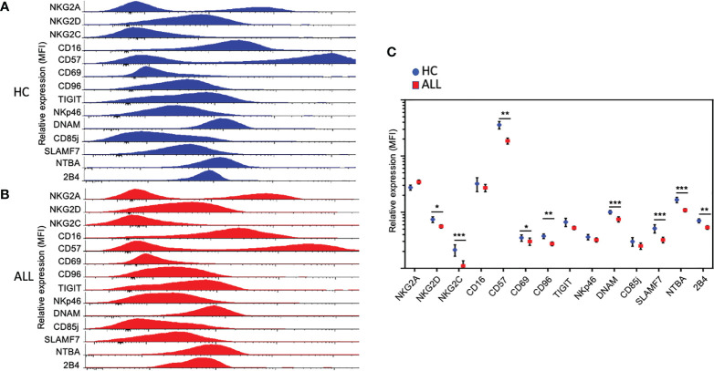
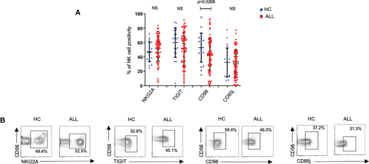
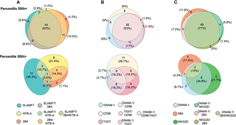
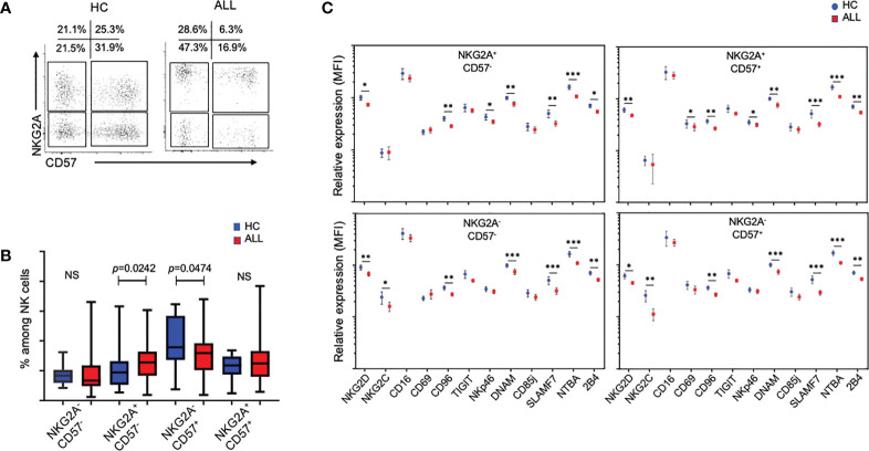
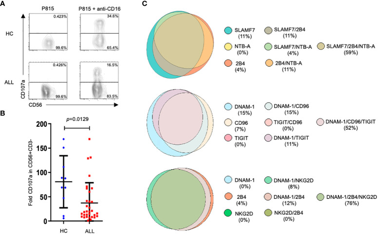
NK cells have unique attributes to react towards cells undergoing malignant transformation or viral infection. This reactivity is regulated by activating or inhibitory germline encoded receptors. An impaired NK cell function may result from an aberrant expression of such receptors, a condition often seen in patients with hematological cancers. Acute lymphoblastic leukemia (ALL) is the most common pediatric cancer worldwide and NK cells have emerged as crucial targets for developing immunotherapies. However, there are important gaps concerning the phenotype and behavior of NK cells during emergence of ALL. In this study we analyze the phenotype and function of NK cells from peripheral blood in pediatric patients with ALL at diagnosis. Our results showed that NK cells exhibited an altered phenotype highlighted by a significant reduction in the overall expression and percent representation of activating receptors compared to age-matched controls. No significant differences were found for the expression of inhibitory receptors. Moreover, NK cells with a concurrent reduced expression in various activating receptors, was the dominant phenotype among patients. An alteration in the relative frequencies of NK cells expressing NKG2A and CD57 within the mature NK cell pool was also observed. In addition, NK cells from patients displayed a significant reduction in the ability to sustain antibody-dependent cellular cytotoxicity (ADCC). Finally, an aberrant expression of activating receptors is associated with the phenomenon of leukemia during childhood.
Keywords: NK cells; acute lymphoblastic leukemia; cancer; immune system; immunooncology.
Copyright © 2022 Valenzuela-Vázquez, Nuñez-Enriquez, Sánchez-Herrera, Medina-Sanson, Pérez-Saldivar, Jiménez-Hernández, Martiín-Trejo, Del Campo-Martínez, Flores-Lujano, Amador-Sánchez, Mora-Ríos, Peñaloza-González, Duarte-Rodríguez, Torres-Nava, Espinosa-Elizondo, Cortés-Herrera, Flores-Villegas, Merino-Pasaye, Almeida-Hernández, Ramírez-Colorado, Solís-Labastida, Medrano-López, Pérez-Gómez, Velázquez-Aviña, Martínez-Ríos, Aguilar-De los Santos, Santillán-Juárez, Gurrola-Silva, García-Velázquez, Mata-Rocha, Hernández-Echáurregui, Sepúlveda-Robles, Rosas-Vargas, Mancilla-Herrera, Jimenez-Morales, Hidalgo-Miranda, Martinez-Duncker, Waight, Hance, Madauss, Mejía-Aranguré and Cruz-Munoz.
Conflict of interest statement
Authors JDW, KWH, and KPM are employed by GlaxoSmithKline. The remaining authors declare that the research was conducted in the absence of any commercial or financial relationships that could be construed as a potential conflict of interest.
Figures

Similar articles
-
NKG2A discriminates natural killer cells with a suppressed phenotype in pediatric acute leukemia.J Leukoc Biol. 2024 Jan 19;115(2):334-343. doi: 10.1093/jleuko/qiad112. J Leukoc Biol. 2024. PMID: 37738462
-
Functional characterization of NK cells in Mexican pediatric patients with acute lymphoblastic leukemia: Report from the Mexican Interinstitutional Group for the Identification of the Causes of Childhood Leukemia.PLoS One. 2020 Jan 17;15(1):e0227314. doi: 10.1371/journal.pone.0227314. eCollection 2020. PLoS One. 2020. PMID: 31951638 Free PMC article.
-
Overexpression of CD158 and NKG2A Inhibitory Receptors and Underexpression of NKG2D and NKp46 Activating Receptors on NK Cells in Acute Myeloid Leukemia.Arch Med Res. 2016 Jan;47(1):55-64. doi: 10.1016/j.arcmed.2016.02.001. Epub 2016 Feb 12. Arch Med Res. 2016. PMID: 26876298
-
Exploitation of natural killer cells for the treatment of acute leukemia.Blood. 2016 Jun 30;127(26):3341-9. doi: 10.1182/blood-2015-12-629055. Epub 2016 May 20. Blood. 2016. PMID: 27207791 Review.
-
Role of Toll-like receptors in natural killer cell function in acute lymphoblastic leukemia.Oncol Lett. 2021 Nov;22(5):748. doi: 10.3892/ol.2021.13009. Epub 2021 Aug 24. Oncol Lett. 2021. PMID: 34539852 Free PMC article. Review.
Cited by
-
Contribution of the TIME in BCP-ALL: the basis for novel approaches therapeutics.Front Immunol. 2024 Jan 17;14:1325255. doi: 10.3389/fimmu.2023.1325255. eCollection 2023. Front Immunol. 2024. PMID: 38299154 Free PMC article. Review.
-
AATF/Che-1 RNA polymerase II binding protein overexpression reduces the anti-tumor NK-cell cytotoxicity through activating receptors modulation.Front Immunol. 2023 Jun 26;14:1191908. doi: 10.3389/fimmu.2023.1191908. eCollection 2023. Front Immunol. 2023. PMID: 37435061 Free PMC article.
-
TLR Agonists Modify NK Cell Activation and Increase Its Cytotoxicity in Acute Lymphoblastic Leukemia.Int J Mol Sci. 2024 Jul 8;25(13):7500. doi: 10.3390/ijms25137500. Int J Mol Sci. 2024. PMID: 39000607 Free PMC article.
-
Exploring NK cell receptor dynamics in paediatric leukaemias: implications for immunotherapy and prognosis.Clin Transl Immunology. 2024 Mar 23;13(3):e1501. doi: 10.1002/cti2.1501. eCollection 2024. Clin Transl Immunology. 2024. PMID: 38525380 Free PMC article.
-
Defects in NK cell immunity of pediatric cancer patients revealed by deep immune profiling.iScience. 2024 Aug 28;27(9):110837. doi: 10.1016/j.isci.2024.110837. eCollection 2024 Sep 20. iScience. 2024. PMID: 39310750 Free PMC article.
References
LinkOut - more resources
Full Text Sources