. 2022 Nov 3:30:438-448.
doi: 10.1016/j.omtn.2022.10.023. eCollection 2022 Dec 13.

Sustainable recovery of MBNL activity in autoregulatory feedback loop in myotonic dystrophy

Affiliations

Sustainable recovery of MBNL activity in autoregulatory feedback loop in myotonic dystrophy

Zuzanna Rogalska et al. Mol Ther Nucleic Acids. .

Abstract

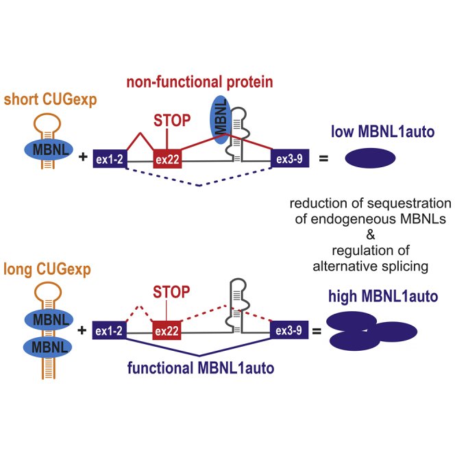
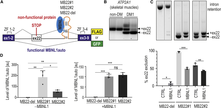
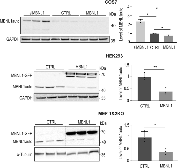
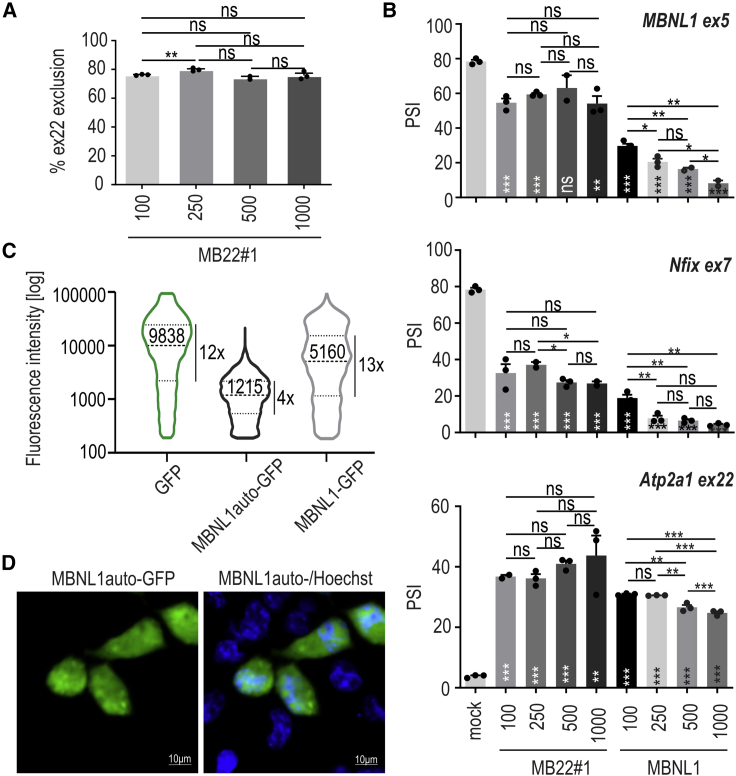
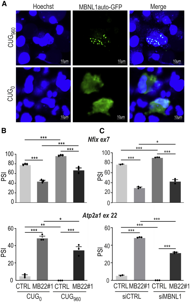
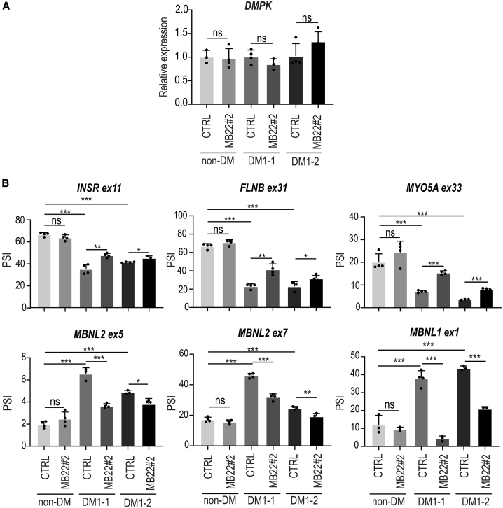
Muscleblind-like proteins (MBNLs) are RNA-binding proteins essential for the developmental regulation of various processes including alternative splicing. Their activity is misregulated in myotonic dystrophy type 1 (DM1), an incurable genetic, neuro-muscular disorder caused by uncontrolled expansion of CTG repeats. Mutant RNAs containing hundreds or thousands of repeats efficiently sequester MBNL proteins. As a consequence, global alternative splicing abnormalities are induced. Importantly, the size of expansion differs significantly not only between patients but also between different parts of the same muscle as a consequence of somatic expansion. One of the potential therapeutic strategies in DM is overexpression of MBNLs. However, gene therapy tools might induce excessive activity of MBNLs, what in turn might change the metabolism of many RNAs. To overcome these limitations, we designed an autoregulated MBNL1 overexpression system. The genetic construct contains an MBNL1-coding sequence separated by the fragment of ATP2A1 pre-mRNA with an MBNL-sensitive alternative exon containing stop codon in the reading frame of MBNL1. Inclusion of this exon leads to the arrangement of an inactive form of the protein, but exclusion gives rise to fully active MBNL1. This approach enables the autoregulation of the amount of overexpressed MBNL1 with high dynamic range which ensures a homogeneous level of this protein in cells treated with the genetic construct. We demonstrated beneficial effects of an autoregulated construct on alternative splicing patterns in DM1 models and cells derived from patients with DM1.

Keywords: DM1; MBNL; MBNL1 overexpression; MT: Delivery Strategies; alternative splicing; expansion of CCUG repeats; expansion of CUG repeats; gene therapy; microsatellites; myotonic dystrophy type 1.


Conflict of interest statement

The authors declare no conflicts of interest.

Figures

None
Graphical abstract
Figure 1
Figure 1
Inclusion of the alternative exon to mRNA encoding MBNL1auto depends on the MBNL-binding RNA regulatory motif (A) The scheme of the genetic construct for autoregulated overexpression of MBNL1auto. The MBNL1 coding sequence was divided into two parts separated by an intron21/exon (ex)22/intron22 sequence from human ATP2A1. Ex1-2 is a sequence of the first two exons of MBNL1, containing the first zinc finger tandem (ZF1–ZF2); ex3-9 is a sequence of cDNA of ex3–ex9 with ZF3 and ZF4. ATP2A1 fragment contains the alternative ex22 flanked from both sides by introns and 7 bp of ex21 and 2 bp of ex23 to maintain correct splicing regulation and to keep the open reading frame of MBNL1auto. The inclusion of ex22, positively regulated by MBNLs, leads to premature translation termination and the arrangement of an inactive form of protein. The MB22 construct contains the wild-type sequence recognized by MBNLs (blue) within intron22 of ATP2A1 (MB22#1). This sequence was replaced by 4xUGCU MBNL-binding motif (MB22#2) or completely removed (MB22-del; orange). GFP and FLAG are tags located in the frame of MBNL1auto. (B) Alternative splicing profile of ATP2A1 ex22 in two normal adult skeletal muscles (non-DM) and two different skeletal muscles from patients with DM1 (DM1) analyzed by RT-PCR. Isoforms with and without ex22 are marked. (C) Results of RT-PCR analysis of ex22 exclusion in cells transfected with MB22 constructs containing different MBNL-sensitive elements (MB22#1, -#2, and -del) and with MBNL1-GFP (MBNL1) or GFP (CTRL) overexpression. The percentage of alternative ex22 exclusion reflects mRNA isoforms coding for the active form of the protein. Bars represent average from n = 3 independent experiments (dots) for each experimental condition with standard deviation (SD). (D) Results of western blot analysis showing the level of MBNL1auto in cells transfected with either MB22-del, -#1, or -#2 (left; −MBNL1) or co-transfected with these three constructs and MBNL1-GFP (right; +MBNL1). Anti-FLAG antibody staining was carried out as FLAG sequence is fused to the C-terminal end of MBNL1auto in each MB22 construct. Bars represent average signal (arbitrary units [a.u.]) from n = 3–4 independent experiments for each group normalized to mCherry. Co-transfection with mCherry expression vector was utilized as a normalization control of transfection. (B and C) Unpaired Student’s t test; ∗p < 0.05; ∗∗p < 0.01; ∗∗∗p < 0.001; ns, non-significant.
Figure 2
Figure 2
Biosynthesis of the MBNL1auto depends on the available pool of MBNLs Western blot analysis of MBNL1auto in COS7, HEK-293, and MEF cells with Mbnl1 and Mbnl2 knockout. Bars represent average signal from n = 3 independent experiments for each group normalized to GAPDH in COS7 and HEK-293 cells and to α-tubulin in MEF cells. Anti-FLAG antibody staining was carried out as FLAG sequence is fused to the C-terminal end of both constructs. Unpaired Student’s t test was used to calculate statistical significance: ∗p < 0.05; ∗∗p < 0.01.
Figure 3
Figure 3
The autoregulatory potential and expression homogeneity of MB22 construct (A) The percentage of alternative ex22 exclusion from mRNA encoding for MBNL1auto after treatment of COS7 cells with different amounts of MB22#1, ranging from 100 to 1,000 ng/mL culture medium; n = 3. (B) The dose-dependent inclusion of alternative exons of three different MBNL-sensitive minigenes after treatment of COS7 cells with MB22#1- or MBNL1-GFP-overexpression construct (ng/mL); n = 3. (A and B) Unpaired Student’s t test; ∗p < 0.05; ∗∗p < 0.01; ∗∗∗p < 0.001; ns, non-significant. Stars placed on bars denote statistical significance compared with the mock. (C) Violin plots showing the distribution of cells with different fluorescent signals of either GFP, MBNL1auto-GFP, or MBNL1-GFP proteins. COS7 cells were transfected with adequate vectors 48 h prior to flow cytometry analysis. Median fluorescent intensity (black solid line) and 25th and 75th percentiles of signal (dashed lines) are shown. Fold change between 25th and 75th percentiles of signal for all analyses is also indicated. Cells with signal below 200 were rejected from analyses based on results for control experiment for mock-transfected cells. GFP and MBNL1-GFP without autoregulatory cassette state as negative controls. Graphs represent values from n = 4 independent biological replicates for each experimental condition; N = 13,942 (GFP), N = 6,760 (MBNL1auto-GFP), and N = 10,793 (MBNL1-GFP) cells. (D) Representative confocal microscopy image showing localization of MBNL1auto-GFP (green) in COS7 cells transfected with MB22#2-GFP. Nuclei were stained with Hoechst (blue); scale bar, 10 μm.
Figure 4
Figure 4
The therapeutic potential of MB22 autoregulated construct in DM models (A) Representative confocal images showing the nuclear foci containing MBNL1auto-GFP in cells expressing a mutant DMPK fragment with CUG960 (top panel). In cells expressing a normal DMPK fragment without CUG repeats, MBNL1auto-GFP is distributed equally (bottom panel); scale bar, 10 μm. (B) Results of RT-PCR analyses showing changes in the regulation of two MBNL-dependent exons from Nfix ex7 and Atp2a1 ex22 minigenes. Splicing changes are expressed as the percent spliced in (PSI). Cells were co-transfected with MB22#1 or control GFP construct (CTRL) and either mutant (CUG960) or normal (CUG0) DMPK-fragment-expressing constructs. (C) As in (B) but for cells treated with either control siRNA (siCTRL) or siRNA targeting the 3′ UTR of MBNL1 (siMBNL1). These cells were then transfected with either GFP or MB22#1 construct. The percentage of mRNA isoform with inclusion of alternative exon was calculated using the inverse of PSI parameter, which demonstrates the portion of mRNA with an included alternative exon. Bars represent average PSI from three independent experiments (with exception of n = 2 for siCTRL + GFP condition) with SD. (B and C) Unpaired Student’s t test was used to calculate statistical significance. ∗p < 0.05; ∗∗p < 0.01; ∗∗∗p < 0.001; ns, non-significant.
Figure 5
Figure 5
Correction of pathogenic missplicing in DM1 cells treated with MB22 lentiviruses (A) Results of quantitative real-time RT-PCR analysis showing relative expression of DMPK (normalized to GAPDH) in three different cell lines: fibroblasts derived from healthy individual (non-DM) and two patients with DM1 (DM1-1 and DM1-2) treated with either control (CTRL) lentiviral vector or lentivirus containing the MB22#2-GFP sequence 12 days from cell transduction. (B) Results of RT-PCR-based analyses of alternative splicing changes in cells described in (A). Changes in the inclusion of positively (top panel) and negatively (bottom panel) regulated MBNL-dependent alternative exons are shown for six transcripts affected in DM1. Splicing changes are expressed as PSI. Bars represent average from 3 to 4 independent experiments (dots); unpaired Student’s t test; ∗p < 0.05; ∗∗p < 0.01; ∗∗∗p < 0.001; ns, non-significant.

Similar articles

Cited by

References

    1. Ho T.H., Charlet-B N., Poulos M.G., Singh G., Swanson M.S., Cooper T.A. Muscleblind proteins regulate alternative splicing. EMBO J. 2004;23:3103–3112. doi: 10.1038/sj.emboj.7600300. - DOI - PMC - PubMed
    1. Batra R., Manchanda M., Swanson M.S. Global insights into alternative polyadenylation regulation. RNA Biol. 2015;12:597–602. doi: 10.1080/15476286.2015.1040974. - DOI - PMC - PubMed
    1. Wang E.T., Cody N.A.L., Jog S., Biancolella M., Wang T.T., Treacy D.J., Luo S., Schroth G.P., Housman D.E., Reddy S., et al. Transcriptome-wide regulation of pre-mRNA splicing and mRNA localization by muscleblind proteins. Cell. 2012;150:710–724. doi: 10.1016/j.cell.2012.06.041. - DOI - PMC - PubMed
    1. Fernandez-Costa J.M., Llamusi M.B., Garcia-Lopez A., Artero R. Alternative splicing regulation by Muscleblind proteins: from development to disease. Biol. Rev. Camb. Philos. Soc. 2011;86:947–958. doi: 10.1111/j.1469-185X.2011.00180.x. - DOI - PubMed
    1. Kanadia R.N., Urbinati C.R., Crusselle V.J., Luo D., Lee Y.J., Harrison J.K., Oh S.P., Swanson M.S. Developmental expression of mouse muscleblind genes Mbnl1, Mbnl2 and Mbnl3. Gene Expr. Patterns. 2003;3:459–462. doi: 10.1016/S1567-133X(03)00064-4. - DOI - PubMed