

A two-year longitudinal study of retinal vascular impairment in patients with amnestic mild cognitive impairment
- PMID: 36420311
- PMCID: PMC9678013
- DOI: 10.3389/fnagi.2022.993621
A two-year longitudinal study of retinal vascular impairment in patients with amnestic mild cognitive impairment
Erratum in
-
Corrigendum: A two-year longitudinal study of retinal vascular impairment in patients with amnestic mild cognitive impairment.Front Aging Neurosci. 2023 Apr 20;15:1197979. doi: 10.3389/fnagi.2023.1197979. eCollection 2023. Front Aging Neurosci. 2023. PMID: 37151846 Free PMC article.
Abstract
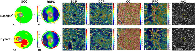
Objective: To evaluate the relation between retinal vascular impairment and cognitive decline in patients with amnestic mild cognitive impairment (aMCI) over time.
Methods: Spectral domain-optical coherence tomography (SD-OCT) and OCT angiography study was performed in aMCI patients over 2 years follow-up and compared to baseline.
Results: Thirty-eight eyes from 19 aMCI patients were evaluated. Structural and vascular OCT measures were reduced at follow-up except for vessel density (VD) of the choriocapillaris, unchanged, and foveal avascular zone, which was increased; no changes in any parameter were found in 18 age-matched healthy controls. Overall, these findings were confirmed when patients were evaluated separately according to progression to dementia. Only non-converters to dementia showed significant VD reduction in the deep capillary plexuses (coeff. β = -4.20; p < 0.001), may be for an initial massive VD depletion becoming less evident with progression of the disease. MMSE reduction was associated with a higher ganglion cell complex reduction (coeff. β = 0.10; p = 0.04) and a higher VD reduction in the radial peripapillary capillary (RPC) plexus (coeff. β = 0.14; p = 0.02) in the whole patient group, while it was associated with a higher VD reduction only in RPC plexus in converters (coeff. β = 0.21; p < 0.001).
Conclusion: Our data shows vascular impairment progression in the inner retina of aMCI patients and support the hypothesis that vascular changes may contribute to the onset and progression of Alzheimer's disease. Other follow-up studies, with a larger number of patients, are needed to better define VD as a potential biomarker.
Keywords: Alzheimer’s disease; OCTA; aMCI; longitudinal study; mini mental state examination.
Copyright © 2022 Chiara, Gilda, Daniela, Antonio, Miriana, Marcello, Elena, Roberta, Ciro and Vincenzo.
Conflict of interest statement
CA has received research grants from Almirall, research grants from ECTRIMS-MAGNIMS and honoraria from Almirall, Biogen, Roche Sanofi-Genzyme and Novartis. MoM has received research grants from ECTRIMS-MAGNIMS, UK MS Society, and Merck; and honoraria from Biogen, Merck, Roche, and Sanofi-Genzyme. The remaining authors declare that the research was conducted in the absence of any commercial or financial relationships that could be construed as a potential conflict of interest.
Figures
References
-
- Albert M. S., DeKosky S. T., Dickson D., Dubois B., Feldman H. H., Fox N. C., et al. . (2011). The diagnosis of mild cognitive impairment due to Alzheimer's disease: recommendations from the National Institute on Aging-Alzheimer's Association workgroups on diagnostic guidelines for Alzheimer's disease. Alzheimers Dement. 7, 270–279. doi: 10.1016/j.jalz.2011.03.008, PMID: - DOI - PMC - PubMed
LinkOut - more resources
Full Text Sources