. 2023 Jan 23;62(4):e202215085.
doi: 10.1002/anie.202215085. Epub 2022 Dec 14.

Dual-Color Photoconvertible Fluorescent Probes Based on Directed Photooxidation Induced Conversion for Bioimaging

Affiliations

Dual-Color Photoconvertible Fluorescent Probes Based on Directed Photooxidation Induced Conversion for Bioimaging

Lazare Saladin et al. Angew Chem Int Ed Engl. .

Abstract

We herein present a new concept to produce dual-color photoconvertible probes based on a mechanism called Directed Photooxidation Induced Conversion (DPIC). As a support of this mechanism, styryl-coumarins (SCs) bearing Aromatic Singlet Oxygen Reactive Moieties (ASORMs) like furan and pyrrole have been synthesized. SCs are bright fluorophores, which undergo a hypsochromic conversion upon visible light irradiation due to directed photooxidation of the ASORM that leads to the disruption of conjugation. SC-P, a yellow emitting probe bearing a pyrrole moiety, converts to a stable blue emitting coumarin with a 68 nm shift allowing the photoconversion and tracking of lipid droplet in live cells. This new approach might pave the way to a new generation of photoconvertible dyes for advanced bioimaging applications.

Keywords: Fluorescent Probes; Lipid Droplets; Photoconversion; Photooxidation; Photoswitching.


Conflict of interest statement

The authors declare no conflict of interest.

Figures

Figure 1
Figure 1
Principle of the Directed Photooxidation Induced Conversion (DPIC) where the conjugation between the coumarin and the Singlet Oxygen Reactive Moieties (ASORMs) is disrupted upon photooxidation of the latter. Structures of the Styryl‐Coumarins (SC), their converted forms after photoirradiation (cSC), and the model of converted form (SC‐V).
Figure 2
Figure 2
Spectroscopic studies and photoirradiation of SCs (5 μM in MeOH). (A) Absorption and emission spectra of SCs. (B–D) Emission spectra of SCs upon irradiation at 488 nm (power density: 33 mW cm−2). Whereas SC‐A undergoes photobleaching upon irradiation (B), SC‐F (C) and SC‐P (D) photoconvert towards new emissive species. (E) 3D HPLC‐UV/Visible‐mass characterization of SC‐P (star) before and after photoconversion. cSC‐P (Hashtag) is a proposed structure of one of the blue emitting photoproducts obtained after conversion based on the mass and absorption spectra below. (F) Normalized excitation (emission at 450 nm) and emission (excitation at 405 nm) spectra of converted forms cSC‐F and cSC‐P along with SC‐V showing similar photophysical properties. Insets are photographs under a UV lamp excitation of cuvette before and after irradiation.
Figure 3
Figure 3
Proposed mechanism for the Directed Photooxidation Induced Conversion. 1O2 is generated upon irradiation and reacts at the ground state leading to a phototransformation (φPt) which can be photoconversion (φPc) or photobleaching (φBl). The constants (in grey) were determined (Table 1) and support the presented mechanism (for definitions see Supporting Information). ΦΔ could not be determined due to the competitive reactivity of SCs and DPBF towards 1O2 (See Figure S11).
Figure 4
Figure 4
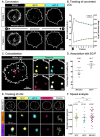
SC‐P labels LDs in Hela cells and can be converted and tracked using confocal imaging. (A) Hela Cells were labelled with SC‐P (1 μM, yellow) and counter stained for plasma membrane and endosomes with MemBright Cy5.5 (grey). Conversion of SC‐P (frame 1) toward cSC‐P (in cyan) can be induced with a 488 nm laser stimulation (green flash) and tracked over time. (B) Evolution of mean fluorescence intensity of SC‐P and cSC‐P after conversion (1 frame per second). (C) Colocalization analysis showed strong association of SC‐P (yellow), c‐SC‐P (cyan) with LDs probe SMCy5.5 (Magenta). (D) Coupling analysis of SC‐P shows strong association with SMCy5.5 (82,66%, p value <10−38) or cSP‐P (94,17% p value <10−171) on 6 independent cells (n=73 SMCy5.5 spots, and n=54 c‐SC‐P spots). (E) Tracking over time of SMCy5.5 (magenta) with SC‐P (yellow) and cSC‐P (cyan) shows correlated trajectories of vesicles over 50 frames. (F) Speed analysis of tracked vesicles reveals no statistical differences between lipid droplets vesicles (SMCy5.5) and SC‐P/cSC‐P spots.

References

    1. Patterson G. H., Lippincott-Schwartz J., Science 2002, 297, 1873–1877. - PubMed
    1. Lukyanov K. A., Chudakov D. M., Lukyanov S., Verkhusha V. V., Nat. Rev. Mol. Cell Biol. 2005, 6, 885–890. - PubMed
    1. Lavis L. D., Biochemistry 2017, 56, 5165–5170. - PubMed
    1. Trinh N., Jolliffe K. A., New E. J., Angew. Chem. Int. Ed. 2020, 59, 20290–20301; - PubMed
    2. Angew. Chem. 2020, 132, 20466–20479.
    1. Li H., Kim D., Yao Q., Ge H., Chung J., Fan J., Wang J., Peng X., Yoon J., Angew. Chem. 2021, 133, 17408–17429. - PubMed

Publication types