

The roles of sub-micron and microscale roughness on shear-driven thrombosis on titanium alloy surfaces
- PMID: 36420933
- PMCID: PMC10010941
- DOI: 10.1111/aor.14467
The roles of sub-micron and microscale roughness on shear-driven thrombosis on titanium alloy surfaces
Abstract
Background: Continuous-flow ventricular assist devices (cfVADs) are implanted in patients with end-stage heart failure to assist with blood circulation. However, VAD implantation is associated with dangerous thrombotic complications. Our goal was to determine the impact of micron and sub-micron scale Ti6Al4V surface roughness on adherent platelet aggregate properties under clinically relevant shear rates.
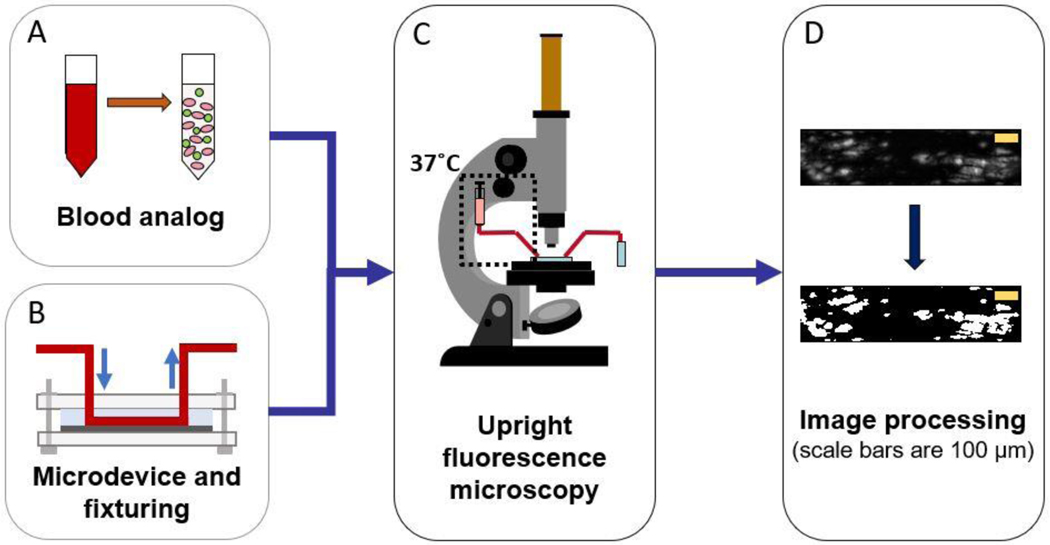
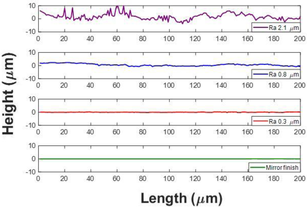
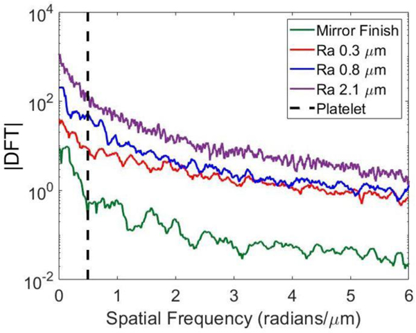
Methods: We used fluorescence microscopy to visualize platelets in real time as they adhered to Ti6Al4V coupons of varying degrees of roughness, including a smooth control, in microfluidic channels and quantified deposition using an image processing algorithm. We systematically characterized roughness using spatial frequencies to generalize results for more blood-biomaterial contact applications.
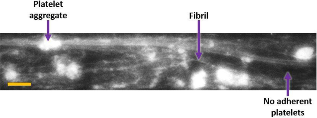
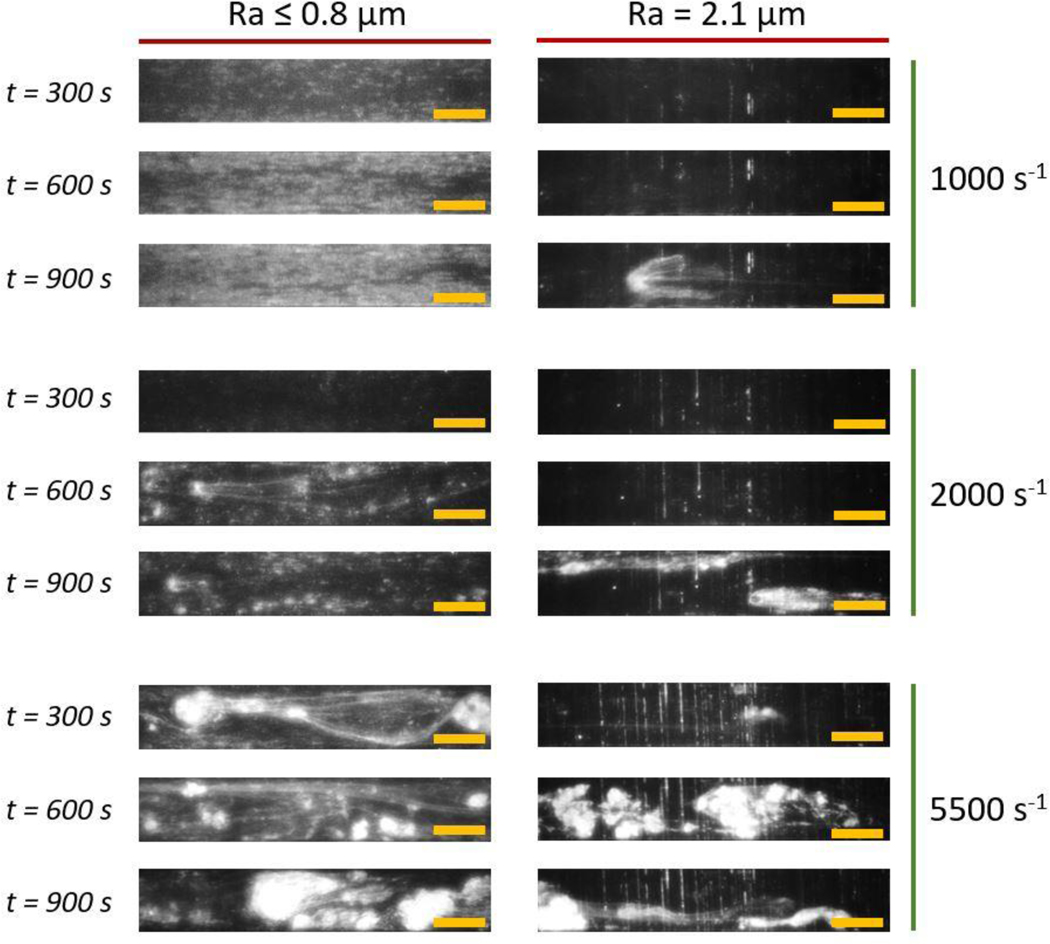
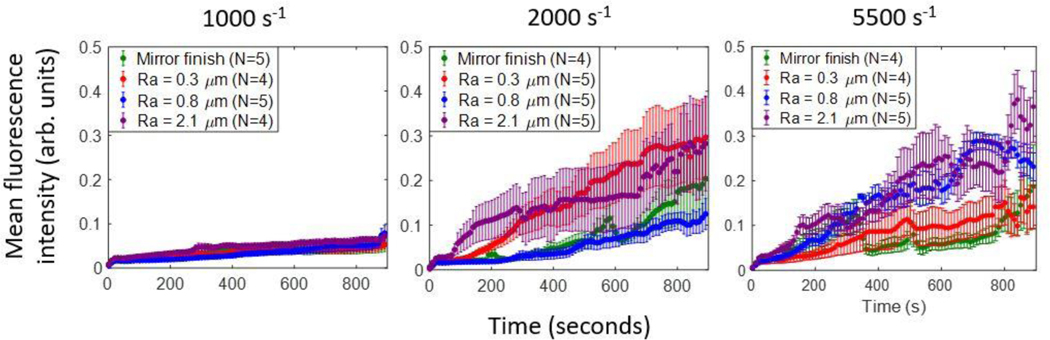
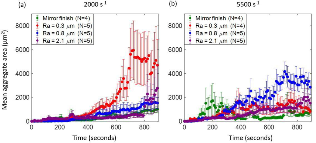
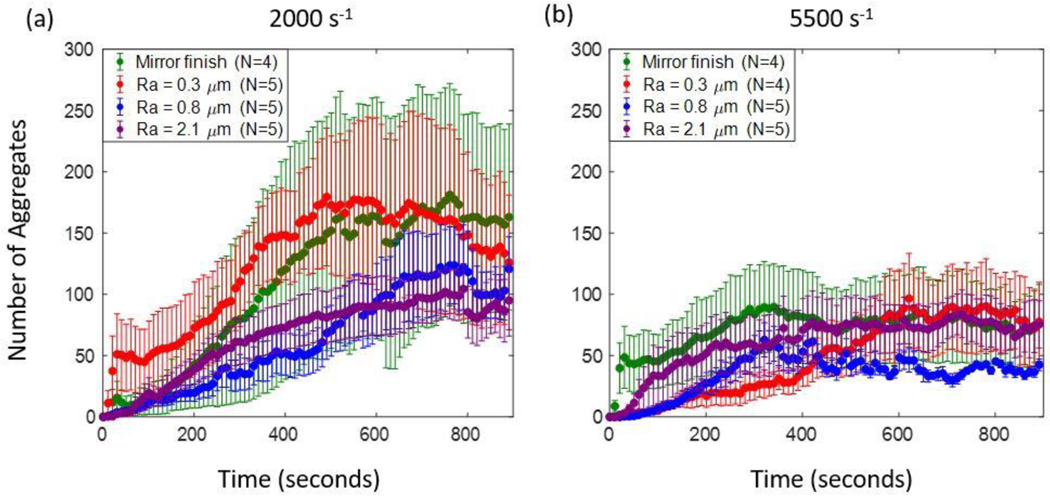
Results: We observed that on the control and sub-micron rough surfaces, at 1000 s-1 , platelets adhered uniformly on the surface. At 2000 s-1 , we observed small and stably adherent platelet aggregates. At 5500 s-1 , platelet aggregates were large, unstable and interconnected via fibrillar structures. On a surface with micron-scale roughness features, at all three shear rates, platelets deposited in the troughs of the roughened surface, and formed aggregates. Thrombus height at 2000 s-1 and 5500 s-1 was greatest on the roughest surface and lowest on the mirror-finished surface, as indicated by the mean fluorescence intensity.
Conclusions: These results demonstrated that at high shear rates, thrombi form regardless of surface topography at the scales applied. At lower shear rates, micron-scale surface features cause thrombus formation, whereas submicron features result in innocuous platelet adhesion. These findings have implications for manufacturing costs and other considerations.
Keywords: Ti6Al4V; adhesion; embolism; microfluidics; platelets; surface roughness; thrombosis; ventricular assist device.
© 2022 International Center for Artificial Organ and Transplantation (ICAOT) and Wiley Periodicals LLC.
Figures
References
-
- Chiu WC, Slepian MJ, Bluestein D. Thrombus formation patterns in the HeartMate II ventricular assist device: clinical observations can be predicted by numerical simulations. ASAIO J [Internet]. 2014. Mar 1 [cited 2022 Jan 22];60(2):237–40. Available from: https://europepmc.org/articles/PMC3992262 - PMC - PubMed
-
- Chernysh IN, Nagaswami C, Kosolapova S, Peshkova AD, Cuker A, Cines DB, et al. The distinctive structure and composition of arterial and venous thrombi and pulmonary emboli. Scientific Reports 2020. 10:1 [Internet]. 2020 Mar 20 [cited 2022 Aug 11];10(1):1–12. Available from: https://www.nature.com/articles/s41598-020-59526-x - PMC - PubMed
-
- Casa LDC, Deaton DH, Ku DN. Role of high shear rate in thrombosis. J Vasc Surg [Internet]. 2015. Apr 1 [cited 2022 Oct 5];61(4):1068–80. Available from: https://www.sciencedirect.com/science/article/pii/S0741521415000221?via%... - PubMed
MeSH terms
Substances
Grants and funding
LinkOut - more resources
Full Text Sources
Medical
