Review
. 2022 Nov 16;12(11):1023.
doi: 10.3390/bios12111023.

Biomedical Applications of Microfluidic Devices: A Review

Affiliations
Review

Biomedical Applications of Microfluidic Devices: A Review

Ghazaleh Gharib et al. Biosensors (Basel). .

Abstract

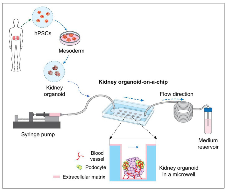
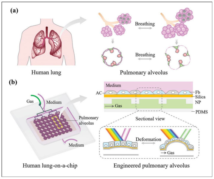
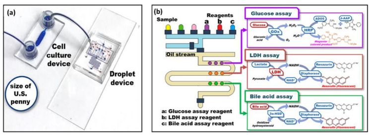
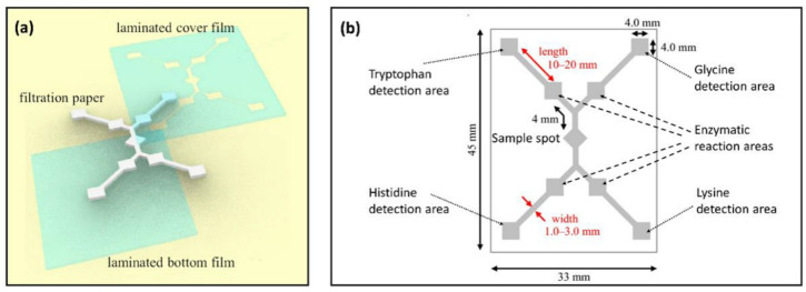
Both passive and active microfluidic chips are used in many biomedical and chemical applications to support fluid mixing, particle manipulations, and signal detection. Passive microfluidic devices are geometry-dependent, and their uses are rather limited. Active microfluidic devices include sensors or detectors that transduce chemical, biological, and physical changes into electrical or optical signals. Also, they are transduction devices that detect biological and chemical changes in biomedical applications, and they are highly versatile microfluidic tools for disease diagnosis and organ modeling. This review provides a comprehensive overview of the significant advances that have been made in the development of microfluidics devices. We will discuss the function of microfluidic devices as micromixers or as sorters of cells and substances (e.g., microfiltration, flow or displacement, and trapping). Microfluidic devices are fabricated using a range of techniques, including molding, etching, three-dimensional printing, and nanofabrication. Their broad utility lies in the detection of diagnostic biomarkers and organ-on-chip approaches that permit disease modeling in cancer, as well as uses in neurological, cardiovascular, hepatic, and pulmonary diseases. Biosensor applications allow for point-of-care testing, using assays based on enzymes, nanozymes, antibodies, or nucleic acids (DNA or RNA). An anticipated development in the field includes the optimization of techniques for the fabrication of microfluidic devices using biocompatible materials. These developments will increase biomedical versatility, reduce diagnostic costs, and accelerate diagnosis time of microfluidics technology.

Keywords: acoustophoresis; biomedical applications; biosensors; cancer diagnosis; cell sorting; dielectrophoresis; disease modeling; electrophoresis; lab-on-a-chip; magnetophoresis; micromixers; optical trapping; organ-on-a-chip; particle enrichment; particle separation; point-of-care; pressure fields; thermal fields.


Conflict of interest statement

The authors declare no conflict of interest.

Figures

Figure 1
Figure 1
The fluorescence intensity maps of micromixer M1 (a), M3 (b), and M7 (c) at Re = 25 [6]. Copyright 2021, Elsevier.
Figure 2
Figure 2
Microfiltration examples: (a) Dead-end membrane-based filtration, adopted with permission from [29], Copyright 2019, Elsevier; (b) Cross-flow membrane-based filtration, adopted with permission from [33] Copyright 2011, Royal Society of Chemistry; (c) Cross-flow pillar-based filtration, adopted with permission from [30], Copyright 2008, Elsevier; and (d) Cross-flow weir-based filtration, adopted with permission from [31], Copyright 2005, Royal Society of Chemistry.
Figure 3
Figure 3
Inertial and secondary flow examples: (a) viscoelastic non-Newtonian spiral device, reprinted with permission from [44], Copyright 2021, Springer Nature; (b) serpentine device, reprinted with permission from [1], Copyright 2016, Springer Nature; (c) successive contraction and extraction channels, reprinted with permission from [38], Copyright 2013, Royal Society of Chemistry; (d) top surface slanted grooves configuration, reprinted with permission from [39], Copyright 2017, IEEE; and (e) Herringbone structure, reprinted with permission from [40], Copyright 2021, Wiley-VCH GmbH.
Figure 4
Figure 4
CTCs isolation based on DLD technique with triangular micro-posts, reprinted with permission from [46], Copyright 2012, AIP Publishing LLC.
Figure 5
Figure 5
Schematic design of different droplet generation geometries: (a) Crossflow, (b) Flow-focusing, and (c) Co-flow; adopted with permission from [53], Copyright 2022, IOP Publishing Ltd.
Figure 6
Figure 6
Synthesized microfluidic-based 0D/1D/2D/3D micro and nano materials. Reproduced with permission [60]. Copyright 2020, John Wiley & Sons.
Figure 7
Figure 7
Microfluid techniques: (a) acoustic radiation force and experimental setup [18], Copyright 2022, Langmuir; (b) Electrophoresis and dielectrophoresis configurations in microfluidic devices [10], Copyright 2019, American Chemical Society; (c) Manipulation of magnetic nanoparticles using Magnetophoresis method [70], Copyright 2020, Langmuir; (d) Visualization of thermal field particle manipulation in a droplet [12], Copyright 2016, Scientific Reports; (e) Optical manipulation of particles inside a microfluidic channel under a certain flow rate [8], Copyright 2021, Sensors and Actuators B: Chemical; and (f) Manipulation of particles by driving pressure field with electric field [71], Copyright 2016, Scientific Reports.
Figure 8
Figure 8
A simple view of an active micromixer device [73], Copyright 2021, ACS Publications.
Figure 9
Figure 9
A cascade system to schematize integrated active type particle separation modules [103], Copyright 2021, ACS Publications.
Figure 10
Figure 10
A view of particle sorting mechanism of white blood cells, red blood cells, and circulating tumor cells (CTCs) [128], Copyright 2018, Springer Open.
Figure 11
Figure 11
Visualization of single-beam gradient force trapping and forces and the effects of the resulting forces on a particle.
Figure 12
Figure 12
(a) A representative model and components of gut-on-a-chip platform. Reprinted with permission from ref. [373], Copyright 2020, Elsevier. (b) The human gut-on-a-chip platform was presented by Jeon et al. to reproduce gastrointestinal structure with co-culture of human and microbial cells. The human Caco-2 cells were utilized to form intestinal lumen, whereas HUVECs were employed to establish vascular lumen in right and left channels, respectively. The channels were separated by collagen type I gels, and the continuous flow of medium was enhanced by osmotic pump. (c) Immunofluorescence staining results indicated that PECAM-1-positive HUVECs and ZO-1-positive Caco-2 cells were positioned in the left and right channels. Reprinted from ref. [377] (open access).
Figure 13
Figure 13
Schematic of human bone marrow-on-a-chip (hBM-on-a-chip) presented by Nelson et al. to investigate hematopoietic stem and progenitor cell behaviour and reaction to pathological stimulus. (a) The developed chip can mimic endosteal BM niche and central perivascular BM niche, which are present in long bones. OB = osteoblasts and mineralized bone-like tissue layer; MSC = mesenchymal or marrow stromal cells, including pericytes; stromal cells = other cells of the BM stroma including CXCL12-abundant reticular cells (CAR), matured hematopoietic cells, and adipose cells; HSPC = hematopoietic stem and progenitor cells; FN = fibronectin; LN = laminin, col I and IV = collagen I and collagen IV; OP = osteopontin; Jag-1 = Jagged 1. (b) Soft lithography was utilized to fabricate a 5-channel PDMS microfluidic platform. An endosteal layer was formed in the central channel with differentiation of MSCs for 21 days. After this, HSPCs, HUVECs, and MSCs were loaded and seeded on top of the endosteal layer for vasculogenesis. Reprinted with permission from ref. [363], Copyright 2021, Elsevier.
Figure 14
Figure 14
A schematic of gut-liver chip presented by Lee et al. to recapitulate hepatic steatosis. (a) Representative figure of gut-liver chip, indicating gut layer top of the membrane and liver layer on the bottom of the membrane. (b) Cross-section of the gut-liver chip. (c) An image of an assembled gut-liver chip (blue ink indicates the liver part, whereas red ink indicates the gut part). (d) This figure shows that absorption of fatty acids by gut cells (enterocytes) and liver cells (hepatocytes) in the gut–liver platform. (e) This illustration demonstrates a lipid accumulation experiment in a microwell plate. Cultured hepatocytes (HepG2) in a well plate were exposed to lipid accumulation, and quantification of lipid accumulation was performed. Reprinted with permission from ref. [383], Copyright 2018, Wiley Online Library.
Figure 15
Figure 15
Schematic of human BBB chip presented by Ahn et al. to reproduce the essential structure and function of human BBB and to investigate nanoparticle distribution in the vascular and perivascular parts. (a) The figure shows the structure of the BBB composed of endothelial cells (ECs), pericytes, and astrocytes with aquaporin-4 (AQP4) expression. (b) Illustration for microengineered human BBB platform. (c) Layer-by-layer schematic of developed BBB platform indicates the upper vascular layer, porous membrane, lower perivascular layer, and glass slide. Reprinted from ref. [385] (open access).
Figure 16
Figure 16
Schematic of heart-on-a-chip device presented by Zhang et al. to investigate in situ electrical stimulation and observation of the function parameters of cardiac tissues. (a) Design of the developed heart-on-a-chip platform consisting of four layers: (i) top layer—a PDMS cover layer containing 4 inlet/outlet channel; (ii) a PDMS channel layer; (iii) a PDMS chamber layer inserted with two platinum wire electrodes; and the (iv) bottom layer—a glass layer coated by four gold electrodes. (b) Three-dimensional illustration of the heart-on-a-chip platform. (c) The side view of the schematic demonstrates cardiac tissue in the chamber. (d) Magnified sketching of the elaborated design of the PDMS channel, representing the channel layer (ii) in (a). (e) Picture of the introduced heart-on-a-chip platform. Reprinted with permission from ref. [386], Copyright 2021, Elsevier.
Figure 17
Figure 17
Schematic shows the developed kidney-organoid-a-chip platform introduced by Lee et al. to investigate the biochemical effect on in vitro development of human pluripotent stem cells (hPSCs)-derived human kidney organoids. Controlled shear force and optimized ECM were utilized to explore biochemical effect. Reprinted from ref. [388] (open access).
Figure 18
Figure 18
Schematic of lung-on-a-chip platform presented by Zhu et al. to develop favourable biomimetic breathing human lung with microphysiological breathing monitoring. (a) Figure of pulmonary alveolus while breathing. (b) Design of the developed breathing human-lung-on-a-chip device consisting of an array of pulmonary-alveolus-like structures. Rhythmic stretch during breathing was mimicked by cyclic airflow. Utilization of structural colours was enhanced to visualize breathing process. Fb = fibrinogen; NP = nanoparticle; AC = alveolar cell. Reprinted with permission from ref. [390], Copyright 2022, Wiley Online Library.
Figure 19
Figure 19
Droplet-based microfluidic platform for cell injury analysis. (a) The device was connected to a cell culturing platform for the simultaneous quantification of biochemical analytes. (b) Representation of the concept of achieving three different enzymatic assays. Reprinted with permission from [424]. Copyright 2019, American Chemical Society.
Figure 20
Figure 20
(a) Representation of the fabrication of a laminated paper-based analytical device (LPAD) for the simultaneous quantification of tryptophan, glycine, histidine, and lysine levels in the range of a few micromolar to 100 uM by reactions of sample-specific aminoacyl–tRNA synthetases. (b) Details of the LPAD. The paper properties and dimensions of channels between detection and reaction areas specify the incubation time for the reaction mixture. Reprinted with permission from [427] (open access).

References

    1. Özbey A., Karimzadehkhouei M., Akgönül S., Gozuacik D., Koşar A. Inertial Focusing of Microparticles in Curvilinear Microchannels. Sci. Rep. 2016;6:38809. doi: 10.1038/srep38809. - DOI - PMC - PubMed
    1. Erdem K., Ahmadi V.E., Kosar A., Kuddusi L. Differential Sorting of Microparticles Using Spiral Microchannels with Elliptic Configurations. Micromachines. 2020;11:412. doi: 10.3390/mi11040412. - DOI - PMC - PubMed
    1. Jiang D., Ni C., Tang W., Huang D., Xiang N. Inertial Microfluidics in Contraction–Expansion Microchannels: A Review. Biomicrofluidics. 2021;15:41501. doi: 10.1063/5.0058732. - DOI - PMC - PubMed
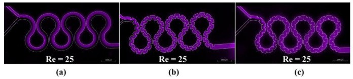
    1. Alijani H., Özbey A., Karimzadehkhouei M., Koşar A. Inertial Micromixing in Curved Serpentine Micromixers with Different Curve Angles. Fluids. 2019;4:204. doi: 10.3390/fluids4040204. - DOI
    1. Zhao Q., Yuan D., Zhang J., Li W. A Review of Secondary Flow in Inertial Microfluidics. Micromachines. 2020;11:461. doi: 10.3390/mi11050461. - DOI - PMC - PubMed

LinkOut - more resources