Review
. 2022 Nov 18;12(11):1045.
doi: 10.3390/bios12111045.

Recent Advances of Organ-on-a-Chip in Cancer Modeling Research

Affiliations
Review

Recent Advances of Organ-on-a-Chip in Cancer Modeling Research

Xingxing Liu et al. Biosensors (Basel). .

Abstract

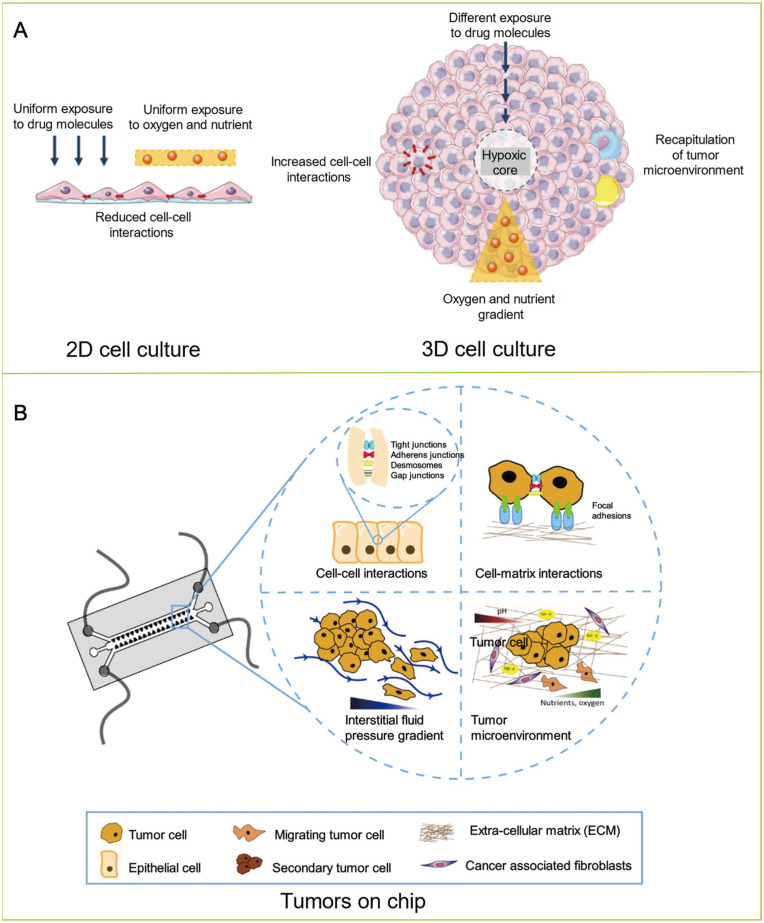
Although many studies have focused on oncology and therapeutics in cancer, cancer remains one of the leading causes of death worldwide. Due to the unclear molecular mechanism and complex in vivo microenvironment of tumors, it is challenging to reveal the nature of cancer and develop effective therapeutics. Therefore, the development of new methods to explore the role of heterogeneous TME in individual patients' cancer drug response is urgently needed and critical for the effective therapeutic management of cancer. The organ-on-chip (OoC) platform, which integrates the technology of 3D cell culture, tissue engineering, and microfluidics, is emerging as a new method to simulate the critical structures of the in vivo tumor microenvironment and functional characteristics. It overcomes the failure of traditional 2D/3D cell culture models and preclinical animal models to completely replicate the complex TME of human tumors. As a brand-new technology, OoC is of great significance for the realization of personalized treatment and the development of new drugs. This review discusses the recent advances of OoC in cancer biology studies. It focuses on the design principles of OoC devices and associated applications in cancer modeling. The challenges for the future development of this field are also summarized in this review. This review displays the broad applications of OoC technique and has reference value for oncology development.

Keywords: cancer modeling; microfluidics; organ-on-a-chip; tumor microenvironment.


Conflict of interest statement

The authors declare no conflicts of interest.

Figures

Figure 4
Figure 4
(A) Vascularized tumor chips. (a) The combination of angiogenesis and vasculogenesis represents the fundamental processes of new blood vessel formation. The newly formed blood vessels can provide nutrients and oxygen for malignant tumor development and metabolic waste removal. Moreover, they are able to have interactions with various types of cells in the vascular niche. The figure shows the processes of different blood vessel formations, including neoarteriogenesis, vascular remodeling, venogenesis, and angiogenesis. Adapted with permission from Ref. [110]. Copyright 2019, MDPI. (b) Schematic of the vascularized tumor spheroid-on-a-chip. The device consists of 5 microchannels in parallel by implanting HUVECs in fibrin gel into the chip (day 0) for self-assembling a vascular network (day 5), wherein tumor spheroids are placed and integrated with the surrounding blood vessels (day 15) to achieve tumor vascularization. Adapted with permission from Ref. [129]. Copyright 2022, ACS. (B) Onco-immuno chips. (a) Schematic representation of the reconstituted immuno-TME on OoC models. Adapted with permission from Ref. [138]. Copyright 2021, Frontiers. (b) Realization of TIME-on-a-chip on a 35 mm Petri dish platform: a ring magnet is used, microfluidic channels printed on a porous membrane are attached to a modified Petri dish and integrated with a microplate containing spheroids for mixing, surrounding the microplate O-rings, providing leak-proof components. Adapted with permission from Ref. [148]. Copyright 2021, IOPScience. (c) Schematic of the OoC for modeling the tumor microdevice. The bottom panel shows a cross-section of the microdevice. Endothelial cells (e.g., HUVECs) are lined in the lumen to generate vascular surrogates, allowing perfusion of culture medium, NK-92 cells, anti-PD-11 antibodies (e.g., atezolizumab), or IDO-1 inhibitors (e.g., epacadostat). Adapted with permission from Ref. [149]. Copyright 2021, AAAS. (C) Hypoxia chips. (a) Schematic of tumor hypoxia in vivo and recapitulating tumor hypoxia in vitro in microfluidic models with diffusion barriers. Reprinted with permission from Ref. [171]. Copyright 2022, ACS. (b) Schematic diagram and image of a 3D culture multiorgan microfluidic platform for precise control of dissolved oxygen concentration. Reprinted with permission from Ref. [170]. Copyright 2021, ACS (D) Tumor metastasis chip. (a) OoC models for mimicking tumor metastasis steps of (A,B) the invasion/intravasation process and (C) the extravasation process. Adapted with permission from Ref. [178]. Copyright 2022, MDPI. (b) A multi-site metastasis-on-a-chip microphysiological system for assessing cancer cells metastatic preference. Reprinted with permission from Ref. [181]. Copyright 2018, Wiley. (c) Schematic and photo of a metastasis-on-a-chip platform with three interconnected culture chambers to study the sympathetic regulation of bone metastasis in breast cancer. Reprinted with permission from Ref. [180]. Copyright 2022, Elsevier.
Figure 5
Figure 5
(A) Design and model of lung-on-a-chip. (a) Cross-sectional view of a lung-on-a-chip microfluidic model with two distinct channels separated by a thin porous membrane. Reprinted with permission from Ref. [190]. Copyright 2022, Elsevier. (b) The model of lung-on-a-chip comprising six wells is used to mimic the lung alveolar barrier, whereby cells are cultured directly at an air–liquid interface for inhalation assays. Reprinted with permission from Ref. [185]. Copyright 2019, Elsevier. (B) Breast OoC. (a) Schematic illustration of the metastatic breast tumors and on-chip steps for cell loading and co-cultivation as well as drug treatment. Adapted with permission from Ref. [196]. Copyright 2016, Nature-Springer. (b) The patients of breast cancer with preexisting cardiac dysfunctions may lead to different incident levels of chemotherapy-induced cardiotoxicity (CIC). This heart breast-cancer-on-a-chip platform with iPSC-derived cardiac tissues and BC tissues could be used for disease modeling and monitoring of cardiotoxicity induced by cancer. Reprinted with permission from Ref. [199]. Copyright 2020, Wiley. (C) Schematic of brain cancer chip for high-throughput drug screening. This chip has a gradient generator with a Christmas-tree-shaped channel system. The channel width is gradually reduced from 300 μm to 100 μm. Moreover, it also has an array of 24 independent culture chambers with 3 inlet banks and 1 outlet bank. Subchannels connect the microwells to the main channel and prevent captured cells from escaping the microwells. Adapted with permission from Ref. [200]. Copyright 2016, Nature-Springer. (D) Schematic of the decellularized liver matrix-based liver OoC, including use of a natural liver to prepare the DLM solution and a 3D schematic diagram of the equipment components, consisting of the microchannels from the top and bottom, the PET membrane, and the air inlet and outlet. Adapted with permission from Ref. [201]. Copyright 2018, Royal Society of Chemistry. (E) Design and characterization of the colorectal OoC system microfluidic chip for precision onco-nanomedicine. Adapted with permission from Ref. [202]. Copyright 2019, AAAS.
Figure 1
Figure 1
Schematic of OoC platforms for mimicking the TME and functions in vitro and their applications of reconstructing the organs on microfluidics for oncology studying.
Figure 2
Figure 2
(A) The TME is a complex ecosystem consisting of various cellular and noncellular components, such as cancer cells, fibroblasts, multiple chemical factors, the extracellular matrix, the vasculature system, and mechanical cues. The progression of a tumor is critically influenced by the interaction between tumor and TME. These factors should be included in the construction of OoC models. Adapted with permission from Ref. [56]. Copyright 2019, MDPI. (B) Bioprinting techniques mostly used for the generation of microfluidics. Adapted with permission from Ref. [71]. Copyright 2018, Royal Society of Chemistry. (C) Microfluidic approaches involved in creating tissues/organs. Adapted with permission from Ref. [71]. Copyright 2018, Royal Society of Chemistry.
Figure 3
Figure 3
(A) The main differences between 2D and 3D cell cultures. Adapted with permission from Ref. [96]. Copyright 2020, MDPI. The monolayer formed by 2D cell culture can uniformly lead to exposure to oxygen, nutrient, and drug molecules. However, 2D cell culture models are unable to properly simulate the architecture and microenvironment of in vivo tumors. Compared to in vivo cells, the cells cultured by 2D cell culture models show fundamental differences in cell proliferation, morphology, differentiation, signal transduction, and metabolism. Three-dimensionally cultured cells are more similar to the in vivo environment in terms of cell proliferation, migration, differentiation, morphology, and transfer. In particular, spheroids are feasible to simulate the central regions of vascularized tumors since the model can produce oxygen gradients and nutrients to form necrotic cores. However, both 2D and 3D cell culture models are performed under static conditions, lacking mechanical factors, such as shear stress, physiological flow, drug exposure, and nutrient delivery. As a result, they hinder the studies of drug sensitivity and toxicity for long-term culture. Copyright 2020, MDPI. (B) The microfluidic platform is an effective tool to investigate a variety of key biological phenomena, from cell–ECM and cell–cell interactions to the flow of stroma within TME. Reprinted with permission from Ref. [98]. Copyright 2022, Elsevier.

References

    1. Siegel R.L., Miller K.D., Jemal A. Cancer statistics, 2018. CA A Cancer J. Clin. 2018;68:7–30. doi: 10.3322/caac.21442. - DOI - PubMed
    1. Siegel R.L., Miller K.D., Jemal A. Cancer statistics, 2020. CA A Cancer J. Clin. 2020;70:7–30. doi: 10.3322/caac.21590. - DOI - PubMed
    1. Sung H., Ferlay J., Siegel R.L., Laversanne M., Soerjomataram I., Jemal A., Bray F. Global cancer statistics 2020: GLOBOCAN estimates of incidence and mortality worldwide for 36 cancers in 185 countries. CA A Cancer J. Clin. 2021;71:209–249. doi: 10.3322/caac.21660. - DOI - PubMed
    1. World Health Organization . WHO Report on Cancer: Setting Priorities, Investing Wisely and Providing Care for All. World Health Organization; Geneva, Switzerland: 2020.
    1. Singh D., Vaccarella S., Gini A., De Paula Silva N., Steliarova-Foucher E., Bray F. Global patterns of Hodgkin lymphoma incidence and mortality in 2020 and a prediction of the future burden in 2040. Int. J. Cancer. 2022;150:1941–1947. doi: 10.1002/ijc.33948. - DOI - PubMed