. 2022 Nov 17;11(11):1674.
doi: 10.3390/biology11111674.

Dimorphilus gyrociliatus (Annelida: Dinophiliformia) Dwarf Male Nervous System Represents a Common Pattern for Lophotrochozoa

Affiliations

Dimorphilus gyrociliatus (Annelida: Dinophiliformia) Dwarf Male Nervous System Represents a Common Pattern for Lophotrochozoa

Elizaveta Fofanova. Biology (Basel). .

Abstract

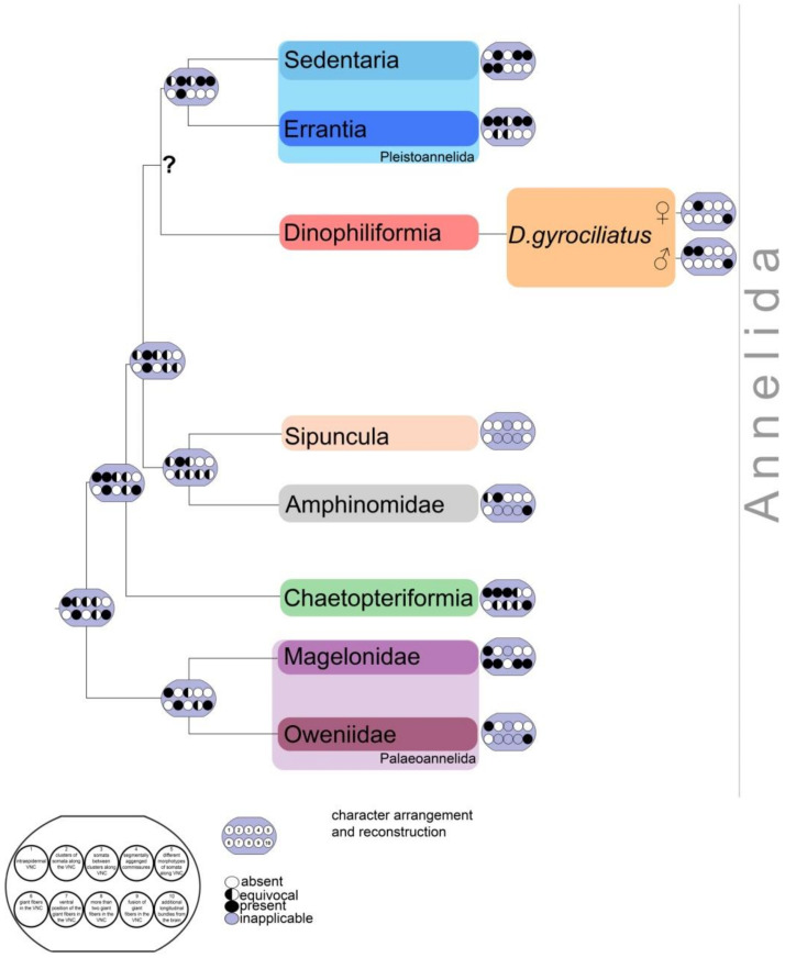
Dinophiliformia is a newly revealed clade within Annelida that is a sister group to Pleistoannelida. Dimorphilus gyrociliatus is a representative of this clade that has fascinated scientists with its high degree of sexual dimorphism. Both males and females are small, worm-like creatures that have specific ciliary structures: anterior ventral, posterior ventral, and dorsal ciliary fields in males, and prototroch, metatroch, and ventral ciliary fields in females. There are data on the morphology and development of the nervous system in Oweniidae, Sipunculida, Pleistoannelida, and even Dinophiliformia. However, data on the neuromorphology and development of D. gyrociliatus dwarf males are limited. Here, we present data on the distribution of cilia, sensory neurons, and the 5-HT-like immunoreactive system in 3D reconstructions and cross-sections. Immunochemical labeling with anti-acetylated tubulin and anti-5-HT antibodies and confocal microscopy were used to visualize the ciliary structures and neurons. The male has three ciliary fields: anterior ventral, posterior ventral, and dorsal. These include frontal ganglia, five commissures, two ventral and two dorsal bundles, and penial nerves. A total of fifty-seven neurons and only five 5-HT-like immunoreactive cells were described. Although the sensory neurons were not 5-HT-like immunoreactive, they had 5-HT innervation, which may indicate the role of 5-HT in perception. There may be homology between male and female ciliary structures. The dwarf male of D. gyrociliatus may have a reduced apical organ consisting of two sensory neurons and a 5-HT-like immunoreactive cell.

Keywords: annelida; apical organ; dwarfism; lophotrochozoa; miniaturization; nervous system; serotonin; trochophore.


Conflict of interest statement

The authors declare no conflict of interest.

Figures

Figure 1
Figure 1
Annelida phylogenetic tree.The diagram is based on recent data. The phylogeny was retrieved from [1,2]. Dinophiliformia is a newly emerged group, sister to Pleistoannelida.
Figure 2
Figure 2
Multiciliated cells of ciliary fields of Dimorphilus gyrociliatus dwarf male. The diagram is based on the results presented in [3,4]. The multiciliated cells distriburion pattern is adapted from [3,4] Copyright © 1988 Alan R. Liss, Inc. Apical end is up, relative size is not to scale. E1–E11 represent multiciliated cells of anterior ventral, posterior ventral and dorsal ciliary fields.
Figure 3
Figure 3
External ciliation in mature Dimorphilus gyrociliatus dwarf male revealed by anti-acetylated alpha-tubulin labeling. On (AC) apical end is up. (A,A′) Ventral view on wholemount. At the anterior part of the body, there is a prominent anterior ventral ciliary field (avc). The posterior ciliary field is located posteriorly the anterior ventral ciliary field. A pair of protonephridia (*) is located symmetrically on the body. At the anterior end prominent cilia of sensory neurons №1 and №16 are visible. (B,B′). Lateral view on wholemount. At the anterior part of the body, the dorsal ciliary field (dc) is present. Posteriorly, two single cilia of sensory neurons №9 are located. A prominent testis (test) fills the most of the inner part of the body. Posteriorly to testis copulatory organ with multiple stylet glad cells (sgd) are located. (C,C′). Dorsal view on wholemount. (D,D′). Anterior view of the wholemount. (E,E′). The posterior surface of wholemount. At the posterior tip cilia of paired sensory neurons №14 and №15 are located. Abbreviations: avc—anterior cilia; dc—dorsal cilia; pvc—posterior cilia; co—copulative organ; sgd—stylet gland cell; sn1sn20—sensory neurons 1–20; test—testis; *—nephridia.
Figure 4
Figure 4
The internal organization of Dimorphilus gyrociliatus dwarf male. (AD) optical cross-sections of the anterior (A,B) and posterior (C,D) parts of the body. (A) Prominent testis (test) and a pair of protonephridia (*), a pair of surface gland cells (gl). (B) symmetrically located seminal vesicles (sv) and unpaired spermioduct (spd). (C) Copulatory organ—glandomuscular organ, includes glandular part, represented by stylet gland cells (sgd) and muscle part (cyan). (D) The same section as (C); without cyan channel, thus only stylet gland cells are visible, without phalloidin.
Figure 5
Figure 5
Type 1a sensory neurons in mature Dimorphilus gyrociliatus dwarf male revealed by anti-acetylated alpha-tubulin labeling. (A,A′) Anterior surface view. (A′) Schematic representation of the anterior surface. (B) The posterior surface view. (B′) Schematic representation of the posterior surface view. (C) The close-up and drawing of sensory neuron №1. (D) Close-up and drawing of sensory neuron №2. Abbreviations: E10, 11—multiciliated cells of the dorsal ciliary field; dc—dorsal cilia; pvc—posterior cilia; sgd—stylet gland cell; sn1sn20—sensory neurons 1–20.
Figure 6
Figure 6
Type 1b sensory neurons in mature Dimorphilus gyrociliatus dwarf male revealed by anti-acetylated alpha-tubulin labeling. (A,A′) A cross-section and schematic representation of the anterior surface. (B,B′) A cross-section and schematic representation of the anterior part of the body. (C,C′) A cross-section and schematic representation of the middle part of the body. (D) The close-up and drawing of sensory neuron №9. (E) The close-up and drawing of sensory neuron №7. (F) The close-up and drawing of sensory neuron №4. (G) The close-up and drawing of sensory neuron №2. (H) The close-up and drawing of sensory neuron №11. (I) The close-up and drawing of sensory neuron №8. (J) The close-up and drawing of sensory neuron №10. Abbreviations: avc—anterior cilia; E10,11—multiciliated cells of the dorsal ciliary field; dc—dorsal cilia; pvc—posterior cilia; test—testis; sv—seminal vesicles; sn1sn20—sensory neurons1–20; *—nephridia.
Figure 7
Figure 7
Type 1c sensory neurons in mature Dimorphilus gyrociliatus dwarf male revealed by anti-acetylated alpha-tubulin labeling. (A) Anterior surface view. (A′) Schematic representation of the anterior surface. (B) Anterior surface view. (B′) Schematic representation of the anterior surface. (C) The posterior surface view. (C′) Schematic representation of the posterior surface view. (D) The close-up and drawing of sensory neuron №6. (E) Close-up and drawing of sensory neuron №3. (F) Clode-up and schematic drawing of sensory neuron №12. (G) Close-up and schematic drawing og sensory neuron №13. Abbreviations: E10, 11—multiciliated cells of the dorsal ciliary field; dc—dorsal cilia; pvc—posterior cilia; sgd—stylet gland cell; sn1–sn20—sensory neurons 1–20.
Figure 8
Figure 8
Type 2 sensory neurons in mature Dimorphilus gyrociliatus dwarf male revealed by anti-acetylated alpha-tubulin labeling. (A) Anterior surface view. (A′) Schematic representation of the anterior surface. (B) The posterior surface view. (B′) Schematic representation of the posterior surface view. (C) The close-up and drawing of sensory neuron №20. (D) Close-up and drawing of sensory neurons №18 and 19. (E) The close-up of sensory neuron №16. (F) The close-up of sensory neuron № 17. Abbreviations: E10, 11—multiciliated cells of the dorsal ciliary field; dc—dorsal cilia; pvc—posterior cilia; sgd—stylet gland cell; sn1–sn20—sensory neurons 1–20.
Figure 9
Figure 9
Tubulin-like immunoreactivity in the mature dwarf male of Dimoprhilus gyrociliatus. (A,A′) Anterior view on wholemount and schematic representation. (B,B′) A cross-section at the middle part of the body and its schematic representation. (C,C′) A cross section of the posterior body region and its schematic representation. (D,D′) A cross section of the posterior body region at the level of stylet gland cells and its schematic representation. Abbreviations: arrowheads indicate neuronal cell bodies; avc—anterior ventral cilia; dc—dorsal commissure; vc—ventral commissure; c—commissure; co—copulatory organ; go—genital opening; test—testis; pvc—posterior cilia; sgd—stylet gland cell; sn1–sn20—sensory neurons1–20; sv—seminal vesicles; vb—ventral bundle; *—nephridia.
Figure 10
Figure 10
5-HT-like immunoreactivity in the mature dwarf male of Dimorphilus gyrociliatus. (A,A′) Ventral view on wholemount. (B,B′) lateral view on wholemount. (C,C′) dorsal view on wholemount. (D,D′) A cross section of the posterior body region as indicated in (C,C′) Abbreviations: arrowheads indicate neuronal cell bodies; avc—anterior ventral cilia; c—commissure, dc—dorsal cilia; test—testis, pn—penial nerve; pvc—posterior cilia; sgd—stylet gland cell; spd—spermioduct; sn1–sn20—sensory neurons1–20; sv—seminal vesicles; vb—ventral bundle; *—nephridia.
Figure 11
Figure 11
5-HT-like immunoreactivity in the mature dwarf male of Dimoprhilus gyrociliatus. (A)Ventral view on wholemount. (B) Sensory neuron№7 and thin 5-HT-like immunoreactive processes. (C) Multiciliated cells of dorsal ciliary field are innervated by 5-HT-like immunoreactive processes. (D,D′,D″) Sensory neurons №9 and unpaired 5-HT-like immunoreactive cell. (E) Sensory neuron №17. (F,F′) The cell bodies of 5-HT-like immunoreactive cells. (G) Sensory neuron №4. (H) Commissure. (I) 5-HT-like immunoreactive processes innervate copulatory organ. Abbreviations: arrowheads indicate neuronal cell bodies; avc—anterior ventral cilia; c—commissure; dc—dorsal cilia; test—testis; pvc—posterior cilia; sgd—stylet gland cell; sn1–sn20—sensory neurons 1–20; sv—seminal vesicles; *—nephridia.
Figure 12
Figure 12
Comparative diagram of the external ciliation and possible homology regions on D. gyrociliatus male and female. The diagram is based on recent data on D. gyrociliatus female [14] and male (this study) external ciliation and male. Female acrotroch and male dorsal cilia, anterior ciliary field in female ad anterior ventral cilia in male, ventral ciliary field in female and posterior ventral cilia in male seems to be homologus structures. The apical end is up. Relative dimensions are not to scale. Abbreviations: act—acrotroch; acz—anterior ciliary field; adz—adoral ciliary zone; avc—anterior cilia; dc—dorsal cilia; mt—metatroch; pvc—posterior cilia; test—testis; co—copulatory organ.
Figure 13
Figure 13
Schematic representation of nervous system morphology in D. gyrociliatus. Apical end is up. Tubulin–blue; 5-HT–magenta. 5-HT-like immunoreactive elements represent a minor part of the whole nervous system.
Figure 14
Figure 14
Schematic diagram of the ventral nervous system anatomy across Annelida phylogenetic tree with addition of Dimorphilus gyrociliatus data. Phylogenetic tree is based on [1,2] the characters pattern and distribution is based on [16]. The legend and character arrangement and reconstruction matrixes were adapted from [16]. The characters are coded in a way: absent (white), present (black) and inapplicable (grey). Note the difference between D. gyrociliatus male and female pattern.
Figure 15
Figure 15
Schematic representation of D. gyrociliatus females nerve development and comparison with dwarf male. The diagram is based on [12]. Females go through three developmental stages: early trochophore, middle trochophore, and late trochophore. The dwarf male 5-HT-like immunoreactive system is similar to the female nervous system during the middle trochophore stage.

Similar articles

References

    1. Martín-Durán J.M., Vellutini B.C., Marlétaz F., Cetrangolo V., Cvetesic N., Thiel D., Henriet S., Grau-Bové X., Carrillo-Baltodano A.M., Gu W., et al. Conservative route to genome compaction in a miniature annelid. Nat. Ecol. Evol. 2021;5:231–242. doi: 10.1038/s41559-020-01327-6. - DOI - PMC - PubMed
    1. Worsaae K., Kerbl A., Domenico M.D., Gonzalez B.C., Bekkouche N., Martínez A. Interstitial Annelida. Diversity. 2021;13:77. doi: 10.3390/d13020077. - DOI
    1. Windoffer R., Westheide W. The Nervous system of the male Dinophilus gyrociliatus (Polychaeta, Dinophilidae): II. electron microscopical reconstruction of nervous anatomy and effector cells. J. Comp. Neurol. 1988;272:475–488. doi: 10.1002/cne.902720403. - DOI - PubMed
    1. Windoffer R., Westheide W. The nervous system of the male Dinophilus gyrociliatus (Annelida: Polychaeta). I. Number, types and distribution pattern of sensory cells. Acta Zool. 1988;69:55–64. doi: 10.1111/j.1463-6395.1988.tb00901.x. - DOI
    1. Carrillo-Baltodano A.M., Seudre O., Guynes K., Martín-Durán J.M. Early embryogenesis and organogenesis in the annelid Owenia fusiformis. EvoDevo. 2021;12:5. doi: 10.1186/s13227-021-00176-z. - DOI - PMC - PubMed

Grants and funding

LinkOut - more resources