. 2022 Nov 16;12(11):1698.
doi: 10.3390/biom12111698.

Redox Regulation of Signaling Complex between Caveolin-1 and Neuronal Calcium Sensor Recoverin

Affiliations

Redox Regulation of Signaling Complex between Caveolin-1 and Neuronal Calcium Sensor Recoverin

Vasiliy I Vladimirov et al. Biomolecules. .

Abstract

Caveolin-1 is a cholesterol-binding scaffold protein, which is localized in detergent-resistant membrane (DRM) rafts and interacts with components of signal transduction systems, including visual cascade. Among these components are neuronal calcium sensors (NCSs), some of which are redox-sensitive proteins that respond to calcium signals by modulating the activity of multiple intracellular targets. Here, we report that the formation of the caveolin-1 complex with recoverin, a photoreceptor NCS serving as the membrane-binding regulator of rhodopsin kinase (GRK1), is a redox-dependent process. Biochemical and biophysical in vitro experiments revealed a two-fold decreased affinity of recoverin to caveolin-1 mutant Y14E mimicking its oxidative stress-induced phosphorylation of the scaffold protein. At the same time, wild-type caveolin-1 demonstrated a 5-10-fold increased affinity to disulfide dimer of recoverin (dRec) or its thiol oxidation mimicking the C39D mutant. The formation of dRec in vitro was not affected by caveolin-1 but was significantly potentiated by zinc, the well-known mediator of redox homeostasis. In the MDCK cell model, oxidative stress indeed triggered Y14 phosphorylation of caveolin-1 and disulfide dimerization of recoverin. Notably, oxidative conditions promoted the accumulation of phosphorylated caveolin-1 in the plasma membrane and the recruitment of recoverin to the same sites. Co-localization of these proteins was preserved upon depletion of intracellular calcium, i.e., under conditions reducing membrane affinity of recoverin but favoring its interaction with caveolin-1. Taken together, these data suggest redox regulation of the signaling complex between recoverin and caveolin-1. During oxidative stress, the high-affinity interaction of thiol-oxidized recoverin with caveolin-1/DRMs may disturb the light-induced translocation of the former within photoreceptors and affect rhodopsin desensitization.

Keywords: apoptosis; caveolin-1; oxidative stress; photoreceptor; recoverin; retina.


Conflict of interest statement

The authors declare no conflicts of interest.

Figures

Figure 1
Figure 1
Schematic representation of domain organization of caveolin-1 and structure of its fragments, Cav and CavE, prepared in this study. SDS-PAGE represents fractions obtained during purification of Cav using Ni-NTA affinity chromatography.
Figure 2
Figure 2
Secondary structure and oligomerization of caveolin-1 fragments. (A) Representative far-UV CD spectra for 5.5 µM Cav and CavE in 10 mM H3BO3-KOH buffer (pH 9.0), 50 mM NaCl, 20 µM DTT at 20 °C. (B) Hydrodynamic diameter of 200 µM Cav and CavE assessed by DLS in 20 mM Tris-HCl buffer (pH 7.5), 100 mM NaCl, 1 mM DTT at 25 °C. The data were determined in three independent measurements.
Figure 3
Figure 3
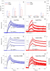
Interaction of reduced monomer (Rec), thiol oxidation mimicking mutant (Rec-C39D), or disulfide dimer (dRec) of recoverin with caveolin-1 fragments. (A,B) The results of pull-down assay of Rec, Rec-C39D, or dRec (1.7 mg/mL) with affinity resins containing immobilized Cav or CavE in 20 mM Tris-HCl buffer (pH 7.5), 100 mM NaCl, 2 mM MgCl2, in the absence (A) or the presence (B) of calcium (1 mM CaCl2 or 2 mM EGTA) at 25 °C. Error bars represent the weight fractions of the bound recoverin forms (in relative units, RU) determined from at least three independent experiments. *—p < 0.05. (CH) Kinetics of the interaction between Rec (C,D), dRec (E,F), or Rec-C39D (G,H) (2.5 μM to 30 μM) with immobilized Cav or CavE in 10 mM HEPES buffer (pH 7.4), 150 mM NaCl, 2 mM DTT (except for dRec studies), 0.05% TWEEN20 in the absence (“Ca2+-free”) or in the presence (“Ca2+-loaded”) of calcium (1 mM CaCl2 or 1 mM EDTA), determined by SPR spectroscopy at 25 °C. Blue and red sensorgrams represent experimental data, while black curves are theoretical fits calculated according to the “heterogeneous ligand” model.
Figure 4
Figure 4
Disulfide dimerization of recoverin in vitro. (A) Kinetics of dRec formation during dialysis of 50 µM reduced recoverin against 10 mM Tris-HCl buffer (pH 7.5), 100 mM NaCl, 1 mM EGTA, containing 1 mM H2O2 for 24 h at 4 °C, with or without (control) preincubation in the presence of 100 µM Cav or 100 µM CavE. (B) Disulfide dimerization of 50 µM reduced recoverin during dialysis (24 h, 4 °C) against 10 mM Tris-HCl buffer (pH 7.5), 100 mM NaCl, containing indicated concentrations of H2O2 in the presence of 1 mM EGTA (“control”) or 4-fold molar excess of ZnCl2 (“zinc”). Weight fractions of dRec were determined by densitometric analysis of SDS-PAGE data from at least three independent experiments and plotted against time (A) or H2O2 concentrations (B).
Figure 5
Figure 5
Disulfide dimerization of recoverin and tyrosine (Y14) phosphorylation of caveolin-1 in MDCK-Rec cells under oxidative stress conditions. (A) Cells were incubated with indicated concentrations of H2O2 in the presence of 0.5 mM vanadate for 10 min and their lysates were subjected to non-reducing Western blotting using antibodies against recoverin (upper panel) or P-caveolin-1 (lower panel). The positions of monomer (“Rec”), disulfide dimer (“dRec”), and disulfide aggregates (“nRec”) of recoverin, as well as P-caveolin-1 (“pCav”) are indicated by arrows. (B,C) Weight fractions of dRec (B) and pCav (C) estimated from Western blotting data from at least three independent experiments. *—p < 0.05 as compared to the data obtained for untreated cells.
Figure 6
Figure 6
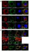
Localization of caveolin-1, P-caveolin-1, and recoverin in MDCK-Rec cells under oxidative stress conditions. Caveolin-1 and P-caveolin-1 are visualized by immunocytochemical analysis using rabbit monoclonal antibodies and goat anti-rabbit Alexa Fluor 555-conjugated IgG (red). Recoverin is visualized using mouse polyclonal antibodies and goat anti-mouse Alexa Fluor 488-conjugated IgG (green). Cell nuclei are stained with DAPI (blue). (A) Normal conditions. White arrow indicates the area of caveolin-1 localization in the Golgi complex. (B) Oxidative stress conditions (10 mM H2O2). White arrows indicate recruitment of recoverin to the plasma membrane. Red arrows point to the sites of co-localization of recoverin with P-caveolin-1. (C) Oxidative stress against the background of the calcium depletion conditions (5 μM BAPTA-AM). Red arrows point to the sites of co-localization of recoverin with P-caveolin-1. The insets with higher magnification (in the middle) demonstrate areas in the cytoplasm with co-localization of recoverin and non-phosphorylated caveolin-1 (indicated by white arrowheads).
Figure 7
Figure 7
The hypothetical function of caveolin-1 complex with Rec/dRec in photoreceptors. Under normal light conditions (low calcium), recoverin forms a complex with caveolin-1 in DRMs of OS, which can be attenuated by tyrosine (Y14) phosphorylation, enabling translocation of recoverin to IS. In oxidative stress, the increased zinc concentration induces the formation of dRec, which retains in OS due to increased affinity to P-caveolin-1. The ability of dRec to constitutively inhibit rhodopsin kinase (GRK1) can slow down rhodopsin desensitization, promote oxidative stress, and induce apoptosis.

Similar articles

Cited by

References

    1. Cohen A.W., Hnasko R., Schubert W., Lisanti M.P. Role of caveolae and caveolins in health and disease. Physiol. Rev. 2004;84:1341–1379. doi: 10.1152/physrev.00046.2003. - DOI - PubMed
    1. Boscher C., Nabi I.R. Caveolin-1: Role in cell signaling. Adv. Exp. Med. Biol. 2012;729:29–50. doi: 10.1007/978-1-4614-1222-9_3. - DOI - PubMed
    1. Del Pozo M.A., Lolo F.N., Echarri A. Caveolae: Mechanosensing and mechanotransduction devices linking membrane trafficking to mechanoadaptation. Curr. Opin. Cell Biol. 2021;68:113–123. doi: 10.1016/j.ceb.2020.10.008. - DOI - PubMed
    1. Porta J.C., Han B., Gulsevin A., Chung J.M., Peskova Y., Connolly S., McHaourab H.S., Meiler J., Karakas E., Kenworthy A.K., et al. Molecular architecture of the human caveolin-1 complex. Sci. Adv. 2022;8:eabn7232. doi: 10.1126/sciadv.abn7232. - DOI - PMC - PubMed
    1. Gu X., Reagan A.M., McClellan M.E., Elliott M.H. Caveolins and caveolae in ocular physiology and pathophysiology. Prog. Retin. Eye Res. 2017;56:84–106. doi: 10.1016/j.preteyeres.2016.09.005. - DOI - PMC - PubMed

Publication types

LinkOut - more resources