Review
. 2022 Nov 20;12(11):1584.
doi: 10.3390/brainsci12111584.

Neurosurgical Treatment of Pain

Affiliations
Review

Neurosurgical Treatment of Pain

Rafael G Sola et al. Brain Sci. .

Abstract

The aim of this review is to draw attention to neurosurgical approaches for treating chronic and opioid-resistant pain. In a first chapter, an up-to-date overview of the main pathophysiological mechanisms of pain has been carried out, with special emphasis on the details in which the surgical treatment is based. In a second part, the principal indications and results of different surgical approaches are reviewed. Cordotomy, Myelotomy, DREZ lesions, Trigeminal Nucleotomy, Mesencephalotomy, and Cingulotomy are revisited. Ablative procedures have a limited role in the management of chronic non-cancer pain, but they continues to help patients with refractory cancer-related pain. Another ablation lesion has been named and excluded, due to lack of current relevance. Peripheral Nerve, Spine Cord, and the principal possibilities of Deep Brain and Motor Cortex Stimulation are also revisited. Regarding electrical neuromodulation, patient selection remains a challenge.

Keywords: chronic pain; cingulotomy; cordotomy; deep brain stimulation (DBS); dorsal root entry zone (DREZ); mesencephalotomy; motor cortex stimulation (MCS); myelotomy; pain management; peripheral nerve stimulation (PNS); spinal cord stimulation (SCS); trigeminal nucleotomy.


Conflict of interest statement

The authors declare no conflict of interest.

Figures

Figure 1
Figure 1
Distribution of afferent pathways in the spinal cord (see text). (Taken from [16]). C.P. = P.C. (Posterior cord). HER = SRT (Spinoreticular tract). HETL = LSTT (Lateral spinothalamic tract). HETA = VSTT (Ventral spinothalamic tract). FDL = DLF (Dorsolateral fasciculus, Lissauer’s tract). I-IX = Laminae of spinal cord gray matter.
Figure 2
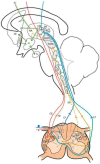
Figure 2
Diagram of the distribution of the main sensory pathways involved in pain perception and control (see text). (Taken from [16]). SGPV = PVG (Periventricular Gray Matter). SGPA = PAG (Periaqueductal Gray Matter). H = H (Hypothalamus). CE = CS (Corticospinal), RE = RS (Reticulospinal), ER = SR (Spinoreticular) and HETL = LSTT (Lateral spinothalamic) Tracts. NP = PN (Pulvinar nucleus). GM = MG (Medial geniculate nucleus).
Figure 3
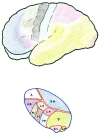
Figure 3
Anatomical–functional correlation between the thalamus and cerebral cortex (see text). LD (Lateral dorsal nucleus). LP (Lateral posterior nucleus). VL (Ventral lateral nucleus). VA (Ventral anterior nucleus). VL = VPL (Ventral posterolateral). VP = VPM (Ventral posteromedial). A = AN (Anterior nuclear group). DM = MD (Medial dorsal nucleus). P (Pulvinar). GL = LG (Lateral geniculate nucleus). GM = MG (Medial geniculate nucleus).
Figure 4
Figure 4
Area distribution according to the neurotransmitters involved (see text). LD (Lateral dorsal nucleus). LP (Lateral posterior nucleus). VL (Ventral lateral nucleus). VA (Ventral anterior nucleus). VL = VPL (Ventral posterolateral). VP = VPM (Ventral posteromedial). A = AN (Anterior nuclear group). DM = MD (Medial dorsal nucleus). P (Pulvinar). GL = LG (Lateral geniculate nucleus).
Figure 5
Figure 5
Melzack and Wall gate control theory. 1: Normal. 2: Increased pain afferents. 3: Increased sensory afferents. (see text). CNS: Central Nervous System. S: Sensory afferents. P: Pain afferents.
Figure 6
Figure 6
Therapeutic possibilities for reducing pain afferents. On the right, surgical interruption of pain pathways; on the left, increasing sensory afferents through neurostimulation. (see text).
Figure 7
Figure 7
DREZ to be altered (see text). C.P. = P.C. (Posterior cord). HER = SRT (Spinoreticular tract). HETL = LSTT (Lateral spinothalamic tract). HETA = VSTT (Ventral spinothalamic tract). FDL = DLF (Dorsolateral fasciculus, Lissauer’s tract).
Figure 8
Figure 8
Lesion zone in the spinothalamic tract (see text). C.P. = P.C. (Posterior cord). HER = SRT (Spinoreticular tract). HETL = LSTT (Lateral spinothalamic tract). HETA = VSTT (Ventral spinothalamic tract). FDL = DLF (Dorsolateral fasciculus, Lissauer’s tract).
Figure 9
Figure 9
Myelotomy (see text). C.P. = P.C. (Posterior cord). HER = SRT (Spinoreticular tract). HETL = LSTT (Lateral spinothalamic tract). HETA = VSTT (Ventral spinothalamic tract). FDL = DLF (Dorsolateral fasciculus, Lissauer’s tract).
Figure 10
Figure 10
Section of the trigeminal spinal tract (TST) at the level approximately 11 mm below the obex (modified from [122]). GN—Gracile Nucleus. CN—Cuneate Nucleus. STrigT—Spinal Trigeminal Tract. STrigN—Spinal Trigeminal Nucleus. AECT—Anterior Spinocerebellar Tract. PECT—Posterior Spinocerebellar Tract. ATLS—Anterolateral System. CG—Central Gray.
Figure 11
Figure 11
Trigeminal nucleotomy (see text). Section approximately 18 mm below the obex (modified from [122]). GF—Gracile Fasciculus. CF—Cuneate Fasciculus. STrigT—Spinal Trigeminal Tract. STrigN—Spinal Trigeminal Nucleus. AECT—Anterior Spinocerebellar Tract. PECT—Posterior Spinocerebellar Tract. LCET—Lateral Corticospinal Tract. GP—Gelatinosa Portion of Spinal Trigeminal Nucleus. MC—Magnocelllular Portion of Spinal Trigeminal Nucleus. RST—Rubrospinal Tract. ALS—Anterolateral System.
Figure 12
Figure 12
Sections at the level of the obex and 2–3 mm below (modified from [122]). GN—Gracile Nucleus. CN—Cuneate Nucleus. RB—Restiform Body. STrigT—Spinal Trigeminal Tract. STrigN—Spinal Trigeminal Nucleus. AECT—Anterior Spinocerebellar Tract. ALS—Anterolateral System. ON—Olivary Nucleus. ML—Medial Lemniscus. P—Pyramid. X—Dorsal Motor Nucleus of Vagus. XII—Hypoglossal Nucleus.
Figure 13
Figure 13
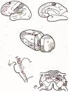
Two types of lesions proposed for stereotactic mesencephalotomy: medial or extralemniscal [128] and lateral [127] (spinothalamic tract) (Diagram taken from [129]). A—Aqueduct. CG—Central Gray. ML—Medial Lemniscus. RN—Red Nucleus. SN—Substantia Nigra. STT—Spinothalamic Tract. STrigT—Spinal Trigeminal Tract.
Figure 14
Figure 14
Mesencephalic section at the level proposed for mesencephalotomy. (Modified from [122]). The actual anatomical complexity of the areas represented in the diagram in Figure 13 is shown. A—Aqueduct. CG—Central Gray. ML—Medial Lemniscus. MLF—Medial Longitudinal Fasciculus. MGM—Medial Geniculate Nucleus. RF—Reticular Formation. RN—Red Nucleus. SN—Substantia Nigra.
Figure 15
Figure 15
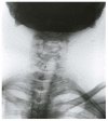
Cervical stimulation in a patient with unstable angina.
Figure 16
Figure 16
DBS at the level of the VPL nucleus in a female patient with Dejerine syndrome, with more disabling pain in the right lower extremity, on the sole of the foot. It prevented her from walking. She has reported good results for more than 5 years. Within the coordinates of the VPL nucleus, an area without hypointensity was located in T2, obtaining a sensory response to stimulation in the pain area.
Figure 17
Figure 17
Control after placing epidural electrodes over the motor cortex.

Similar articles

Cited by

References

    1. Bonica J.J. The need of a taxonomy. Pain. 1979;6:247–252. - PubMed
    1. Leriche R. The Surgery of Pain. Williams & Wilkins; Baltimore, MD, USA: 1939.
    1. Loonen A.J.M., Ivanova S.A. Circuits regulating pleasure and happiness: The evolution of reward-seeking and misery-fleeing behavioral mechanisms in vertebrates. Front. Neurosci. 2015;9:394. doi: 10.3389/fnins.2015.00394. - DOI - PMC - PubMed
    1. De Ridder D., Adhia D., Vanneste S. The anatomy of pain and suffering in the brain and its clinical implications. Neurosci. Biobehav. Rev. 2021;130:125–146. doi: 10.1016/j.neubiorev.2021.08.013. - DOI - PubMed
    1. Bonica J. Pain. Raven Press; New York, NY, USA: 1980.

LinkOut - more resources