. 2022 Nov 15;12(11):1114.
doi: 10.3390/metabo12111114.

Expression Analysis of Molecular Chaperones Hsp70 and Hsp90 on Development and Metabolism of Different Organs and Testis in Cattle (Cattle-yak and Yak)

Affiliations

Expression Analysis of Molecular Chaperones Hsp70 and Hsp90 on Development and Metabolism of Different Organs and Testis in Cattle (Cattle-yak and Yak)

Yan Cui et al. Metabolites. .

Abstract

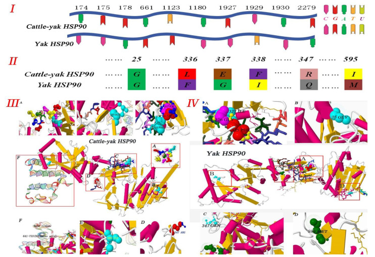
Hsp70 and Hsp90 play an important role in testis development and spermatogenesis regulation, but the exact connection between Hsp70 and Hsp90 and metabolic stress in cattle is unclear. Here, we focused on the male cattle−yak and yak, investigated the expression and localization of Hsp70 and Hsp90 in their tissues, and explored the influence of these factors on development and metabolism. In our study, a total of 54 cattle (24 cattle−yaks and 30 yaks; aged 1 day to 10 years) were examined. The Hsp90 mRNA of the cattle−yak was first cloned and compared with that of the yak, and variation in the amino acid sequence was found, which led to differences in protein spatial structure. Using real-time quantitative PCR (RT-qPCR) and Western blot (WB) techniques, we investigated whether the expression of Hsp70 and Hsp90 mRNA and protein are different in the cattle−yak and yak. We found a disparity in Hsp70 and Hsp90 mRNA and protein expression in different non-reproductive organs and in testicular tissues at different stages of development, while high expression was observed in the testes of both juveniles and adults. Moreover, it was intriguing to observe that Hsp70 expression was significantly high in the yak, whereas Hsp90 was high in the cattle−yak (p < 0.01). We also examined the location of Hsp70 and Hsp90 in the testis by immunohistochemical (IHC) and immunofluorescence (IF) techniques, and the results showed that Hsp70 and Hsp90 were positive in the epithelial cells, spermatogenic cells, and mesenchymal cells. In summary, our study proved that Hsp70 and Hsp90 expressions were different in different tissues (kidney, heart, cerebellum, liver, lung, spleen, and testis), and Hsp90 expression was high in the testis of the cattle−yak, suggesting that dysplasia of the cattle−yak may correlate with an over-metabolism of Hsp90.

Keywords: Hsp70 and Hsp90; cattle; different organs; expression; metabolism.


Conflict of interest statement

The authors declare no conflict of interest.

Figures

Figure 1
Figure 1
Physicochemical and evolutionary tree analysis of Hsp90 in cattle–yak. (A) The physical and chemical properties of Hsp90 in cattle–yak. Modle-template alignment, secondary protein structure and protein characteristics. (B) Homology analysis of amino acids and nucleotides.
Figure 2
Figure 2
Physicochemical and evolutionary tree analysis of Hsp90 in yak. (A) The physical and chemical properties of Hsp90 in yak. Modle-template alignment, secondary protein structure and protein characteristics. (B) Homology analysis of amino acids and nucleotides.
Figure 3
Figure 3
Analysis of Hsp90 protein structure in cattle–yak and yak. (I) and (II) Hsp90 nucleotide and amino acid sequence analysis showing the sites and types of mutations in cattle–yak and yak. (III) Hsp90 protein structure of cattle–yak. (A) The initiation terminal in cattle–yak is much more than in yak and the following amino acids are shown: MET, PRO, GLU, GLU, THR, GLN, ALA, GLN, ASP, and PRO, PRO. (B), (D), (E), (F) Mutation sites and amino acids are as follows: 25 (GLU), 36 (THR), 37 (PHE), 38 (TYR), 347 (ARG), 595 (ILE). (C) More than 92 amino acids are at the terminal. (IV) Hsp90 protein structure in yak. (A), (B), (C), (D) Mutation sites and amino acids are as follows: 14 (GLY), 25 (THR), 26 (PHE), 27 (TYR), 336 (GLN), 584 (MET).
Figure 4
Figure 4
Gene expression in different tissues of cattle–yak and yak. (I) The results of RT-PCR. Lanes: (1) lung; (2) cerebellum; (3) kidney; (4) liver; (5) heart; and (6) spleen. Lanes: (A) newborn; (B) childhood; (C) juvenile; (D) adult; and (E) senile. (II) The gene expression levels of Hsp70 and Hsp90 in non-reproductive tissues from cattle–yak and yak. (III) Gene expression levels of Hsp70 and Hsp90 in testicular tissues from male cattle (cattle–yak and yak). Note: The curved lines represent expression trends of Hsp70 and Hsp90 proteins in different organs and different developmental stages in testis. Different colors represent different expressions of Hsp70 and Hsp90 in the organs and testicular tissue from cattle–yak and yak.
Figure 5
Figure 5
Hsp70 and Hsp90 expression of in diffrent tissues. The arrow represents the site of positive expression. IHC(A,B,C,D,E) and IF(a,b,c,d,e). (I) Cattle-yak: (A) Control section from yak spleen, without immunoreactions (negative control). (B), (D) Positive staining for Hsp90 was observed in the terminal bronchioles, distal convoluted tubule, and proximal convoluted tubule of lungs and kidney. (C), (E) Positive staining for Hsp70 was observed in the cardiac muscle fibers and red pulp of heart and spleen. (a), (c), (e) Positive staining for Hsp70 was observed in the distal convoluted tubule, proximal convoluted tubule, cardiac muscle fibers and hepatocytes in kidney, heart, and liver. (b), (d) Positive staining for Hsp90 was observed in the terminal bronchioles, cerebellar medulla, granular layer and Purkinje cell layer of lungs and cerebellum. (II) Yak: (A) Control section from yak heart, without immunoreactions (negative control). (B), (D) Positive staining for Hsp70 was observed in the terminal bronchioles, cerebellar medulla, granular layer and Purkinje cell layer of lungs and cerebellum. (C), (E) Positive staining for Hsp90 was observed in hepatocytes, distal convoluted tubule, and proximal convoluted tubule of liver and kidney. (a), (c), (e) Positive staining for Hsp90 was observed in the cardiac muscle fibers, hepatocytes, distal convoluted tubule, and proximal convoluted tubule of heart, liver, and kidney. (b), (d) Positive staining for Hsp70 was observed in the red pulp, cerebellar medulla, granular layer, and Purkinje cell layer of the spleen and cerebellum. Note: Terminal bronchiole (TB); hepatocyte (Hc); central vein (CV); cardiac muscle fibers (CMF); distal convoluted tubule (DCT); Purkinje cell layer (PCL); renal glomerulus (RG); biofilm (Bf); trabecula (Tc); red pulp (RP); white pulp (WP); cerebellar medulla (CM); molecular layer (ML); granular layer (GL); and proximal convoluted tubule (PCT). (III) The result of optical density analysis value. The curved lines represent expression trends of HSP proteins in different organs, and different color represents different proteins.
Figure 6
Figure 6
Hsp70 and Hsp90 expression in testicular tissue of cattle–yak and yak. (I) Schematic diagram of labeled bands and their sizes. (II) Different colors represent expressions of Hsp70 and Hsp90 in different testicular tissues from cattle–yak and yak.
Figure 7
Figure 7
IHC and IF expression of Hsp70 and Hsp90 in testicular tissues from cattle–yak and yak. The arrow represents the site of positive expression. IHC(A,B,C,D,E) and IF(a,b,c,d,e). (I) Cattle-yak: (A) Control section from testicular tissue of adult yak, without immunoreactions (negative control). (B), (D) Positive staining for Hsp70 was observed in spermatogonium and primary spermatocyte of newborn and juvenile testis. (C), (E) Positive staining for Hsp90 was observed in mesenchymal, primary spermatocyte, and secondary spermatocyte cells of testes from childhood and adulthood. (a) Control section from testicular tissue of adult yak, without immunoreactions (negative control). (b), (d) Positive staining for Hsp90 was observed in the mesenchymal cells and primary spermatocyte of newborn and adult testis. (c), (e) Positive staining for Hsp70 was observed in spermatogonium, Sertoli cells, and seminiferous tubule of testes from childhood and adulthood. (II) Yak: (A), (C), (E) Positive staining for Hsp90 was observed in spermatogonium, secondary spermatocyte, and mesenchymal cells of newborn, juvenile, and senile testis. (B), (D) Positive staining for Hsp70 was observed in primary spermatocyte, Sertoli cells, spermoblast, and spermatogonium of testes from childhood and adulthood. (a), (c), (e) Positive staining for Hsp70 was observed in the spermatogonium, sperm and mesenchymal cells of newborn, juvenile, and senile testis. (b), (d) Positive staining for Hsp90 was observed in the mesenchymal cells and sperm of testes from childhood and adulthood. Note: Primary spermatocyte (PS); secondary spermatocyte (SS); spermatogonium (Sg); spermoblast (Sb); sperm (S); Sertoli cells (SC); mesenchymal cells (MC); myoid cells (MC); seminiferous tubule (ST). (III) The results of optical density analysis value. The curved lines represent expression trends of HSP proteins in different testicular tissues, and different colors represent different proteins.
Figure 8
Figure 8
Hsp70 and Hsp90 schematic diagram of action mechanism in testicular tissues. The highly expressed Hsp90 could block development of primary spermatocyte in testis of cattle–yak. The highly expressed Hsp70 and decreased Hsp90 could promote sustained development from spermatocyte to mature sperm in testis of yak.

References

    1. Wiener G., Jianlin H., Ruijun L. The Yak. FAO Regional Office for Asia and the Pacific; Bangkok, Thailand: 2003.
    1. Zou S.N., Liu P.G., Yu S.J., Cui Y., He J.F., Afedo S.Y., Zhang H.Z., Niayale R., Zhao K. Hsp60 expression and localization in different tissues and testis development of male cattle (Cattle-yak and Yak) Folia Morphol. 2021;80:857–869. doi: 10.5603/FM.a2020.0126. - DOI - PubMed
    1. Liu P.G., Yu S.J., Cui Y., He J.F., Zhang Q., Sun J., Huang Y.F., Yang X.Q., Cao M.X., Liao B., et al. Regulation by Hsp27/P53 in testis development and sperm apoptosis of male cattle (cattle-yak and yak) J. Cell. Physiol. 2018;234:650–660. - PubMed
    1. Shen H., Fan X.R., Zhang Z., Xi H.M., Ji R., Liu Y.H., Yue M.S., Li Q.H., He J.P. Effects of elevated ambient temperature and local testicular heating on the expressions of heat shock protein 70 and androgen receptor in boar testes. Acta Histochem. 2019;121:297–302. doi: 10.1016/j.acthis.2019.01.009. - DOI - PubMed
    1. Wells A.D., Malkovsky M. Heat shock proteins, tumor immunogenicity and antigen presentation: An integrated view. Immunol. Today. 2000;21:129–132. doi: 10.1016/S0167-5699(99)01558-3. - DOI - PubMed

LinkOut - more resources