Morphometric examination of trigeminal nerve and its adjacent structures in patients with trigeminal neuralgia: a case-control study
- PMID: 36422504
- PMCID: PMC10395698
- DOI: 10.55730/1300-0144.5503
Morphometric examination of trigeminal nerve and its adjacent structures in patients with trigeminal neuralgia: a case-control study
Abstract
Background: Morphological differences that can lead the trigeminal nerve to neurovascular conflict and a new solitary pontine lesion are associated with the pathogenesis of trigeminal neuralgia (TN). In this case-control study, we aimed to contribute to the current discussions about the pathogenesis of TN by investigating the anatomical structures that may have an effect on the morphometric parameters of the trigeminal nerve.
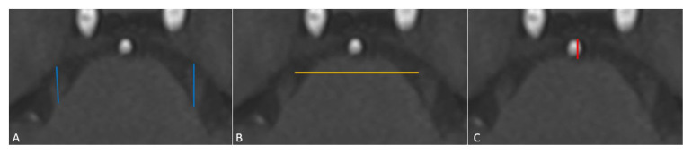
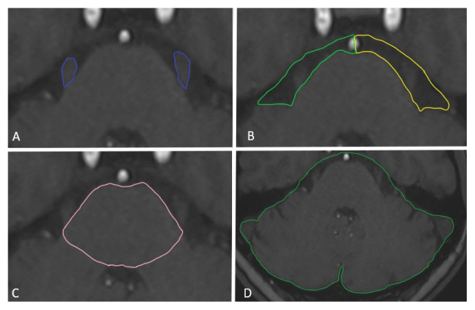
Methods: This study included 25 patients with TN followed up for pain in the Department of Algology, Faculty of Medicine, and 25 age- and gender-matched controls. We performed morphometric measurements including the length and volume of the trigeminal nerve, cerebellopontine cistern, pons, and posterior fossa in the MRIs of these individuals. Comparative analyses were performed for the mean of the affected and unaffected sides of the TN patients and the right, left, and both sides of the control group.
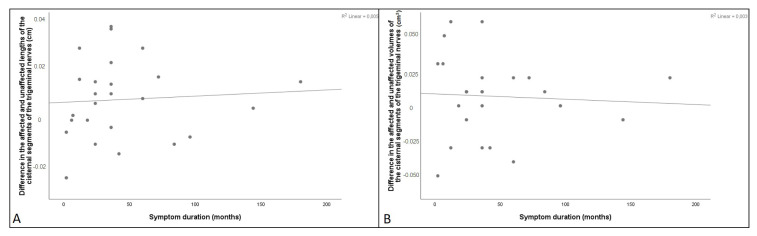
Results: In patients with TN, on the affected side, length and volume of the trigeminal nerve and cerebellopontine cistern volume were found smaller than controls (p < 0.05). Pons volume was higher in patients with TN compared to controls (p < 0.05). The length of the affected nerve was significantly related to prepontine cistern length and cerebellopontine cistern volume (p < 0.05).
Discussion: The cerebellopontine cistern volume has a significant impact on the morphometric characteristics of the trigeminal nerve. Especially, whether the increase in the volume of pons causes a decrease in the volume of cerebellopontine cistern should be clarified with further research.
Keywords: Trigeminal neuralgia; anatomy; brain stem; facial pain; morphometry.
Figures


Similar articles
-
Sex-dependent posterior fossa anatomical differences in trigeminal neuralgia patients with and without neurovascular compression: a volumetric MRI age- and sex-matched case-control study.J Neurosurg. 2019 Feb 1;132(2):631-638. doi: 10.3171/2018.9.JNS181768. Print 2020 Feb 1. J Neurosurg. 2019. PMID: 30717058
-
Significance of the Cerebellopontine Cistern Cross-Sectional Area and Trigeminal Nerve Anatomy in Trigeminal Neuralgia: An Anatomical Study Using Magnetic Resonance Imaging.Turk Neurosurg. 2020;30(2):271-276. doi: 10.5137/1019-5149.JTN.27735-19.2. Turk Neurosurg. 2020. PMID: 32091126
-
Nerve Atrophy and a Small Trigeminal Pontine Angle in Primary Trigeminal Neuralgia: A Morphometric Magnetic Resonance Imaging Study.World Neurosurg. 2017 Aug;104:575-580. doi: 10.1016/j.wneu.2017.05.057. Epub 2017 May 19. World Neurosurg. 2017. PMID: 28532907
-
[Trigeminal nerve lipoma presenting with trigeminal neuralgia: case report and literature review].Zh Vopr Neirokhir Im N N Burdenko. 2021;85(6):102-110. doi: 10.17116/neiro202185061102. Zh Vopr Neirokhir Im N N Burdenko. 2021. PMID: 34951767 Review. Russian.
-
Is There a Magnetic Resonance Imaging-Discernible Cause for Trigeminal Neuralgia? A Structured Review.World Neurosurg. 2017 Feb;98:89-97. doi: 10.1016/j.wneu.2016.10.104. Epub 2016 Oct 27. World Neurosurg. 2017. PMID: 27989975 Free PMC article. Review.
Cited by
-
A combined radiomics and anatomical features model enhances MRI-based recognition of symptomatic nerves in primary trigeminal neuralgia.Front Neurosci. 2024 Oct 24;18:1500584. doi: 10.3389/fnins.2024.1500584. eCollection 2024. Front Neurosci. 2024. PMID: 39513045 Free PMC article.
References
-
- Ruiz-Juretschke F, González-Quarante LH, García-Leal R, Martínez de Vega V. Neurovascular Relations of the Trigeminal Nerve in Asymptomatic Individuals Studied with High-Resolution Three-Dimensional Magnetic Resonance Imaging. The Anatomical Record. 2019;302(4):639–645. doi: 10.1002/ar.23818. - DOI - PubMed
MeSH terms
LinkOut - more resources
Full Text Sources
Medical