. 2022 Nov 12;15(11):1393.
doi: 10.3390/ph15111393.

Three-Dimensional Microtumor Formation of Infantile Hemangioma-Derived Endothelial Cells for Mechanistic Exploration and Drug Screening

Affiliations

Three-Dimensional Microtumor Formation of Infantile Hemangioma-Derived Endothelial Cells for Mechanistic Exploration and Drug Screening

Yanan Li et al. Pharmaceuticals (Basel). .

Abstract

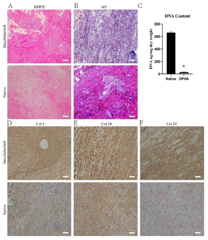
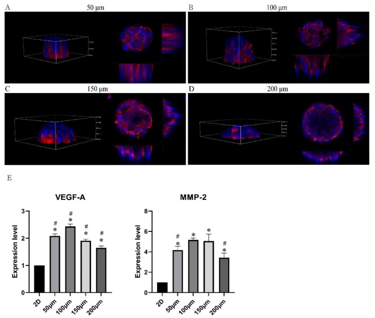
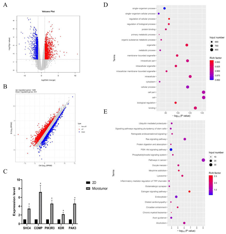
Infantile hemangioma (IH) is the most prevalent type of vascular tumor in infants. The pathophysiology of IH is unknown. The tissue structure and physiology of two-dimensional cell cultures differ greatly from those in vivo, and spontaneous regression often occurs during tumor formation in nude mice and has severely limited research into the pathogenesis and development of IH. By decellularizing porcine aorta, we attempted to obtain vascular-specific extracellular matrix as the bioink for fabricating micropattern arrays of varying diameters via microcontact printing. We then constructed IH-derived CD31+ hemangioma endothelial cell three-dimensional microtumor models. The vascular-specific and decellularized extracellular matrix was suitable for the growth of infantile hemangioma-derived endothelial cells. The KEGG signaling pathway analysis revealed enrichment primarily in stem cell pluripotency, RAS, and PI3KAkt compared to the two-dimensional cell model according to RNA sequencing. Propranolol, the first-line medication for IH, was also used to test the model's applicability. We also found that metformin had some impact on the condition. The three-dimensional microtumor models of CD31+ hemangioma endothelial cells were more robust and efficient experimental models for IH mechanistic exploration and drug screening.

Keywords: drug screening; extracellular matrix; infantile hemangioma; microtumor model; three-dimensional.


Conflict of interest statement

The authors declare no conflict of interest.

Figures

Figure 1
Figure 1
Characteristics of the decellularized porcine heart aortic scaffolds. (A) Hematoxylin and eosin staining and (B) Masson’s trichrome (MT) staining of native and decellularized porcine heart aortic scaffolds. (C) Relative DNA content in native and decellularized porcine heart aortic scaffolds. (D) Collagen I, (E) collagen Ⅲ, and (F) collagen IV staining of native and decellularized porcine heart aortic scaffolds. * p < 0.05 versus the native group. Scale bar = 100 μm.
Figure 2
Figure 2
Characteristics of the decellularized extracellular matrix hydrogel derived from cultures of decellularized porcine heart aortic scaffolds. Live/dead staining of (A) CD31+ HemECs cultured on different hydrogels on days 1, 3, and 5. The cell adhesion rate of (B) CD31+ HemECs on different hydrogels at 10 min after cell seeding. The proliferation rate of (C) CD31+ HemECs on different hydrogels at 24 h after cell seeding. * p < 0.05 versus the ECM group; # p < 0.05 versus the untreated group. Scale bar = 100 μm.
Figure 3
Figure 3
Fabrication of micropattern arrays and the formation of CD31+ HemEC microtumors. Micropattern arrays (A) in bright field and (B) in fluorescence light from PDMS seals with 50, 100, 150, and 200 μm diameters. (C) The CD31+ HemECs cultured on micropattern arrays with different diameters at 6 h after cell seeding. (D) The CD31+ HemECs formed microtumors with different diameters at day 3 after cell seeding. (E) Live/dead staining of CD31+ HEC microtumors. Scale bars = 50 μm.
Figure 4
Figure 4
Characteristics of the CD31+ HemEC microtumors. (AD) Three-dimensional view of CD31+ HemEC microtumors in micropattern arrays with 50, 100, 150, and 200 μm diameters by confocal microscopy. (E) Gene expression levels of MMP-2 and VEGF-A in CD31+ HemEC microtumors with different diameters. * p < 0.05 compared to the 2D culture; # p < 0.05 compared to the 100 μm diameters.
Figure 5
Figure 5
Results of the RNA sequencing. (A) Volcano plot; (B) the differential expressions of genes between the CD31+ HemEC microtumors and the 2D cell model. High relative expression is indicated by red, whereas low relative expression is indicated by blue. (C) Gene expression levels of focal adhesion-related genes (SHC4, COMP, PIK3R3, KDR, and PAK3). (D,E) The top 20 enriched GO and KEGG signaling pathways for significantly upregulated genes. * p < 0.05 compared to the 2D culture.
Figure 6
Figure 6
Features of microtumors detected by immunocytochemistry of (A) GLUT-1 (specific immunity indicator of IH), (B) HIF-1α (hypoxia marker), (C) Ki67 (proliferation marker), and (D) MCL-1 (antiapoptotic marker) in CD31+ HemEC microtumors and monolayers. Immunocytochemistry of (E) N-cadherin in CD31+ HEC microtumors and monolayers. (F) Cytosolic pH of CD31+ HemEC microtumors and monolayers. Scale bars = 100 μm.
Figure 7
Figure 7
Drug screening of propranolol (A) and metformin (B) in CD31+ HemEC microtumors with live/dead staining. Scale bars = 100 μm.

References

    1. Moisan F., Oucherif S., Kaulanjan-Checkmodine P., Prey S., Rousseau B., Bonneu M., Claverol S., Gontier E., Lacomme S., Dousset L., et al. Critical role of Aquaporin-1 and telocytes in infantile hemangioma response to propranolol beta blockade. Proc. Natl. Acad. Sci. USA. 2021;118:e2018690118. doi: 10.1073/pnas.2018690118. - DOI - PMC - PubMed
    1. Anderson K.R., Schoch J.J., Lohse C.M., Hand J.L., Davis D.M., Tollefson M.M. Increasing incidence of infantile hemangiomas (IH) over the past 35 years: Correlation with decreasing gestational age at birth and birth weight. J. Am. Acad. Dermatol. 2016;74:120–126. doi: 10.1016/j.jaad.2015.08.024. - DOI - PMC - PubMed
    1. Cotter C., Solman L., Flohr C. A national audit of oral propranolol for the treatment of infantile haemangiomas. Br. J. Dermatol. 2021;186:189–191. doi: 10.1111/bjd.20738. - DOI - PubMed
    1. Itinteang T., Tan S.T., Brasch H., Day D.J. Primitive mesodermal cells with a neural crest stem cell phenotype predominate proliferating infantile haemangioma. J. Clin. Pathol. 2010;63:771–776. doi: 10.1136/jcp.2010.079368. - DOI - PubMed
    1. de Jong S., Itinteang T., Withers A.H., Davis P.F., Tan S.T. Does hypoxia play a role in infantile hemangioma? Arch. Dermatol. Res. 2016;308:219–227. doi: 10.1007/s00403-016-1635-x. - DOI - PubMed

LinkOut - more resources