. 2022 Nov 19;14(11):806.
doi: 10.3390/toxins14110806.

Extracellular Vesicles from Bothrops jararaca Venom Are Diverse in Structure and Protein Composition and Interact with Mammalian Cells

Affiliations

Extracellular Vesicles from Bothrops jararaca Venom Are Diverse in Structure and Protein Composition and Interact with Mammalian Cells

Larissa Gonçalves-Machado et al. Toxins (Basel). .

Abstract

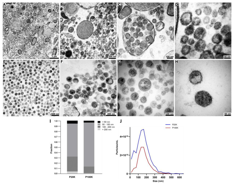
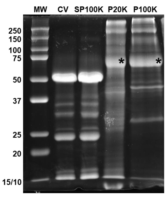
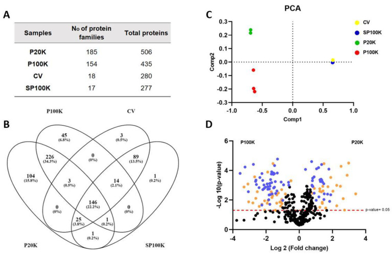
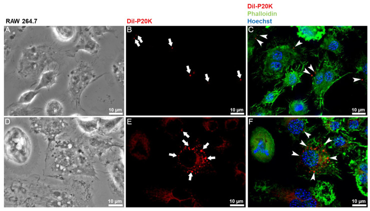
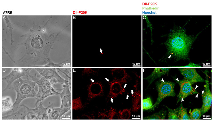
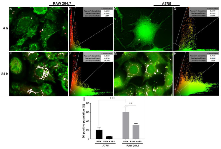
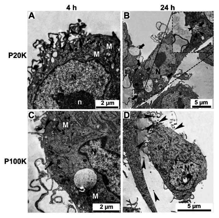
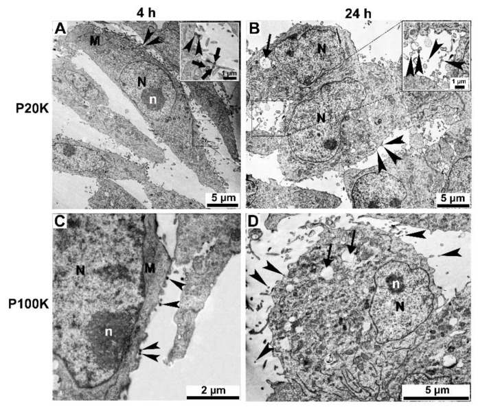
Snake venoms are complex cocktails of non-toxic and toxic molecules that work synergistically for the envenoming outcome. Alongside the immediate consequences, chronic manifestations and long-term sequelae can occur. Recently, extracellular vesicles (EVs) were found in snake venom. EVs mediate cellular communication through long distances, delivering proteins and nucleic acids that modulate the recipient cell's function. However, the biological roles of snake venom EVs, including possible cross-organism communication, are still unknown. This knowledge may expand the understanding of envenoming mechanisms. In the present study, we isolated and characterized the EVs from Bothrops jararaca venom (Bj-EVs), giving insights into their biological roles. Fresh venom was submitted to differential centrifugation, resulting in two EV populations with typical morphology and size range. Several conserved EV markers and a subset of venom related EV markers, represented mainly by processing enzymes, were identified by proteomic analysis. The most abundant protein family observed in Bj-EVs was 5'-nucleotidase, known to be immunosuppressive and a low abundant and ubiquitous toxin in snake venoms. Additionally, we demonstrated that mammalian cells efficiently internalize Bj-EVs. The commercial antibothropic antivenom partially recognizes Bj-EVs and inhibits cellular EV uptake. Based on the proteomic results and the in vitro interaction assays using macrophages and muscle cells, we propose that Bj-EVs may be involved not only in venom production and processing but also in host immune modulation and long-term effects of envenoming.

Keywords: 5′-nucleotidase; extracellular vesicles; snake venom.


Conflict of interest statement

The authors declare no conflict of interest.

Figures

Figure 1
Figure 1
Morphological characterization of extracellular vesicles from Bj-EVs. Ultrathin sections of (AD) P20K and (EH) P100K were obtained by high voltage electron microscopy (Fei Tecnai Spirit, 120kV). (I) EV size distribution was determined by morphometric analysis of individual Bj-EVs. A total of 705 and 174 particles were analyzed from P20K and P100K, respectively, using negative staining images obtained by transmission electron microscopy. (J) Size distribution of Bj-EVs by nanoparticle tracking analysis (NTA) using a Zeta View (Particle Metrix). P20K = Bj-EVs pelleted at 20,000× g; P100K = Bj-EVs pelleted at 100,000× g.
Figure 2
Figure 2
Protein profile of B. jararaca crude venom and its fractions. SDS-PAGE of B. jararaca crude venom and fractions (15 µg of protein) under reducing conditions, stained with Coomassie Brilliant Blue G-250. The asterisks highlight the protein bands digested and identified by mass spectrometry, described in Figure S3. CV = crude venom; SP100K = supernatant of P100K, or venom depleted of vesicles; P20K = vesicles pelleted at 20,000× g; P100K = vesicles pelleted at 100,000× g.
Figure 3
Figure 3
Shotgun Proteomics of B. jararaca crude venom and fractions. (A) Overview of the number of protein families and individual proteins identified in the samples. The full description of the identified protein families is listed in Supplementary Data S1 and S2. (B) Venn diagram showing common proteins among the crude venom and its fractions. (C) Principal component analysis (PCA) shows the global quantitative differences between the crude venom and its fractions. (D) Volcano plot showing the quantitative differences between P20K and P100K fractions. Blue dots represent highly significant quantitative differences between P20K and P100K. Orange dots are related to low abundance signals of proteins, filtered out by the L-stringency in the T-Fold node from PatternLab for Proteomics software, (q-value 0.05, F-stringency 0.10, L-stringency 0.40). CV = crude venom; SP100K = supernatant of P100K, or the venom depleted of vesicles; P20K = vesicles pelleted at 20,000× g; P100K = vesicles pelleted at 100,000× g.
Figure 4
Figure 4
Fluorescence microscopy of RAW 264.7 macrophages treated with Dil-labeled vesicles. Macrophages were incubated with 5 µg of Dil-labeled P20K EV fraction for four hours (AC) and 24 h (DF). Images represent a maximum projection of images from different focal planes after the 3D deconvolution process. Cells maintained at the same condition but without P20K treatment (control) are shown in Figure S5. Red = P20K-EVs (Dil); green = actin (phalloidin); blue = nucleus (Hoechst). White arrows and arrowheads highlight internalized vesicles.
Figure 5
Figure 5
Fluorescence microscopy of A7R5 muscle cells treated with Dil-labeled vesicles. Muscle cells were incubated with 5 µg of Dil-labeled P20K EV fraction for four hours (AC) and 24 h (DF). Images represent a maximum projection of images from different focal planes after the 3D deconvolution process. Cells maintained under the same conditions but without P20K treatment are shown in Figure S5. Red = P20K-EVs (Dil); green = actin (phalloidin); blue = nucleus (Hoechst). White arrows and arrowheads highlight internalized vesicles.
Figure 6
Figure 6
EV uptake in macrophages (RAW 264.7) and muscle cells (A7R5). (AH) Pearson’s correlation analysis. White areas in A, C, E, and G and yellow areas in B, D, F, and H correspond to the overlap of P20K with the cytosolic region. (I) Fluorescence quantification of cells treated for 24 h with Dil-labeled P20K or Dil-labeled P20K pre-incubated with antibothropic antivenom (ABS). Statistical evaluation was performed by Tukey’s multiple comparisons test. *** p = 0.0003; ** p = 0.0025 red = P20K-EVs (Dil); green = actin (phalloidin).
Figure 7
Figure 7
Ultrathin sections of RAW 264.7 macrophages treated with Bj-EVs. Macrophages were treated with P20K (A,B) and P100K (C,D). The arrowheads point to the EVs interacting with the cell surface or inside the cytosol. After four hours of interaction, several membrane projections can be observed (A,C). In addition, large vacuoles containing EVs (B, arrows) and alterations in the nucleus morphology and chromatin condensation (B and D, large arrow) were observed. Images were obtained by transmission electron microscopy in scanning electron microscopy (STEM-IN-SEM). N, nucleus; n, nucleolus; M, mitochondria.
Figure 8
Figure 8
Ultrathin sections of A7R5 muscle cell line treated with Bj-EVs. Muscle cells were treated with P20K (A,B) and P100K (C,D). The arrowheads point to the EVs interacting with the cell surface. The inset on panel A shows a membrane projection. Arrows display large vacuoles (D) or vacuoles containing EVs (B). After 24 h of treatment, it is possible to observe abnormalities in the nuclear envelope morphology and chromatin condensation (B,D). Images were obtained by transmission electron microscopy in scanning electron microscopy (STEM-IN-SEM). N, nucleus; n, nucleolus; M, mitochondria.

References

    1. Warshawsky H., Haddad A., Gonçalves R.P., Valeri V., De Lucca F.L. Fine structure of the venom gland epithelium of the south american rattlesnake and radioautographic studies of protein formation by the secretory cells. Am. J. Anat. 1973;138:79–119. doi: 10.1002/aja.1001380106. - DOI - PubMed
    1. Oron U., Bdolah A. Intracellular transport of proteins in active and resting secretory cells of the venom gland of Vipera palaestinae. J. Cell Biol. 1978;78:488–502. doi: 10.1083/jcb.78.2.488. - DOI - PMC - PubMed
    1. Villar-Briones A., Aird S.D. Organic and peptidyl constituents of snake venoms: The picture is vastly more complex than we imagined. Toxins. 2018;10:392. doi: 10.3390/toxins10100392. - DOI - PMC - PubMed
    1. Zancolli G., Casewell N.R. Venom systems as models for studying the origin and regulation of evolutionary novelties. Mol. Biol. Evol. 2020;37:2777–2790. doi: 10.1093/molbev/msaa133. - DOI - PubMed
    1. Casewell N.R., Wüster W., Vonk F.J., Harrison R.A., Fry B.G. Complex cocktails: The evolutionary novelty of venoms. Trends Ecol. Evol. 2013;28:219–229. doi: 10.1016/j.tree.2012.10.020. - DOI - PubMed

Publication types