Review
. 2023 Jul 1;103(3):1827-1897.
doi: 10.1152/physrev.00030.2021. Epub 2022 Nov 24.

Pathophysiology and pathogenic mechanisms of pulmonary hypertension: role of membrane receptors, ion channels, and Ca2+ signaling

Affiliations
Review

Pathophysiology and pathogenic mechanisms of pulmonary hypertension: role of membrane receptors, ion channels, and Ca2+ signaling

Angela Balistrieri et al. Physiol Rev. .

Abstract

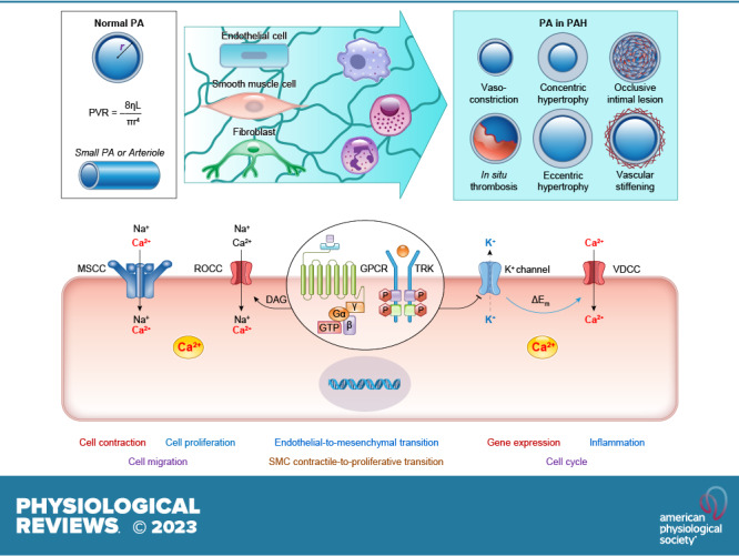
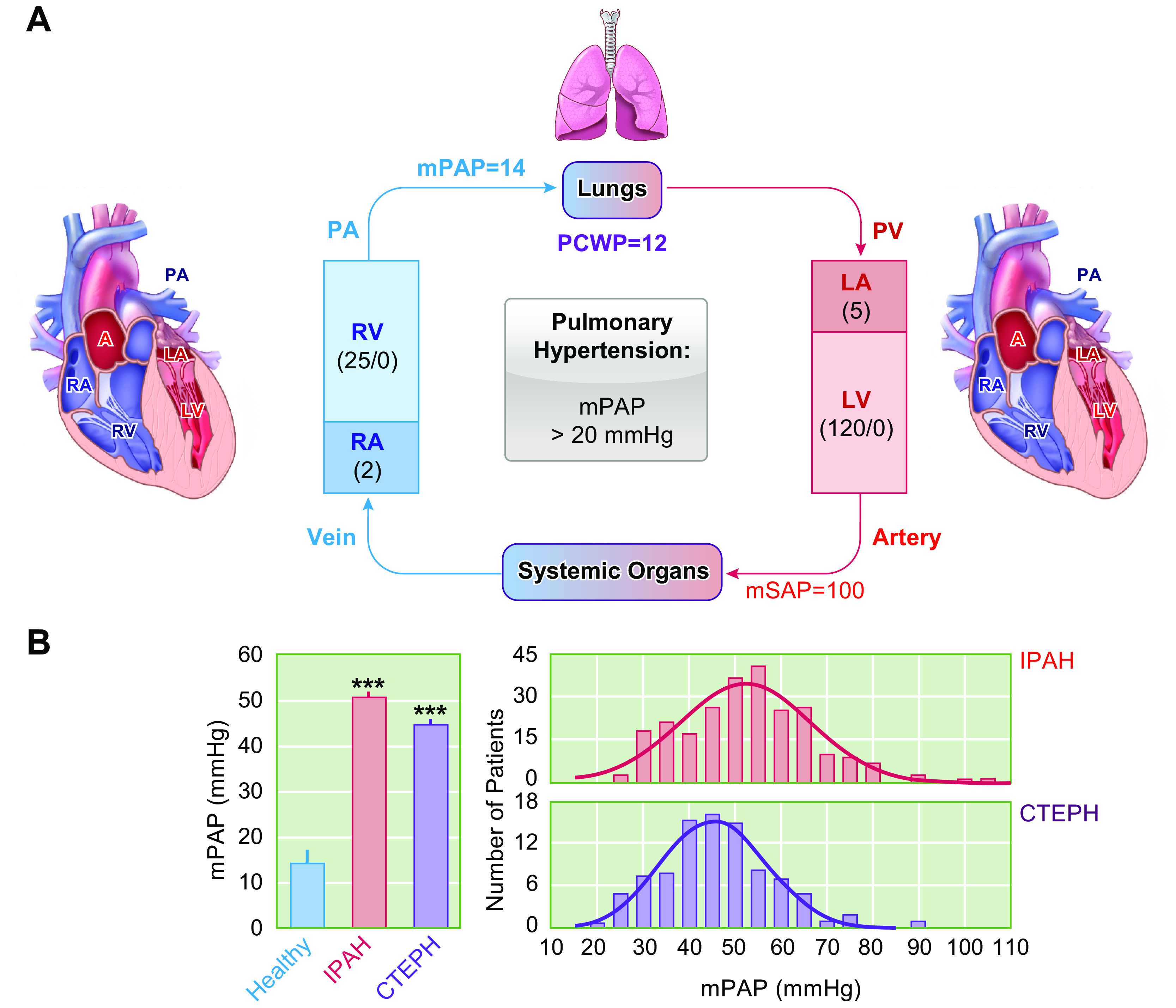
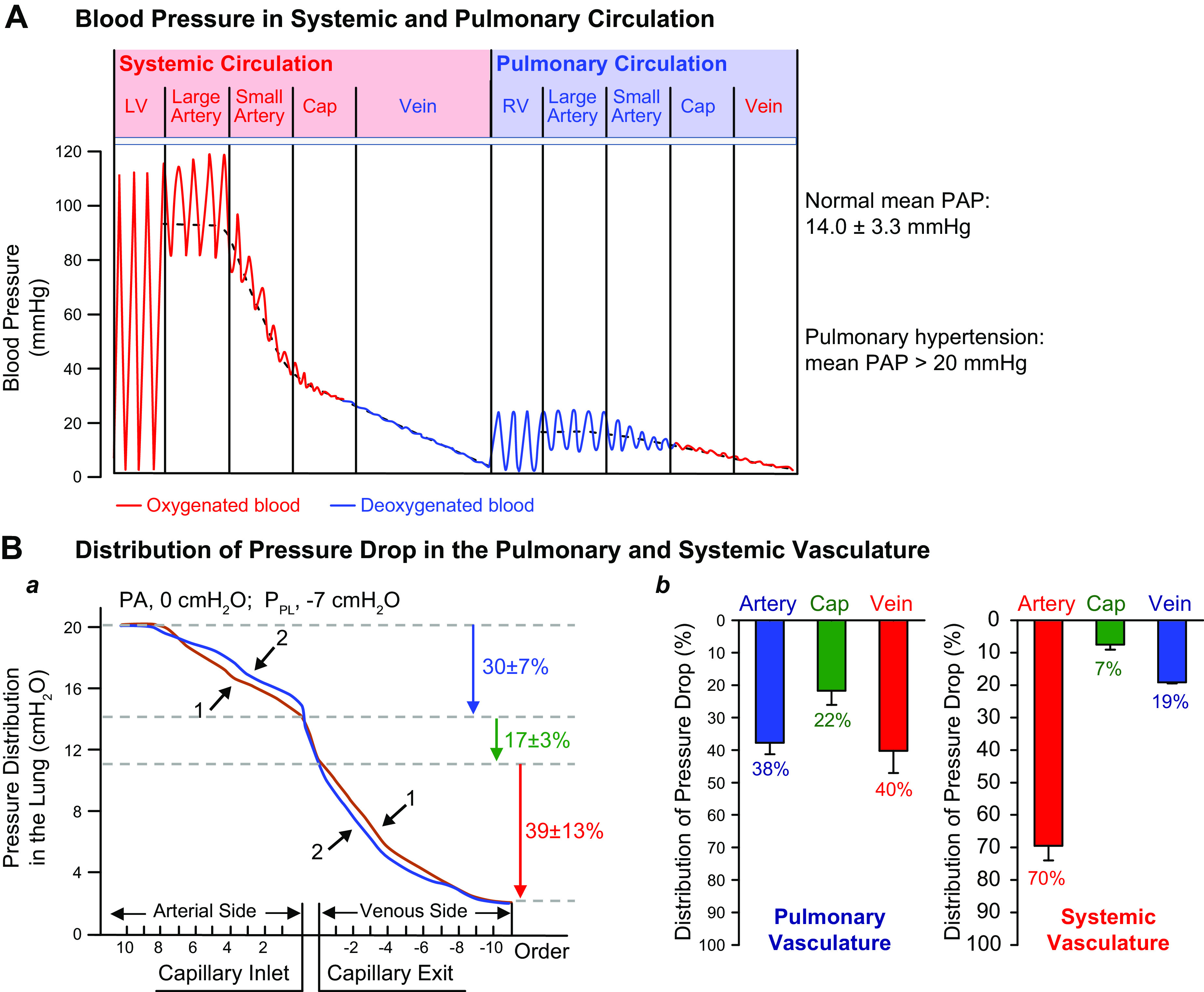
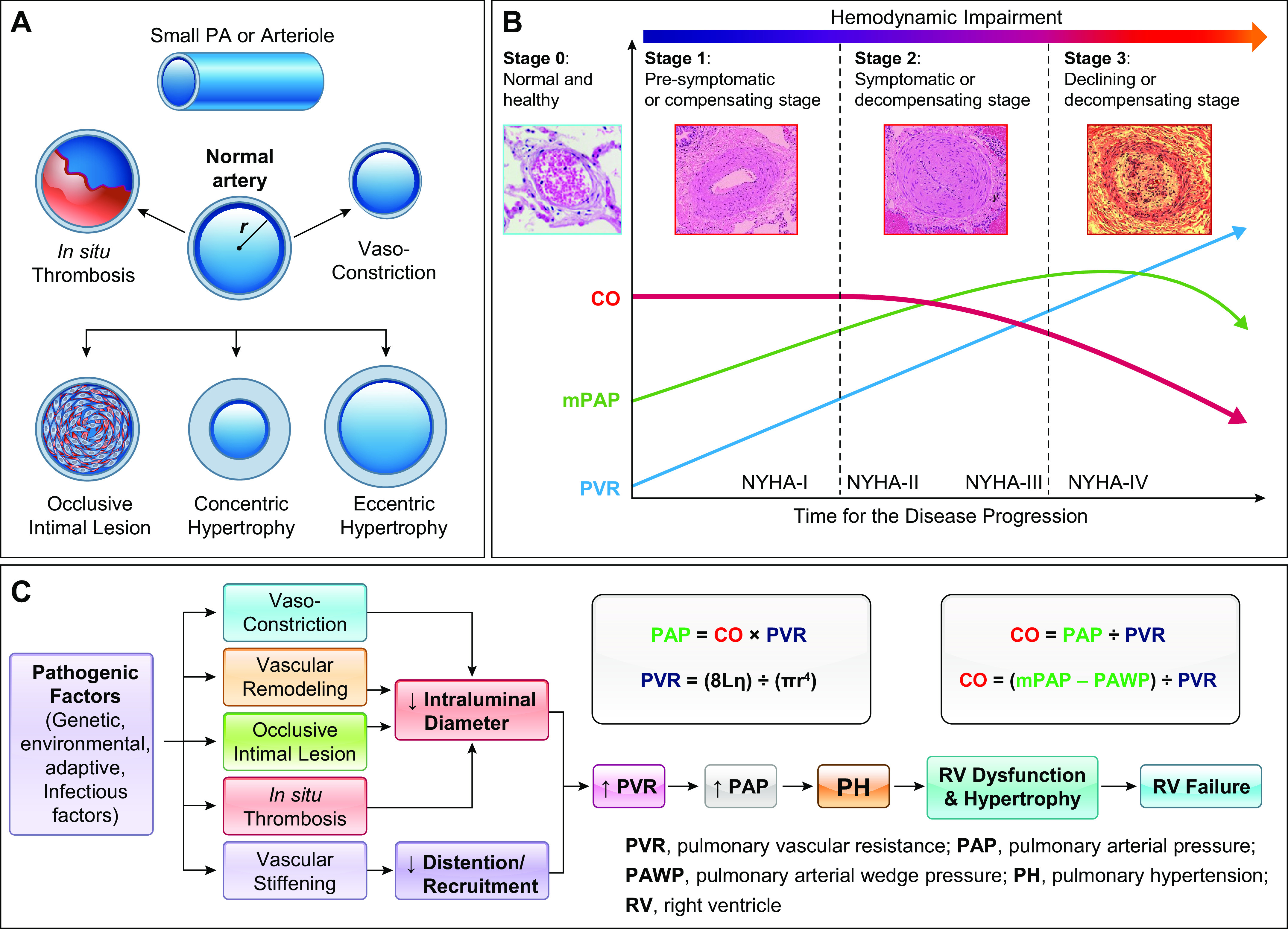
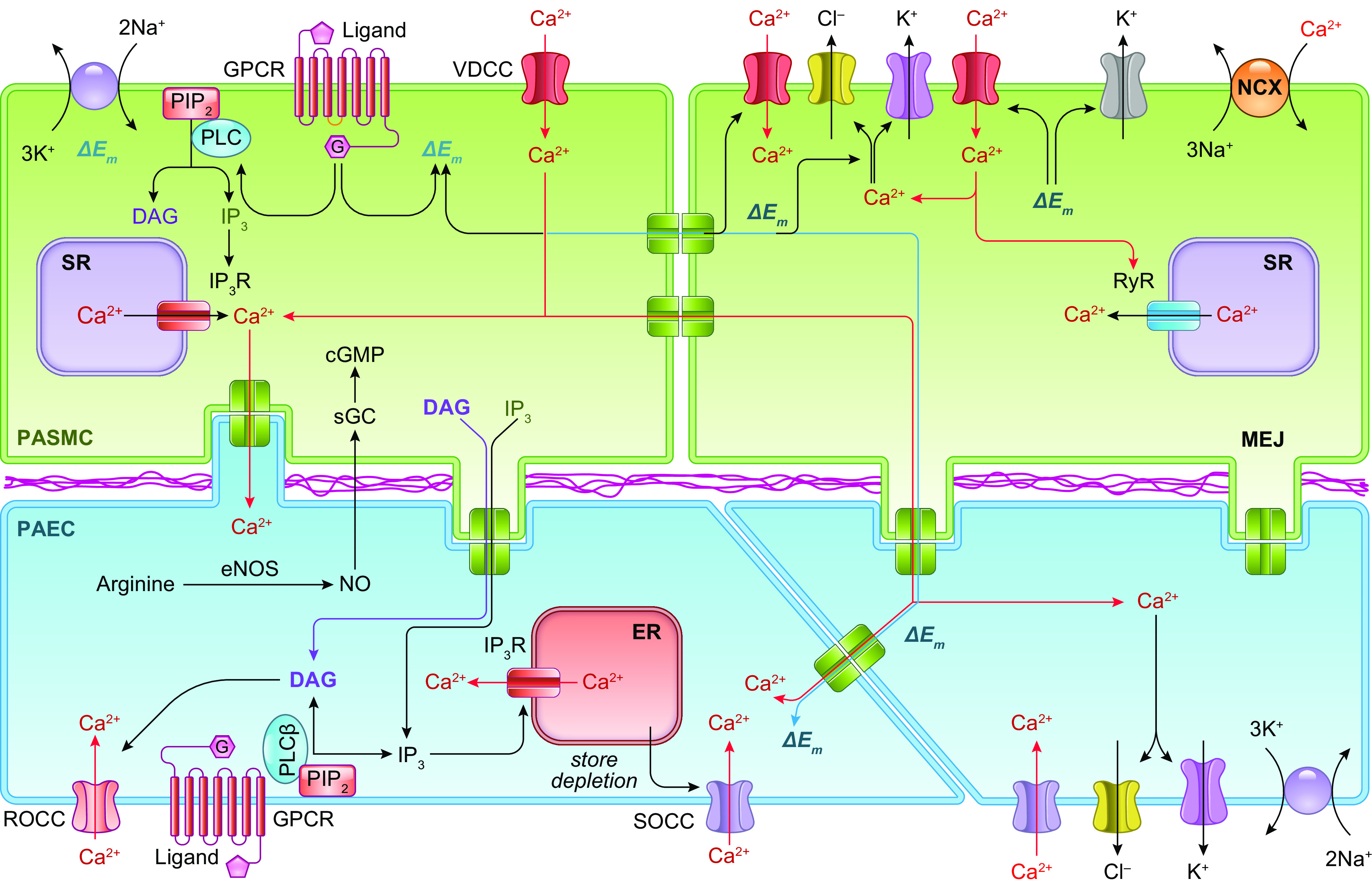
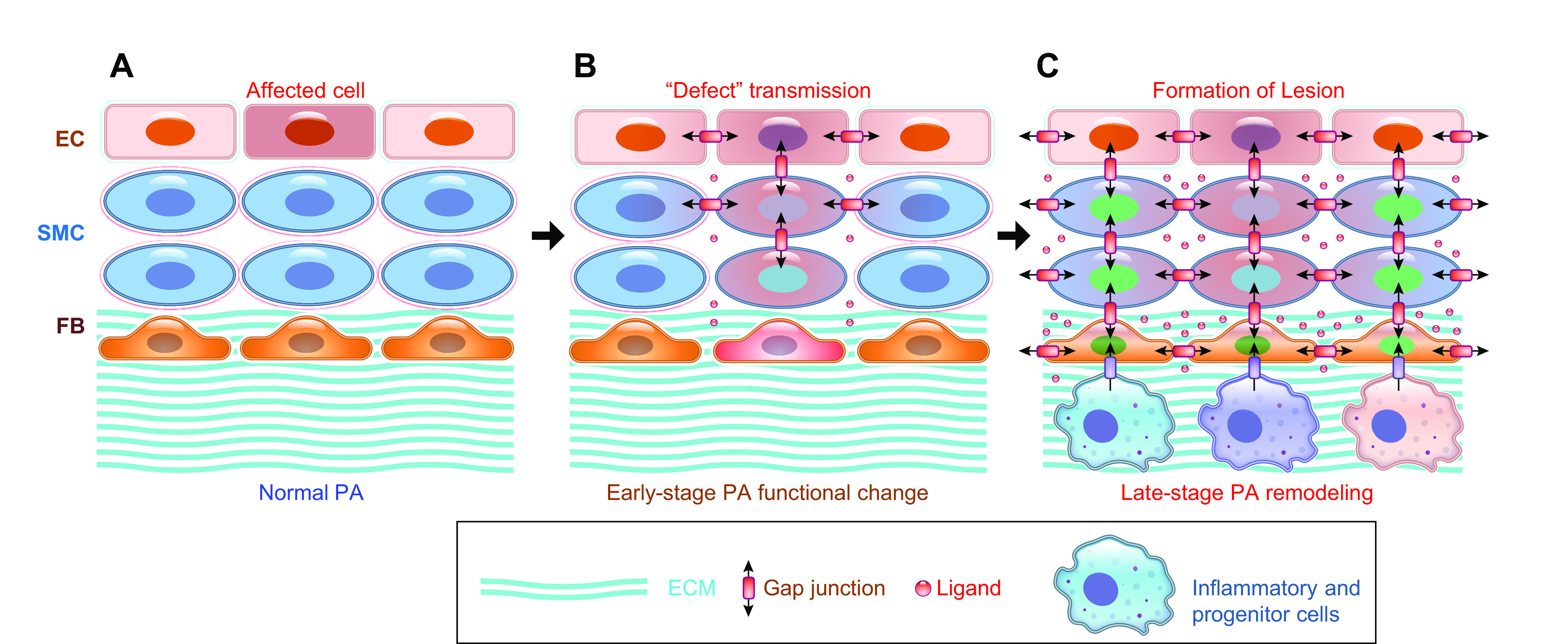
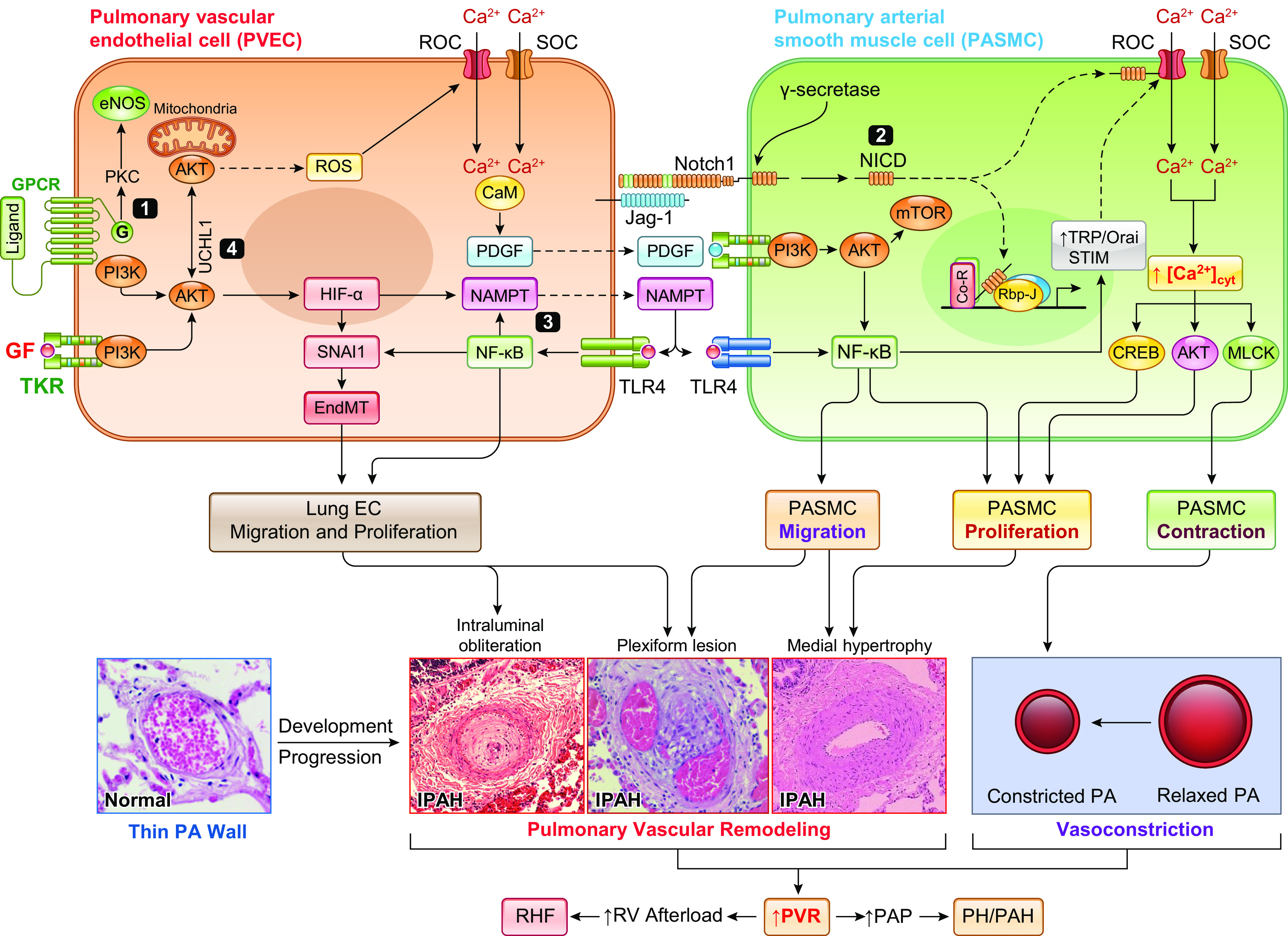
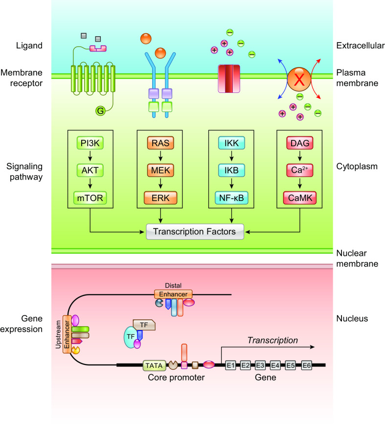
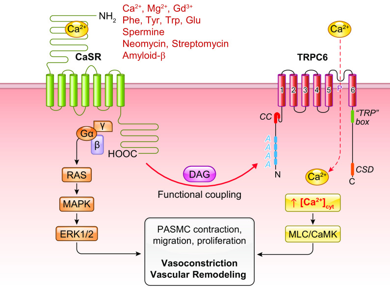
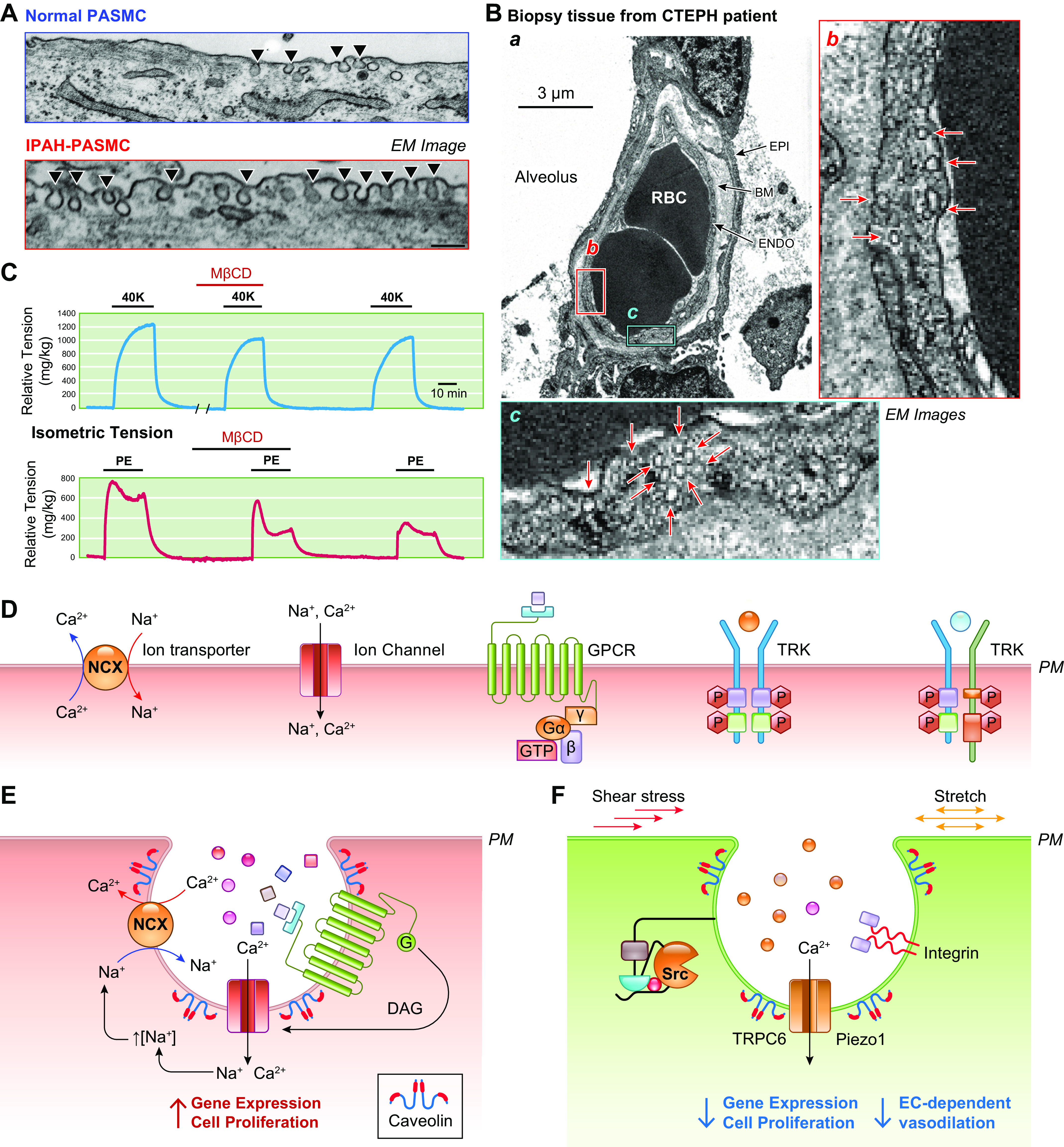
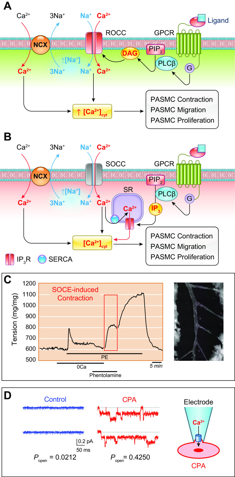
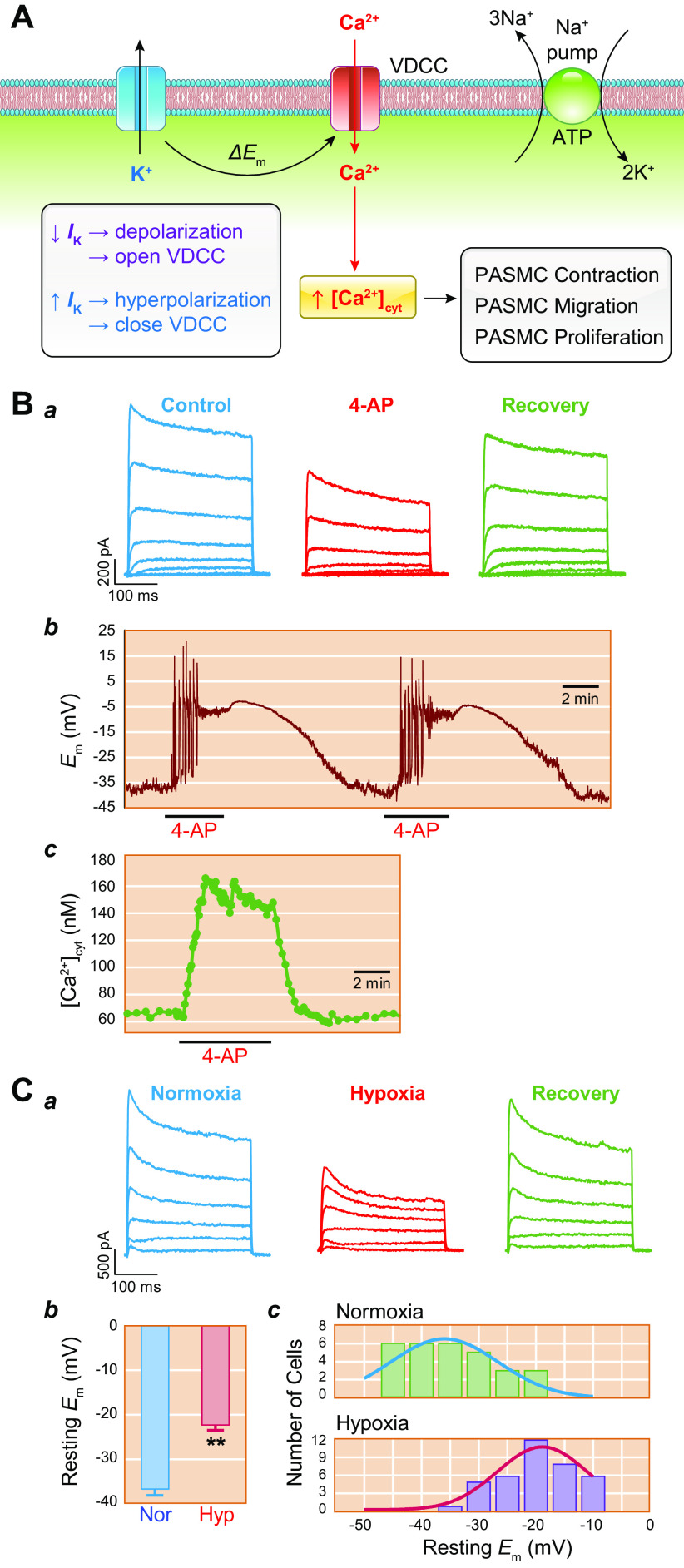
The pulmonary circulation is a low-resistance, low-pressure, and high-compliance system that allows the lungs to receive the entire cardiac output. Pulmonary arterial pressure is a function of cardiac output and pulmonary vascular resistance, and pulmonary vascular resistance is inversely proportional to the fourth power of the intraluminal radius of the pulmonary artery. Therefore, a very small decrease of the pulmonary vascular lumen diameter results in a significant increase in pulmonary vascular resistance and pulmonary arterial pressure. Pulmonary arterial hypertension is a fatal and progressive disease with poor prognosis. Regardless of the initial pathogenic triggers, sustained pulmonary vasoconstriction, concentric vascular remodeling, occlusive intimal lesions, in situ thrombosis, and vascular wall stiffening are the major and direct causes for elevated pulmonary vascular resistance in patients with pulmonary arterial hypertension and other forms of precapillary pulmonary hypertension. In this review, we aim to discuss the basic principles and physiological mechanisms involved in the regulation of lung vascular hemodynamics and pulmonary vascular function, the changes in the pulmonary vasculature that contribute to the increased vascular resistance and arterial pressure, and the pathogenic mechanisms involved in the development and progression of pulmonary hypertension. We focus on reviewing the pathogenic roles of membrane receptors, ion channels, and intracellular Ca2+ signaling in pulmonary vascular smooth muscle cells in the development and progression of pulmonary hypertension.

Keywords: Ca2+ signaling; ion channel; membrane receptor; pulmonary arterial hypertension; pulmonary circulation.


Conflict of interest statement

No conflicts of interest, financial or otherwise, are declared by the authors.

Figures

None
Graphical abstract
FIGURE 1.
FIGURE 1.
Schematic diagram of the pulmonary circulation and systemic circulation (A) and distribution of mean pulmonary arterial pressure (mPAP) in normal subjects and patients with idiopathic pulmonary arterial hypertension (IPAH) and chronic thromboembolic pulmonary hypertension (CTEPH) (B). A: the venous blood (or deoxygenated blood) is pumped from the right ventricle (RV) to the lungs via the pulmonary artery (PA). After taking in O2 from (and releasing CO2 to) alveoli to the capillary, the reoxygenated blood circulates to the left atria (LA) and is then delivered to systemic organs via the left ventricle (LV) through the systemic circulation. After releasing O2 to (and taking in CO2 from) organs and tissues, the deoxygenated blood circulates to the right atria (RA). The numbers in parentheses indicate the average pressure values in mmHg in healthy human subjects. For example, the mPAP is 14 mmHg, and the mean systemic arterial pressure (mSAP) is 100 mmHg. The pulmonary capillary wedge pressure (PCWP), also called pulmonary arterial wedge pressure (PAWP) or pulmonary artery occlusion pressure (PAOP), is ∼12 mmHg (4–12 mmHg in normal subjects). The RV systolic and diastolic pressure is 25 and 0 mmHg, whereas the LV systolic and diastolic pressure is 120 and 0 mmHg. The RA pressure and LA pressure are 2 and 5 mmHg, respectively. B: average mPAP in healthy control subjects (Healthy) and patients with IPAH and CTEPH (left) and distribution of mPAP in patients with IPAH and CTEPH (right). ***P <0.001 vs. Healthy control.
FIGURE 2.
FIGURE 2.
Distribution of pressure changes in the pulmonary circulation. A: blood pressure measured in the systemic circulation system including the left ventricle (LV), large artery, small artery, capillary (Cap), and vein and the pulmonary circulation system including the right ventricle (RV), large and small pulmonary artery, lung capillary, and vein. B: distribution of pressure in the lung (in cmH2O) from the arterial side to the capillary and to the venous side (a). The pressure drop in the pulmonary artery accounts for 30 ± 7% of the total pressure drop, whereas the pressure drop in the capillary and vein accounts for 17 ± 3% and 39 ± 13%, respectively. The pressure drop in the artery, capillary, and vein is compared between the pulmonary (b, left) and systemic (b, right) circulation systems. The bar graphs are based on data from Refs. –.
FIGURE 3.
FIGURE 3.
A: typical cast of a small segment of the arterial tree in human lungs shows the complex structure of the vascular tree. B–D: the 3 proposed schemes describing this complex structure are illustrated by the Weibel model (B), the Strahler model (C), and the diameter-defined Strahler system (D). Generation numbers are indicated on each branch. Note that in the Weibel model the largest vessel is designated as a vessel of generation 1. After each bifurcation, the generation number of the offsprings is increased by 1. The exact opposite is true in the Strahler model and the diameter-defined Strahler system, in which the smallest noncapillary blood vessels are defined as order 1. In the Strahler model, when 2 vessels of the same order meet, the order number of the confluent vessel is increased. When 2 vessels of different orders meet, the order number of the confluent vessels remains the same as the larger of the 2. In the diameter-defined Strahler system, when 2 vessels of different order and diameter meet one another, the order number of the confluent vessel is increased only if its diameter is larger than either of the 2 segments by a certain amount. Otherwise, the order number of the confluent segment is not increased. E: lumen diameter (closed circles in blue; left) and length (closed squares in dark cyan; right) of each segment of pulmonary arteries and distribution of total cross-sectional area (closed circles in red; left) and number of branches (closed squares in dark red; right) of all segments of each order of pulmonary arteries in human lungs. Diameter and length of each of the individual pulmonary arterial branches decline exponentially from orders 1 to 15. The number of pulmonary arterial branches increases exponentially from orders 1 to 15. F: distribution of vascular resistance in different orders of pulmonary arteries showing that resistance increases exponentially from order 1 [the largest pulmonary artery (PA)] to order 15 (the smallest PA). Reproduced from Huang et al. (36). The vessels are subjectively classified into large (diameter > 0.6 mm), medium-sized (diameter = 0.6–0.2 mm), and small (diameter < 0.2 mm) based on their arterial diameter. G–I: MICROFIL-filled mouse lung via the right ventricle (RV) (G), high-resolution computerized tomography (CT) scan image of the mouse lung (H), and ex vivo angiogram of mouse lung (I) show peripheral pulmonary vascular branches.η, viscosity.
FIGURE 4.
FIGURE 4.
Schematic diagrams showing the patterns of pathological changes in the pulmonary artery (PA) and arteriole (A), the disease progression or the hemodynamic changes and pulmonary vascular remodeling (B), and the proposed mechanisms for increased pulmonary vascular resistance (PVR) and pulmonary arterial pressure (PAP) (C) in patients and animals with pulmonary hypertension. A: intraluminal radius (r) in a small PA is changed or reduced by in situ thrombosis, vasoconstriction, occlusive intimal lesions, and concentric hypertrophy, whereas PA wall stiffness can be increased by eccentric hypertrophy. B: normal cardiac output (CO), mean PAP (mPAP), and PVR are maintained in normal subjects (stage 0) because of a thin PA wall and a relaxed PA. At the early stage (stage 1) of disease initiation, pulmonary vasoconstriction is probably the major cause for increasing PVR and mPAP, whereas CO maintains normal. During the disease progression (stage 2), pulmonary vascular remodeling becomes the major contributor to the increased PVR and mPAP, whereas CO starts declining. At the late stage of the disease, combined pulmonary vascular remodeling and occlusive vascular lesions further increase PVR, whereas mPAP declines because of right ventricular dysfunction and right heart failure. NYHA-I to -IV, New York Heart Association functional classification I–IV. C: flow charts showing the key pathophysiological and pathological changes directly involved in the development and progression of pulmonary hypertension (PH) and right ventricle (RV) failure. The equations for calculating PAP and PVR are also listed to correlate vasoconstriction, concentric vascular remodeling, in situ thrombosis, and vascular wall stiffening to the increased PVR and PAP. L, total length of the pulmonary vasculature; r, intraluminal radius; η, blood viscosity; π, a constant, = 3.14.
FIGURE 5.
FIGURE 5.
An increase in cytosolic free Ca2+ concentration ([Ca2+]cyt) in pulmonary arterial smooth muscle cells (PASMCs) is a trigger for pulmonary vasoconstriction and an important stimulus for cell proliferation. A: pulmonary vasoconstriction, determined by measuring isometric tension in freshly isolated pulmonary artery (PA) (a), is almost abolished by removal of extracellular Ca2+ from superfusate (Ca2+ free) when 40 mM K+-containing solution (40K+) (which induces membrane depolarization and opens voltage-dependent Ca2+ channels) and phenylephrine (Phen; an agonist that activates α-adrenergic receptor and opens receptor-operated Ca2+ channels) (b) are used as stimuli for vasoconstriction. The selective constrictive effect of hypoxia on PA, but not on mesenteric artery (MA), is shown in c. B: PASMC proliferation assay showing increasing cell growth curves of PASMCs in growth media containing serum/growth factors with 1.6 mM Ca2+ (Control) and in media with EGTA (a Ca2+ chelator that decreases free [Ca2+] from 1.6 mM to the nanomolar range). Chelation of extracellular free Ca2+ significantly inhibits serum/growth factor-mediated PASMC proliferation. C: schematic diagram showing the proposed mechanisms for Ca2+-mediated PASMC contraction and proliferation, pulmonary vasoconstriction, and vascular remodeling. A rise in [Ca2+]cyt due to Ca2+ influx through various Ca2+ channels and Ca2+ release from the intracellular Ca2+ stores, the sarcoplasmic (SR) or endoplasmic (ER) reticulum, activates myosin light chain (MLC) kinase (MLCK) by binding to calmodulin (CaM). MLCK-mediated phosphorylation of MLC (MLC-P) is a major step for smooth muscle contraction. The Ca2+-sensitive signaling proteins and transcription factors then propel cells to go through the cell cycle for proliferation. Ca2+/CaM activates at least 4 steps in the cell cycle: transition from G0 to G1, transition of G1 to S, transition of G2 to M, and mitosis itself. D: hematoxylin and eosin (H&E) staining showing cross section of PA in a normal subject (Control, top) and 2 different patients with idiopathic pulmonary arterial hypertension (IPAH). The images show significant adventitial and medial hypertrophy in PA from 1 IPAH patient (middle) and occlusive vascular lesion in another IPAH patient (bottom). MLCP, myosin light chain phosphatase; mTOR, mammalian target of rapamycin; PI3K, phosphatidylinositol 3-kinase.
FIGURE 6.
FIGURE 6.
Schematic diagram depicting the progression of pulmonary vascular remodeling caused by changes in adventitial fibroblasts (FBs) and macrophages (MΦs), medial smooth muscle cells (SMCs), and intimal endothelial cells (ECs) (A) and the pathogenic interaction among different cells through the autocrine, paracrine, and juxtacrine mechanisms (B). A: normal pulmonary artery (PA) is thin and composed of the intima or the endothelium (with a monolayer of ECs), the media (mainly contains SMCs and may contain SMC-like pericytes), and the adventitia [mainly contains FBs, MΦs, progenitor cells, and extracellular matrix (ECM)] (a). The intima and media are separated by the internal elastic membrane (IEM), whereas the media is separated by the external elastic membrane (EEM) from the adventitia. Because of the stimulation of pathogenic triggers and self-defects (e.g., somatic mutation, genetic manifestation), increased SMC/FB proliferation results in concentric PA wall thickening (b), whereas EC injury, phenotypical change [e.g., endothelium-to-mesenchymal transition (EndMT)], and SMC migration contribute to the development occlusive intimal lesions (c). All cell types in the PA contribute to the development and progression of the pathological changes that narrow the lumen and increase pulmonary vascular resistance (PVR) and pulmonary arterial pressure (PAP). B: diagram showing cell interactions through different mechanisms including autocrine (a), paracrine (b), juxtacrine (c), and endocrine (d) signaling. All cells can use the autocrine signaling mechanism for self-regulation or -stimulation. Adjacent cells can regulate each other through paracrine and juxtacrine signaling mechanism in the same layers (e.g., EC↔EC, SMC↔SMC, FB↔FB), whereas EC and SMC (or SMC and FB) can also interact with each other through the myoendothelial junction as well as the internal (and external) elastic lamina.
FIGURE 7.
FIGURE 7.
Canonical Notch signaling pathway (A) and Notch interaction with Ca2+ signaling (B). A: Notch ligands, such as Jagged (Jag) 1–2 and Delta-like (DLL) 1–4, in the signal-sending cell [pulmonary arterial smooth muscle cell (PASMC) or endothelial cell (PAEC)] bind to the extracellular domain of the Notch receptor (Notch1–3) in the signal-receiving cell (SMC or EC), inducing a conformational change in Notch that exposes the extracellular ADAM cleavage site for S2 cleavage. A subsequent internal membrane proteolytic cleavage (S3 cleavage) by γ-secretase releases the Notch intracellular domain (NICD) to the cytoplasm. NICD then translocates into the nucleus and interacts with CSL (CBF1, suppressor of hairless, lag-1) or RBPJ and Mastermind (MAM) on the target DNA (the promoter of target genes). In the absence of NICD, CSL/RBPJ recruits corepressors to turn off gene transcription. When NICD binds CSL/RBPJ and MAM, corepressors are replaced by coactivators to turn on gene transcription. A critical group of Notch target genes includes the Hes (hairy/enhancer of split) and Hey (Hes-related repressor Herp, Hesr, Hrt, CHF, gridlock) genes. PM, plasma membrane. B: NICD in the cytoplasm may directly interact with the store-operated (SOCC) and receptor-operated (ROCC) cation channels to increase Ca2+ influx. The cytoplasmic NICD may also interact with STIM protein in the sarcoplasmic (SR) or endoplasmic (ER) reticulum membrane, promote STIM translocation to the plasma-SR/ER membrane junction (or puncta) to recruit Orai channels in the plasma membrane to form SOCC, and ultimately enhance store-operated Ca2+ entry. The canonical Notch signaling pathway may also directly or indirectly stimulate transcription of TRPC6, Orai1/2, and STIM1/2 genes to upregulate ROCC and SOCC.
FIGURE 8.
FIGURE 8.
Longitudinal and transverse signaling via gap junction or gap junction channels in pulmonary arterial smooth muscle cells (PASMCs) and endothelial cells (PAECs). Gap junction is formed by a connexon (which consists of 6 connexins) in 1 cell (SMC or EC) that forms a channel with another connexon (also consists of 6 connexins) in an adjacent cell (SMC or EC). Changes in membrane potential and cytosolic free Ca2+ concentration [Ca2+]cyt in 1 cell can be quickly and efficiently communicated to an adjacent cell through the gap junction channel formed by 2 connexons. In addition, the gap junction channels also allow intracellular second messengers, for example, diacylglycerol (DAG), inositol (1,3,5)-trisphosphate (IP3), and cAMP/cGMP to be transferred from 1 cell to another adjacent cell. The changes in membrane potential and [Ca2+]cyt due to Ca2+ influx or release in 1 SMC can be communicated to an adjacent EC through gap junction channels in the myoendothelial junction. Em, membrane potential; ΔEm, change in membrane potential (e.g., membrane depolarization); eNOS, endothelial nitric oxide synthase; ER, endoplasmic reticulum; G, G protein; GPCR, G protein-coupled receptor; IP3R, IP3 receptor; MEJ, myoendothelial junction; NCX, Na+/Ca2+ exchanger; NO, nitric oxide; PIP2, phosphoinositol phosphate 2; PLC, phospholipase C; ROCC, receptor-operated Ca2+ channel; sGC, soluble guanylate cyclase; SOCC, store-operated Ca2+ channel; SR, sarcoplasmic reticulum; VDCC, voltage-dependent Ca2+ channel.
FIGURE 9.
FIGURE 9.
Juxtacrine signaling among pulmonary vascular endothelial cells (ECs), smooth muscle cells (SMCs), and fibroblasts (FBs) may contribute to spread “pathogenic signals.” A: schematic diagram showing the proposed scenario in which a single cell in normal pulmonary artery (PA) may become an affected cell or a “diseased” cell by, for example, somatic mutation and stimulation through localized pathogenic factors. B: the “diseased cell” then spreads the pathogenic signals to adjacent cells through juxtacrine signaling mechanisms to affect other cells in close vicinity. C: the pathogenic communication or interaction among the affected cells eventually forms the local lesion that can further develop and expand and, ultimately, affect the function and structure of the segment of the pulmonary vasculature, especially in the presence of inflammatory cells and pathogenic progenitor cells. ECM, extracellular matrix.
FIGURE 10.
FIGURE 10.
Potential pathogenic role of paracrine and juxtacrine interactions between pulmonary vascular endothelial cells (PAECs) and smooth muscle cells (PASMCs) in the development of pulmonary hypertension (PH). Aberrant phosphorylation of endothelial nitric oxide synthase (eNOS) due to G protein-coupled receptor (GPCR)-mediated activation of protein kinase C (PKC) results in eNOS uncoupling and reactive oxygen species (ROS) generation leading to increased EC proliferation via an eNOS-Akt-mitochondrial hypoxia-inducible factor (HIF) signaling axis (345, 346). The phosphatidylinositol 3-kinase (PI3K)/Akt/HIF/SNAI1 signal axis or the constitutively upregulated HIF-2α due to reduced PHD2 (63, 348, 349) results in endothelium-to-mesenchymal transition (EndMT), upregulates Notch ligand Jag-1, and increases the synthesis and production of platelet-derived growth factor (PDGF) and nicotinamide phosphoribosyltransferase (NAMPT) in PAECs. EndMT converts slow-growing PAECs to highly proliferative myofibroblasts (myoFBs), causing obliterative intimal lesions. Juxtacrine activation of Notch signaling via PAEC-PASMC interaction functionally activates and transcriptionally upregulates receptor-operated Ca2+ channels (ROCs) and store-operated Ca2+ channels (SOCs) to increase cytosolic free Ca2+ concentration ([Ca2+]cyt) in PASMCs, leading to PASMC contraction and pulmonary vasoconstriction and to PASMC proliferation and concentric pulmonary medial hypertrophy. HIF-mediated upregulation of PDGF and NAMPT (254, 255) in PAECs activates their receptors [PDGF receptor (PDGFR) and Toll-like receptor 4 (TLR4), respectively] in PASMCs through a paracrine mechanism, which results in PAMSC migration and proliferation through the PI3K/Akt/mammalian target of rapamycin (mTOR) (–354) and NF-κB (123, 355) signaling pathways. Increased PASMC migration and proliferation contribute to the development and progression of concentric pulmonary vascular remodeling, arteriole muscularization, and occlusive vascular lesions. Enhanced NAMPT also activates TLR4/NF-κB signaling in PAECs via autocrine mechanism to enhance inflammation-associated PAEC proliferation and migration, whereas UCHL1-mediated regulation of AKT degradation further contributes to enhancing PAEC proliferation by promoting HIF and NF-κB signaling cascades (356, 357). Both NAMPT-NF-κB and Akt1/mTOR signaling also upregulate ROCs and SOCs involved in receptor- and store-operated Ca2+ entry, increase [Ca2+]cyt, and further induce PASMC contraction, migration, and proliferation. Ultimately, concentric PA wall thickening, sustained pulmonary vasoconstriction, and obliterative lung vascular lesions all contribute to increasing pulmonary vascular resistance (PVR) and pulmonary arterial pressure (PAP) in patients with pulmonary arterial hypertension (PAH). IPAH, idiopathic PAH; MCLK, myosin light chain kinase; NICD, Notch intracellular domain; RHF, right heart failure; TKR, tyrosine kinase receptor.
FIGURE 11.
FIGURE 11.
Cell signaling. Extracellular ligands (e.g., vasoconstrictive agonists, mitogenic and inflammatory factors) bind to and activate specific receptors [e.g., G protein-coupled receptors (GPCRs) and tyrosine kinase receptors (TKRs) or receptors that are ion channels]. The intracellular domains (e.g., G proteins or kinases) interact with intracellular signaling proteins and produce/activate transcription factors in the cytosol. Then, ligand/receptor-mediated transcription factors (TFs) translocate into the nucleus to bind to the promoters and enhancers of respective genes to stimulate or repress transcription of genes. There are different intracellular signaling cascades that transduce extracellular signals to the nucleus to control and regulate transcription of specific genes. DAG, diacylglycerol; mTOR, mammalian target of rapamycin; PI3K, phosphatidylinositol 3-kinase.
FIGURE 12.
FIGURE 12.
Functional interaction of membrane receptor with ion channels in the plasma membrane via second messengers is important to link extracellular stimuli to the regulation of cell functions (e.g., contraction, migration, proliferation, apoptosis, and differentiation). This schematic diagram depicts the interaction of Ca2+-sensing receptor (CaSR), a unique G protein-coupled receptor (GPCR), with receptor-operated and mechanosensitive cation channels of TRPC6 via the second messenger diacylglycerol (DAG). CaSR can be activated by cations (e.g., Ca2+, Mg2+, Gd3+); amino acids like phenylalanine (Phe), tyrosine (Tyr), tryptophan (Trp), and glutamic acid (Glu); antibiotics like neomycin and streptomycin; and amyloid-β peptide. Activation of CaSR not only stimulates TRPC6 channels to induce extracellular Ca2+-mediated intracellular Ca2+ increase to induce pulmonary arterial smooth muscle cell (PASMC) contraction, migration, and proliferation but also activates the RAS/MAPK signaling pathway to reinforce its mitogenic effect. [Ca2+]cyt, cytosolic free Ca2+ concentration. CSD, caveolin scaffolding domain; MLC, myosin light chain.
FIGURE 13.
FIGURE 13.
Caveola-mediated clustering of membrane receptors and ion channels in pulmonary arterial smooth muscle cells (PASMCs) is implicated in the development of pulmonary vasoconstriction and remodeling in pulmonary arterial hypertension (PAH) and pulmonary hypertension (PH). A: electron microscopy (EM) images showing caveola (indicated by arrowheads) in the surface membrane of PASMCs from a normal subject (top) and a patient with idiopathic PAH (IPAH) (bottom). The number of caveolae in the plasma membrane of IPAH PASMCs is significantly more than in the membrane of normal control PASMCs; the increased caveola number in IPAH PASMCs is associated with upregulation of caveolin-1/2 (398). B: EM images showing the lung capillary endothelial cell (ENDO) and alveolar epithelial cell (EPI) of the biopsy tissue from a patient with chronic thromboembolic pulmonary hypertension (CTEPH) (a; O. Mathieu-Costello, unpublished observations). Many caveola (indicated by arrows) are found in the different areas of the endothelial cell membrane (b in red box and c in cyan box), as shown in enlarged images (b and c) corresponding to the boxes in the main image. C: isometric tension measured in isolated rat pulmonary artery (PA) challenged with 40 mM K+ (40K)-containing physiological salt solution (PSS) (top) or phenylephrine (PE)-containing PSS (bottom), in the presence or absence of methyl-β-cyclodextrin (MβCD) (402, 403). MβCD, which disrupts caveolae in the plasma membrane by removing cholesterol, had negligible effect on 40K-mediated PA contraction (top) but significantly inhibited PE-mediated PA contraction (bottom). D: schematic diagram depicting membrane receptors, ion channels, and transporters in the plasma membrane (PM) in the absence of caveolae. E: schematic diagram depicting a cluster of G protein-coupled receptor (GPCR), cation channel (e.g., TRPC6), and Na+/Ca2+ exchanger (NCX) in the membrane of caveolae formed by caveolin (Cav) as well as accumulated extracellular ligands in the caveola sac. F: schematic diagram showing mechanosensitive receptor (e.g., Src), integrin, and cation channel (e.g., Piezo1 or TRPC6) in the caveola membrane. Formation of caveolae and relocation of mechanosensitive membrane proteins in caveolae play an important mechanoprotection role in preventing cell membrane from shear stress and/or membrane stretch. DAG, diacylglycerol; NCX, Na+/Ca2+ exchanger. BM, basement membrane; RBC, red blood cell; TRK, tyrosine receptor kinase.
FIGURE 14.
FIGURE 14.
Proposed mechanisms of receptor-operated Ca2+ entry (ROCE) and store-operated Ca2+ entry (SOCE). A and B: upon ligand binding to receptor [e.g., G protein-coupled receptor (GPCR)], phospholipase C (PLCβ for GPCR activation) cleaves phosphatidylinositol 4,5-bisphosphate (PIP2) to produce diacylglycerol (DAG) and inositol (1,4,5)-trisphosphate (IP3). DAG activates receptor-operated Ca2+ channels (ROCCs) like TRPC6, allowing Ca2+ influx that is often termed as ROCE (A). IP3 activates IP3 receptor (IP3R), a Ca2+ release channel, in the sarcoplasmic reticulum (SR) membrane, allowing Ca2+ release or mobilization to the cytosol. The subsequent store depletion opens store-operated Ca2+ channels (SOCCs) (formed by Orai/STIM and TRPC) in the plasma membrane and induces SOCE (B). Ca2+ influx through ROCC/TRPC6 and/or SOCC/Orai/STIM-TRPC is a major contributor to raising cytosolic free Ca2+ concentration [Ca2+]cyt in pulmonary arterial smooth muscle cells (PASMCs) stimulated by agonists. Na+ influx through ROCC/TRPC6 and/or SOCC/Orai/STIM-TRPC would cause a localized increase in cytosolic [Na+], which triggers Na+/Ca2+ exchanger (NCX) (the reverse mode) to outwardly transport Na+ and inwardly transport Ca2+, which further increases [Ca2+]cyt and eventually results in PASMC contraction, migration, and proliferation. C: changes of the isometric tension in isolated pulmonary arterial (PA) ring before, during, and after application of phenylephrine (PE, an α-adrenergic receptor agonist) in 1.8 mM Ca2+-containing solution or Ca2+-free solution (0Ca) with or without phentolamine (an α-receptor blocker). The data show 3 components of pulmonary vasoconstriction induced by Ca2+ release from intracellular stores [or SR/endoplasmic reticulum (ER)], SOCE, and ROCE. Inset: isolated mouse PA ring. D: single-channel cation currents recorded in the cell-attached membrane patch (inset) of PASMCs before (Control) and during extracellular application of cyclopiazonic acid (CPA), an inhibitor of SERCA that depletes intracellularly stored Ca2+ in the SR/ER and induces SOCE. CPA-mediated store depletion rapidly induces an inward cation current with a slope conductance at the range of 5–15 pS in PASMCs. Popen, steady-state open probability.
FIGURE 15.
FIGURE 15.
Proposed mechanisms of voltage-dependent Ca2+ entry. A: membrane potential (Em) is regulated by Na+ pump (or Na+-K+-ATPase) and K+ channels in the plasma membrane. Decreased K+ currents (IK) due to blockade or dysfunction of K+ channels and/or downregulation of K+ channel expression (and inhibited Na+ pump by, for example, ouabain) lead to membrane depolarization that opens voltage-dependent Ca2+ channels (VDCCs), increases Ca2+ influx (or voltage-dependent Ca2+ entry), raises cytosolic free Ca2+ concentration [Ca2+]cyt, and eventually induces pulmonary arterial smooth muscle cell (PASMC) contraction, migration, and proliferation. B: representative records showing whole cell voltage-gated K+ (KV) currents (a), membrane potential (Em, b) and [Ca2+]cyt (c) in PASMCs before (Control), during (4-AP) and after (Recovery) extracellular application of 4-aminopyridine (4-AP), a relatively selective blocker of KV channels. 4-AP-mediated decrease in whole cell KV currents (a) depolarizes the cell and induces Ca2+ action potentials (b) and induces a sustained increase in [Ca2+]cyt due apparently to Ca2+ influx through VDCC (c). C: representative records showing whole cell KV currents (a), elicited by depolarizing the cell from a holding potential of −70 mV to a series of test potentials ranging from −60 to +80 mV (in 20-mV increments) in PASMCs before [normoxia (Nor), Po2 = 155 Torr], during [hypoxia (Hyp)], and after (Recovery, Po2 = 155 Torr) acute superfusion of hypoxic solution (Po2 = 15 Torr). Acute hypoxia-mediated decrease in whole cell KV currents is associated with membrane depolarization (b and c) in PASMCs. Resting Em is measured with the patch-clamp technique in the current-clamp mode (b), whereas the histogram of Em recorded in PASMCs under normoxic (c, top) and hypoxic (c, bottom) conditions is constructed from 29 normoxic cells and 38 hypoxic cells. **P <0.01 vs. Nor.
FIGURE 16.
FIGURE 16.
Large-conductance Ca2+-activated K+ (KCa) currents in pulmonary arterial smooth muscle cells (PASMCs). A: representative single-channel K+ currents in cell-attached membrane patch elicited by depolarizing the patch to a series of test potentials in a PASMC bathed in physiological salt solution (left). The current-voltage (I-V) relationship curve (right), which shifts to the right because of the membrane potential (−43 mV), shows a slope conductance (g) of 249 pS. B, top: representative single-channel KCa currents in a cell-attached membrane patch of PASMC before (Control), during (NO), and after (Recovery) extracellular application of the nitric oxide (NO) donor S-nitroso-N-acetylpenicillamine. Bottom: the steady-state open probability (NPopen) of the currents.
FIGURE 17.
FIGURE 17.
Schematic diagram showing structure of K+ channels. Planar membrane topologies of single K+ channel subunits for a voltage-gated (KV) K+ channel (A), a Ca2+-activated (BKCa) K+ channel (B), a 2-pore domain (K2P) K+ channel (C), and a ATP-sensitive K+ (KATP) channel (D), which is composed of inward-rectifier (KIR) K+ channel and sulfonylurea receptor (SUR). The pore-forming loop is indicated (P) and the voltage sensor (+) in the fourth transmembrane domain (TMD4) for KV (A) and BKCa (B) channels, which are homotetramers or heterotetramers with 4 β-subunits (left and center). C: membrane topology of a K2P channel subunit featuring 2 pore regions, P1 and P2, and 4 transmembrane spanning domains and cytoplasmic NH2 and COOH termini. The functional K2P channels are thus dimers. D: the KATP channels are heterooctamers formed by 4 pore-forming KIR subunits (e.g., KIR6.x) and 4 SURs. Right: the channel genes associated with pulmonary arterial hypertension (PAH) and the decreased (↓) whole cell K+ currents through different K+ channels found in PASMCs from PAH patients or related to the mutations/single-nucleotide polymorphisms (SNPs) identified from PAH patients. For BKCa channels, knockout (KO) of KCNMB1, a β-subunit of the large-conductance KCa channel, enhances experimental pulmonary hypertension (PH) in mice.

References

    1. Simonneau G, Montani D, Celermajer DS, Denton CP, Gatzoulis MA, Krowka M, Williams PG, Souza R. Haemodynamic definitions and updated clinical classification of pulmonary hypertension. Eur Respir J 53: 1801913, 2019. doi: 10.1183/13993003.01913-2018. - DOI - PMC - PubMed
    1. Humbert M, Kovacs G, Hoeper MM, Badagliacca R, Berger RM, Brida M, Carlsen J, Coats AJ, Escribano-Subias P, Ferrari P, Ferreira DS, Ghofrani HA, Giannakoulas G, Kiely DG, Mayer E, Meszaros G, Nagavci B, Olsson KM, Pepke-Zaba J, Quint JK, Rådegran G, Simonneau G, Sitbon O, Tonia T, Toshner M, Vachiery JL, Vonk Noordegraaf A, Delcroix M, Rosenkranz S; the ESC/ERS Scientific Document Group. 2022 ESC/ERS Guidelines for the diagnosis and treatment of pulmonary hypertension. Eur Respir J 61: 2200879, 2023. doi: 10.1183/13993003.00879-2022. - DOI - PubMed
    1. Humbert M, Kovacs G, Hoeper MM, Badagliacca R, Berger RM, Brida M, et al. 2022 ESC/ERS Guidelines for the diagnosis and treatment of pulmonary hypertension. Eur Heart J 43: 3618–3731, 2022. doi: 10.1093/eurheartj/ehac237. - DOI - PubMed
    1. Remillard CY. Characterization of hemodynamics in patients with idiopathic and thromboembolic pulmonary hypertension. Clin Med Insights Circ Respir Pulm Med 2: 59–68, 2008. doi: 10.4137/CCRPM.S696. - DOI
    1. Hassoun PM. Pulmonary arterial hypertension. N Engl J Med 385: 2361–2376, 2021. doi: 10.1056/NEJMra2000348. - DOI - PubMed

Publication types