. 2022 Dec 8;185(25):4811-4825.e17.
doi: 10.1016/j.cell.2022.11.006. Epub 2022 Nov 10.

Intranasal pediatric parainfluenza virus-vectored SARS-CoV-2 vaccine is protective in monkeys

Affiliations

Intranasal pediatric parainfluenza virus-vectored SARS-CoV-2 vaccine is protective in monkeys

Cyril Le Nouën et al. Cell. .

Abstract

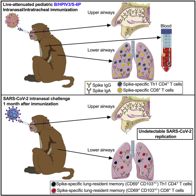
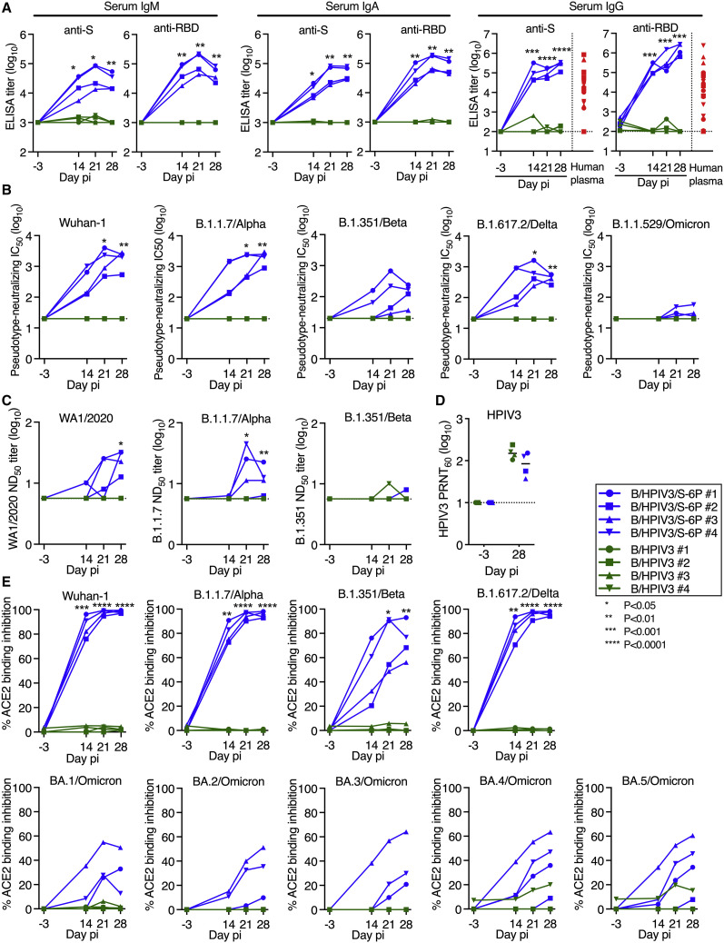
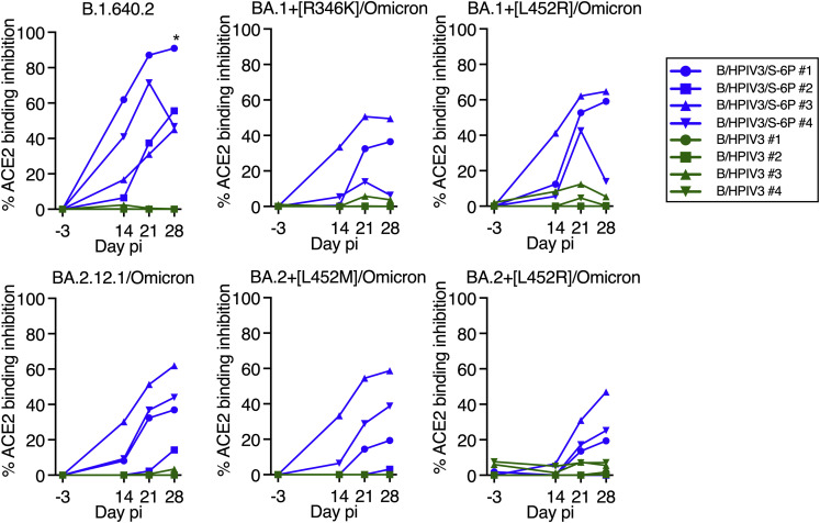
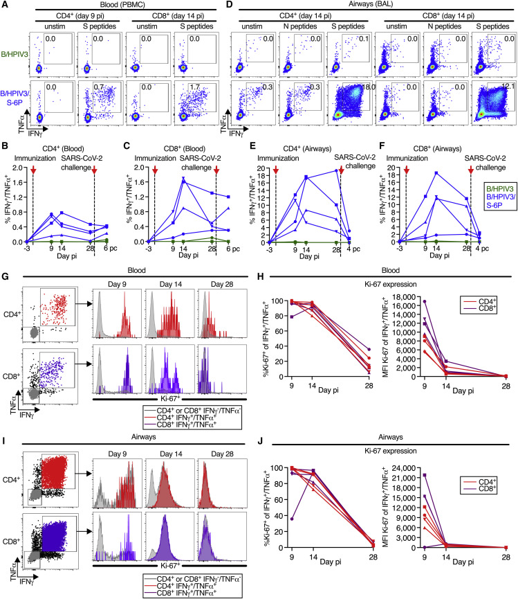
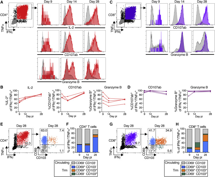
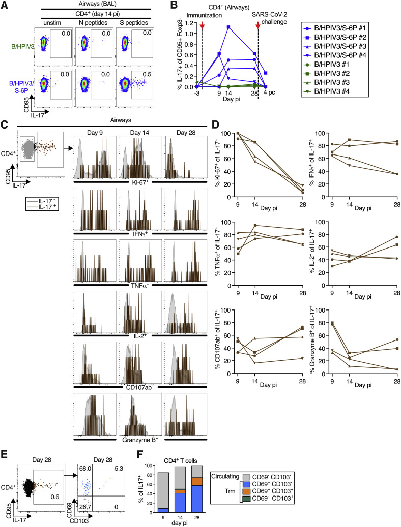
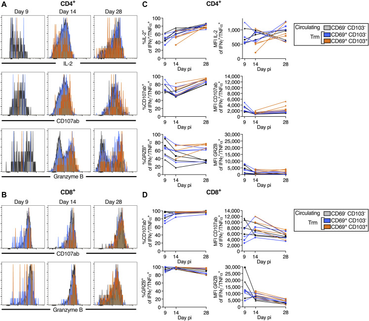
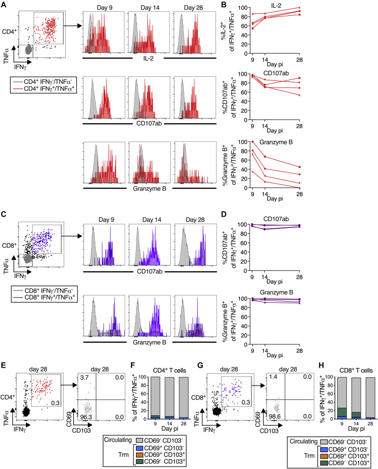
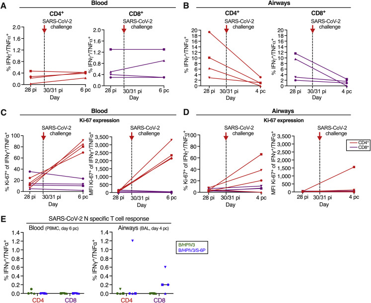
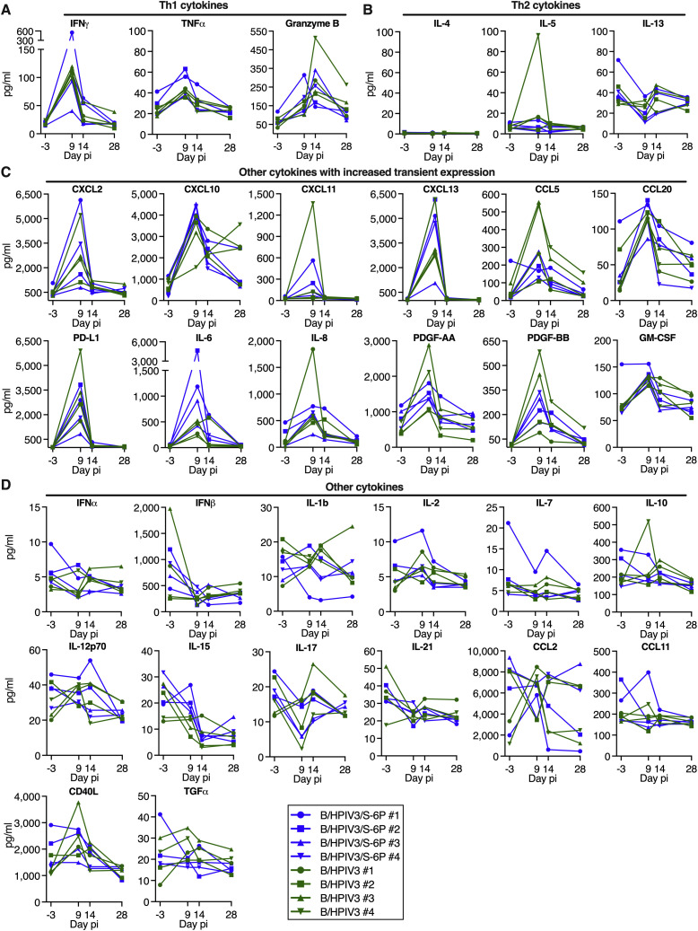
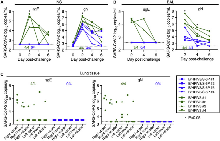
Pediatric SARS-CoV-2 vaccines are needed that elicit immunity directly in the airways as well as systemically. Building on pediatric parainfluenza virus vaccines in clinical development, we generated a live-attenuated parainfluenza-virus-vectored vaccine candidate expressing SARS-CoV-2 prefusion-stabilized spike (S) protein (B/HPIV3/S-6P) and evaluated its immunogenicity and protective efficacy in rhesus macaques. A single intranasal/intratracheal dose of B/HPIV3/S-6P induced strong S-specific airway mucosal immunoglobulin A (IgA) and IgG responses. High levels of S-specific antibodies were also induced in serum, which efficiently neutralized SARS-CoV-2 variants of concern of alpha, beta, and delta lineages, while their ability to neutralize Omicron sub-lineages was lower. Furthermore, B/HPIV3/S-6P induced robust systemic and pulmonary S-specific CD4+ and CD8+ T cell responses, including tissue-resident memory cells in the lungs. Following challenge, SARS-CoV-2 replication was undetectable in airways and lung tissues of immunized macaques. B/HPIV3/S-6P will be evaluated clinically as pediatric intranasal SARS-CoV-2/parainfluenza virus type 3 vaccine.

Keywords: SARS-CoV-2; intranasal vaccine; live-attenuated viral vector; mucosal immmunization; nasal spray vaccine; next-generation COVID vaccine; parainfluenza virus; pediatric; pediatric COVID vaccine.


Conflict of interest statement

Declaration of interests X.L., C.L., C.L.N., S.M., and U.J.B. are inventors on the provisional patent application number 63/180,534, entitled “recombinant chimeric B/HPIV3 expressing SARS-CoV-2 spike protein and its use,” filed by the United States Department of Health and Human Services.

Figures

None
Graphical abstract
Figure 1
Figure 1
Genome organization of B/HPIV3/S-6P; timeline of the rhesus macaque study, vaccine replication following intranasal/intratracheal immunization of rhesus macaques (A) Diagram of the B/HPIV3/S-6P genome, with BPIV3 (N, P, M, and L; blue) and HPIV3 genes (F and HN; red). The full-length SARS-CoV-2 S ORF (codons 1–1,273) was codon optimized and inserted as additional gene (orange) between the N and P ORFs. The S sequence includes 6 stabilizing proline substitutions (S-6P) and RRAR-to-GSAS substitutions to ablate the S1/S2 cleavage site. Each gene begins and ends with PIV3 gene-start and gene-end transcription signals (light and dark bars). (B) Experimental timeline for the immunization of groups of 4 macaques with the B/HPIV3/S-6P vaccine candidate or the empty B/HPIV3 vector used as a control. Challenge with the SARS-CoV-2 WA1/2020 isolate was performed on day 30 or 31 post-immunization. Pre- and post-challenge sampling schedules are summarized; pi, post-immunization; pc, post challenge. Details are described in STAR Methods. (C and D) Replication of B/HPIV3/S-6P and B/HPIV3 in upper (C) and lower (D) airways of rhesus macaques. Two groups of 4 macaques were immunized intranasally and intratracheally with 6.3 log10 PFU of B/HPIV3/S-6P (blue) or B/HPIV3 (green). Nasopharyngeal swabs (upper airways; daily from days 0 to 10 and on day 12 pi), tracheal lavages (lower airways; every other day from days 2 to 8 and on day 12 pi), and bronchoalveolar lavages (lower airways; day 9 pi) were performed as described in (B). Vaccine virus titers were determined by immunoplaque assay (STAR Methods); expressed as log10 PFU/mL (limit of detection: 0.7 log10 PFU/mL for nasopharyngeal swabs; 1 log10 PFU/mL for tracheal lavages [dotted line]). (E and F) The stability of S expression by B/HPIV3/S-6P in macaques was evaluated by dual-staining immunoplaque assay on Vero cells from nasopharyngeal swab (E) and tracheal lavage (F) samples collected at the peak of vaccine shedding (days 5 through 7). Plaques were immunostained with an HPIV3-specific rabbit hyperimmune serum to detect B/HPIV3 antigens, and a goat hyperimmune serum to the secreted SARS-CoV-2 S to detect co-expression of the S protein, followed by infrared-dye secondary antibodies. Fluorescent staining for PIV3 proteins and SARS-CoV-2 S was visualized in green and red, respectively, generating yellow plaques when merged. The percentage of yellow plaques expressing both HPIV3 and S proteins was determined. Each macaque is indicated by a symbol; lines represent medians (p < 0.05, ∗∗p < 0.01; two-way ANOVA, Sidak multiple comparison test).
Figure S1
Figure S1
Vital signs of macaques after immunization with B/HPIV3 or B/HPIV3/S-6P and SARS-CoV-2 challenge, related to Figures 1B–1D Macaques in groups of 4 were immunized with B/HPIV3/S-6P or with B/HPIV3 (empty vector control). On day 30 post-immunization (pi), animals were challenged in a BSL3 facility with the SARS-CoV-2 WA1/2020 isolate. Animals were euthanized on day 36 pi (day 6 post challenge). The body weight, rectal temperature, respiration rate, oxygen saturation rate, and heart rate were monitored on the indicated day pi. Timing of immunization and SARS-CoV-2 challenge is indicated by dashed lines and red arrows. pi, post-immunization; pc, post challenge. B/HPIV3/S-6P-immunized animals are represented in blue, and B/HPIV3-immunized animals are represented in green. Each animal is represented by a unique symbol.
Figure 2
Figure 2
Intranasal/intratracheal immunization with B/HPIV3/S-6P induces mucosal antibody responses to SARS-CoV-2 S in the upper and lower airways Rhesus macaques (n = 4 per group) were immunized with B/HPIV3/S-6P or B/HPIV3 (control) by the intranasal/intratracheal route (Figure 1B). To determine the mucosal antibody response in the upper airways, nasal washes (NWs) were performed before immunization and on days 14, 21, and 28. To analyze the antibody response in the lower airways, bronchoalveolar lavages (BALs) were collected before immunization and on days 9, 14, and 28 pi. (A and B) S- and receptor binding domain (RBD)-specific mucosal IgA and IgG titers on indicated days pi in the upper (A) and lower (B) airways, determined by time-resolved dissociation-enhance lanthanide fluorescence immunoassay (DELFIA-TRF). Endpoint titers are expressed in log10 for mucosal IgA and IgG to a secreted prefusion-stabilized form (aa 1–1,208; S-2P14) of the S protein (left panels) or to a fragment of the S protein (aa 328–531) containing SARS-CoV-2 RBD (right panels). The limit of detection is 1.6 log10 (dotted line). B/HPIV3/S-6P-immunized macaques are shown in blue, while B/HPIV3-immunized macaques are in green, with each macaque represented by a symbol. p < 0.05 (two-way ANOVA, Sidak multiple comparison test).
Figure 3
Figure 3
B/HPIV3/S-6P induces serum binding antibody responses to SARS-CoV-2 S and neutralizing antibody responses to VoCs in macaques Sera were collected from macaques before immunization and on days 14, 21, and 28 pi. (A) Endpoint ELISA titers of serum IgM, IgA, and IgG to S-2P (left panels) or RBD (right panels), expressed in log10. Twenty-three plasma samples from COVID-19 convalescent individuals were evaluated in parallel for IgG to S-2P or the RBD (red symbols). The limits of detection are 3 log10 for IgM and IgA and 2.0 log10 for IgG. (B) Serum neutralizing titers to pseudoviruses bearing spike proteins from SARS-CoV-2 Wuhan-1 (matching S-6P), B.1.1.7/Alpha, B.1.351/Beta, B.1.617.2/Delta, or B.1.1.529/Omicron. The 50% inhibitory concentration (IC50) titers of sera were determined. The detection limit is 1.3 log10. (C) The 50% SARS-CoV-2 serum neutralizing titers (ND50) were determined on Vero E6 cells against vaccine-matched WA1/2020 or viruses from lineages B.1.1.7/Alpha or B.1.351/Beta. The limit of detection is 0.75 log10. (D) Serum HPIV3 neutralizing antibody titers pi, determined by 60% plaque reduction neutralization test (PRNT60). The detection limit is 1 log10. (E) Percentage of ACE2 binding inhibition to SARS-CoV-2 S proteins from Wuhan-1, B.1.1.7/Alpha, B.1.351/Beta, B.1.617.2/Delta, or indicated Omicron variants by serum antibodies from immunized macaques, relative to no-serum controls. ACE2 binding inhibition to additional S proteins is shown in Figure S2. Each macaque is represented by a symbol. p < 0.05, ∗∗p < 0.01, ∗∗∗p < 0.001, ∗∗∗∗p < 0.0001, two-way ANOVA, Sidak multiple comparison test.
Figure S2
Figure S2
ACE2 binding inhibition to SARS-CoV-2 spike proteins by the sera of immunized macaques, related to Figure 3E The neutralizing activity of each serum to inhibit ACE2 binding to S from the variant under monitoring B.1.640.2 Omicron BA.2.12.1 or Omicron BA.1 and BA.2 subvariants that contain the indicated additional mutation is represented as percent inhibition relative to no-serum controls. pi, post-immunization. B/HPIV3/S-6P-immunized animals are represented in blue, and B/HPIV3-immunized animals are represented in green. Each animal is represented by a unique symbol.
Figure S3
Figure S3
Gating strategy of the CD4+ and CD8+ T cells isolated from BAL of macaques, related to Figures 4 and 5 Representative flow cytometry dot plots of cells isolated from a BAL sample, visualizing the typical gating strategy used to identify the CD4+ and CD8+ T cell populations described in Figures 4, 5, S4, S5, and S7. The same gating strategy was applied to identify and analyze the CD4+ and CD8+ T cells from PBMC isolated from the blood (Figures 4, S6, and S7). Live cells were first gated based on a live/dead staining and forward scatter area. Live lymphocytes were identified based on forward and side scatter areas. Then, singlets were selected using a first gate based on forward scatter height and forward scatter area followed by a second gate based on side scatter height and side scatter area. An additional live/dead gating was performed to discard any remaining dead cells. The live single CD3+ IFNγ+ T cells were next gated using CD3 and IFNγ. As CD3 expression can be downregulated on activated T cells, a wide CD3 gate was applied. IFNγ+ CD4+ or CD8+ T cells were next identified using a CD4 or CD8 antibody. Non-naive, non-regulatory CD4+ or CD8+ T cells were finally gated using CD95 and Foxp3, respectively. The phenotypic analyses described in Figures 4, 5, and Figure S4, Figure S5, Figure S6, Figure S7 were performed on live single CD3+ CD4+ CD95+ Foxp3 or live single CD3+ CD8+ CD95+ Foxp3 T cells.
Figure 4
Figure 4
Intranasal/intratracheal immunization with B/HPIV3/S-6P induces S-specific CD4+ and CD8+ T cell responses in blood and lower airways (A–F) Frequencies of S-specific CD4+ and CD8+ T cells from the blood (A–C) or BAL (D–F). Mononuclear cells collected on indicated days pi were stimulated with overlapping SARS-CoV-2 S or (BAL only) N peptides or left unstimulated and processed for flow cytometry. Phenotypic analyses were performed on non-naive non-regulatory (CD95+/Foxp3) CD4+ or CD8+ T cells (see Figure S3 for gating); frequencies are relative to that population. (A and D) Dot plots showing IFNγ and TNFα expression by CD4+ or CD8+ T cells from blood (A) or BAL (D) of representative B/HPIV3 (top) or B/HPIV3/S-6P-immunized (bottom) macaques. (B, C, E, and F) Background-corrected frequencies of S-specific IFNγ+/TNFα+ CD4+ (B and E) or CD8+ (C and F) T cells from blood (B and C) or BAL (E and F) on indicated days. (G–J) Expression of proliferation marker Ki-67 by IFNγ+/TNFα+ CD4+ (red) or CD8+ (purple) T cells from the blood (G and H) or BAL (I and J) of B/HPIV3/S-6P-immunized macaques (n = 4, each represented by different symbols). IFNγ/TNFα cells in gray. (G and I) Gating and histograms showing Ki-67 expression. (H and J) Frequency and median fluorescence intensity (MFI) of Ki-67 in IFNγ+/TNFα+ T cells from the blood (H) and BAL (J). BAL, bronchoalveolar lavage.
Figure 5
Figure 5
B/HPIV3/S-6P-elicited S-specific CD4+ and CD8+ T cells in lower airways (LAs) transition to tissue-resident memory phenotype T cells obtained by bronchoalveolar lavage (BAL) were stimulated with overlapping S peptides prior to flow cytometry analysis. (A–D) S-specific T cells in LA are functional. (A and C) Representative dot plots showing gating on S-specific IFNγ+/TNFα+ CD4+ (red), CD8+ (purple), and IFNγ/TNFα (gray) T cells (for complete gating, see Figure S3); histograms showing expression of IL-2 (CD4+ T cells only), CD107ab and granzyme B by IFNγ+/TNFα+ T cells collected on indicated days pi. (B and D) Frequencies of IL-2+, CD107ab+, and granzyme B+ of IFNγ+/TNFα+ S-specific CD4+ (B) or CD8+ (D) T cells from 4 BHPIV3/S-6P-immunized macaques on indicated days. (E–H) Transition to memory phenotype. (E and G) Representative dot plots showing gating on S-specific IFNγ+/TNFα+ T cells (left panels, % indicated). CD69 and CD103 were used to differentiate circulating (CD69/CD103, gray) and tissue-resident memory (Trm; CD69+/CD103 [blue], CD69+/CD103+ [orange], and CD69/CD103+ [green]) S-specific IFNγ+/TNFα+ T cells from LA (right panels, % indicated). (F and H) The median % of circulating and each of the 3 Trm S-specific IFNγ+/TNFα+ CD4+ (F) or CD8+ (H) T cell subsets present on indicated days in BAL of 4 B/HPIV3/S-6P-immunized macaques are stacked.
Figure S4
Figure S4
B/HPIV3/S-6P immunization induced a small population of S-specific IL-17+ CD4+ T cells in lower airways (LAs) that transitioned to tissue-resident memory phenotype, related to Figures 4 and 5 T cells obtained by bronchoalveolar lavage (BAL) were stimulated with overlapping N or S peptides and processed for flow cytometry. Phenotypic analyses were performed on non-naive non-regulatory (CD95+/Foxp3) CD4+ T cells (see Figure S3 for gating) and frequencies are relative to that population. (A and B) Frequencies of S-specific IL-17+ CD4+ T cells from BAL. (A) Dot plots showing IL-17 expression by CD95+ cells of representative B/HPIV3 (top) or B/HPIV3/S-6P-immunized (bottom) macaques. (B) Background-corrected frequencies of S-specific IL-17+ CD4+ on indicated days. Timing of immunization and SARS-CoV-2 challenge are indicated with red arrow and dotted line. (C and D) Phenotype of the S-specific IL-17+ CD4+ T cells in LA. (C) Representative dot plots showing gating on S-specific CD95+/IL-17+ (brown), and CD95+/IL-17 (gray) CD4+ T cells. Histograms show expression of Ki-67, IFNγ, TNFα, IL-2, CD107ab, and granzyme B by IL-17+ CD4+ T cells on indicated days pi. (D) Frequencies of Ki-67+, IFNγ+, TNFα+, IL-2+, CD107ab+, and granzyme B+ of S-specific IL-17+ CD4+ T cells from 4 BHPIV3/S-6P-immunized macaques on indicated days. (E and F) S-specific IL-17+ CD4+ T cells transition to memory phenotype. (E) Representative dot plots showing gating on S-specific CD95+/IL-17+ CD4 T+ cells (left panel, % of IL-17+ cells indicated). CD69 and CD103 were used to differentiate circulating (CD69/CD103, gray) and tissue-resident memory (Trm; CD69+/CD103 [blue], CD69+/CD103+ [orange], CD69/CD103+ [green]) S-specific CD4+ T cells from LA (right panel, % indicated). (F) The median % of circulating and each of the 3 Trm S-specific IL-17+ CD4+ T cells present on indicated days in BAL of 4 B/HPIV3/S-6P-immunized macaques are stacked.
Figure S5
Figure S5
Comparable phenotype of circulating (CD69 CD103) and tissue-resident memory (CD69+ CD103 and CD69+ CD103+) S-specific IFNγ+/TNFα+ CD4+ and CD8+ T cells in the airways, related to Figure 5 (A and B) Histograms representing IL-2 expression (A only), CD107ab, and granzyme B expression by S-specific circulating and tissue-resident memory (Trm) IFNγ+/TNFα+ CD4+ (A) or CD8+ (B) T cells obtained from BAL on indicated days pi. (C and D) % and level of expression (MFI, median fluorescence intensity) of IL-2 (CD4+ T cells only), CD107ab and granzyme B by the S-specific circulating and Trm IFNγ+/TNFα+ CD4+ (C) and CD8+ T cells (D) in the 4 B/HPIV3/S-6P-immunized macaques. Due to the low frequency of CD69+ CD103+ T cells on day 9 pi, the frequencies and MFIs of IL-2, CD107ab and granzyme B by this subset are only indicated on days 14 and 28 pi. In (C) and (D), each macaque is indicated by a symbol.
Figure S6
Figure S6
Phenotype of SARS-CoV-2 S-specific CD4+ and CD8+ T cells in the blood of the B/HPIV3/S-6P immunized macaques, related to Figures 4 and 5 (A) Dot blot of the CD4+ T cells from blood of a representative B/HPIV3/S-6P-immunized macaque describing the gating of S-specific IFNγ+/TNFα+ cells (red). The levels of expression of IL-2, CD107ab and granzyme B by the IFNγ+/TNFα+ CD4+ T cells from the same macaque are shown as histograms on the indicated day pi with the IFNγ/TNFα CD4+ T cells (gray) used for reference. (B) % of IFNγ+/TNFα+ CD4+ T cells in the blood of the 4 B/HPIV3/S-6P-immunized macaques that expressed IL-2, CD107ab, or granzyme B on the indicated day pi. (C) Dot blot of the CD8+ T cells in the blood of a representative B/HPIV3/S-6P-immunized macaque describing the gating of the S-specific IFNγ+/TNFα+ cells (purple). The level of expression of CD107ab and granzyme B by the IFNγ+/TNFα+ CD4+ T cells from the same macaque are shown as histograms on the indicated day pi with the IFNγ/TNFα CD4+ T cells (gray) used for reference. (D) % CD107ab+ or granzyme B+ of IFNγ+/TNFα+ CD8+ T cells on the indicated days pi in the blood of the 4 B/HPIV3/S-6P-immunized macaques. Each macaque is represented by a different symbol. (E and G) Representative dot plots showing gating on S-specific IFNγ+/TNFα+ T cells (left panels). CD69 and CD103 were used to differentiate circulating (CD69 CD103, gray) and tissue-resident memory (Trm; CD69+ CD103 [blue], CD69+ CD103+ [orange], and CD69 CD103+ [green]) S-specific IFNγ+/TNFα+ T cells isolated from blood (right panels, % indicated). (F and H) The median % of the circulating and each of the 3 Trm S-specific IFNγ+/TNFα+ CD4+ (F) or CD8+ (H) T cell subsets present in blood of 4 B/HPIV3/S-6P-immunized macaques on indicated days are stacked.
Figure S7
Figure S7
SARS-CoV-2 specific CD4+ and CD8+ T cell responses 4 days after SARS-CoV-2 challenge, related to Figure 4 (A and B) Background-corrected frequencies of S-specific IFNγ+/TNFα+ CD4+ or CD8+ T cells from blood (A) or airways (B) on days 28 post-infection and 4 post-challenge (airways) or 28 post-infection and 6 post-challenge (blood). These frequencies are similar to the frequencies shown in Figures 4B, 4C, 4E, and 4F for the blood and airways, respectively. (C and D) % and median fluorescence intensity (MFI) of proliferation marker Ki-67 by IFNγ+/TNFα+ CD4+ (red) or CD8+ (purple) T cells from blood (C) or airways (D) of the 4 B/HPIV3/S-6P-immunized macaques, each represented by a different symbol; pi, post-immunization; pc, post-challenge. (E) Background-corrected frequencies of N-specific IFNγ+/TNFα+ CD4+ or CD8+ T cells from blood (left panel) or airways (right panel) of B/HPIV3 (green)- and B/HPIV3/S-6P (blue)-immunized macaques on day 4 post challenge. Each macaque is represented by a different symbol. When appropriate, the median value from n = 4 macaques is indicated.
Figure S8
Figure S8
Cytokines in BAL of B/HPIV3- and B/HPIV3/S-6P-immunized macaques, related to Figures 4 and 5 Macaques were immunized with B/HPIV3 or B/HPIV3/S-6P as described in Figure 1B. BAL collected before immunization (day −3) and on days 9, 14, and 28 after immunization with B/HPIV3 or B/HPIV3/S-6P were concentrated 10×, and the concentration of 36 cytokines from each BAL sample was determined using the NHP XL cytokine Luminex premixed kit. Th1-related cytokines (A), Th2-related cytokines (B), cytokines that showed transient increased expression (C), and cytokines that did not exhibit increased expression (D) are shown. Four additional cytokines (BDNF, VEGF, FGF basic, and G-CSF) that did not exhibit increased expression are not shown. Each macaque is represented by a different symbol. Cytokine concentration is expressed in pg/mL.
Figure 6
Figure 6
Absence of detectable SARS-CoV-2 challenge virus replication in the upper and lower airways and lung tissues of B/HPIV3/S-6P-immunized macaques Rhesus macaques immunized with a single intranasal/intratracheal dose of B/HPIV3/S-6P or B/HPIV3 control (n = 4 per group) were challenged intranasally/intratracheally on day 30 pi with 5.8 TCID50 of SARS-CoV-2. (A and B) Evaluation of challenge virus shedding by qRT-PCR. (A) Nasal swabs (NSs) and (B) bronchoalveolar lavage (BAL) fluid were collected on days 0 (NS only), 2, 4, and 6 post-challenge (pc) and SARS-CoV-2 subgenomic E mRNA (sgE, indicative of active SARS-CoV-2 replication) and genomic N RNA (gN, indicative of the presence of challenge virus) were quantified by RT-qPCR. (C) Challenge virus detection in lung tissues. Animals were euthanized on day 6 pc, and RNA was extracted from indicated areas of lung tissue to quantify SARS-CoV-2 sgE mRNA and gN RNA by qRT-PCR. (A–C) Copies/ml of SARS-CoV-2 sgE mRNA and gN RNA in NS (A) and BAL (B) and copies/g in lung tissues (C) are indicated for macaques immunized with B/HPIV3/S-6P (blue) or B/HPIV3 (green), each identified by a symbol. The numbers of animals in each group with detectable RNA are indicated above the x axis (A and B) or at the top of the graph (C). The limit of detection was 2.6 log10 copies/mL of NS or BAL fluid and 3.3 log10 copies/g of lung tissue. p < 0.05, two-way ANOVA, Sidak multiple comparison test.

Similar articles

Cited by

References

    1. Rankin D.A., Talj R., Howard L.M., Halasa N.B. Epidemiologic trends and characteristics of SARS-CoV-2 infections among children in the United States. Curr. Opin. Pediatr. 2021;33:114–121. doi: 10.1097/MOP.0000000000000971. - DOI - PMC - PubMed
    1. Funk A.L., Florin T.A., Kuppermann N., Tancredi D.J., Xie J., Kim K., Neuman M.I., Ambroggio L., Plint A.C., Mintegi S., et al. Outcomes of SARS-CoV-2-positive youths tested in emergency departments: the global PERN-COVID-19 study. JAMA Netw. Open. 2022;5:e2142322. doi: 10.1001/jamanetworkopen.2021.42322. - DOI - PMC - PubMed
    1. Gerber J.S., Offit P.A. COVID-19 vaccines for children. Science. 2021;374:913. doi: 10.1126/science.abn2566. - DOI - PubMed
    1. Verdoni L., Mazza A., Gervasoni A., Martelli L., Ruggeri M., Ciuffreda M., Bonanomi E., D'Antiga L. An outbreak of severe Kawasaki-like disease at the Italian epicentre of the SARS-CoV-2 epidemic: an observational cohort study. Lancet. 2020;395:1771–1778. doi: 10.1016/S0140-6736(20)31103-X. - DOI - PMC - PubMed
    1. Riphagen S., Gomez X., Gonzalez-Martinez C., Wilkinson N., Theocharis P. Hyperinflammatory shock in children during COVID-19 pandemic. Lancet. 2020;395:1607–1608. doi: 10.1016/S0140-6736(20)31094-1. - DOI - PMC - PubMed

Supplementary concepts