

Effects of occlusal splint and exercise therapy, respectively, for the painful temporomandibular disorder in patients seeking for orthodontic treatment: a retrospective study
- PMID: 36424568
- PMCID: PMC9685899
- DOI: 10.1186/s12903-022-02538-y
Effects of occlusal splint and exercise therapy, respectively, for the painful temporomandibular disorder in patients seeking for orthodontic treatment: a retrospective study
Abstract
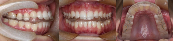
Objective: To evaluate the effect of hard stabilization splints (HSS), counselling and exercise therapies, respectively, for the painful temporomandibular disorder (TMD) in patients seeking for orthodontic treatment through magnetic resonance imaging (MRI) and clinical examination.
Materials and methods: Eighty-seven TMD patients were divided into two groups according to their therapies: the HSS group (n = 43) comprising of patients treated with HSS, counselling and masticatory muscle exercises; the control group (n = 44) comprising of patients treated with counselling and masticatory muscle exercises alone. All patients had orthodontic therapies after the first treatment phase. The joint pain and clicking of all patients were recorded via clinical examination. MRIs of HSS groups were taken before (T0), after the first phase (T1), and after the orthodontic treatment (T2). Parameters indicating the condyles and articular discs were evaluated. Clinical symptom (pain and clicking) changes among T0, T1 and T2 time point were detected in the two groups respectively. The significant differences between HSS and control groups, as well as between male and female were tested at T1 and T2. Position changes of condyles and discs in HSS group among T0, T1 and T2 were detected in male and female respectively.
Results: After the first treatment phase, there was no difference in the decrease of facial pain between the two group, as well as between male and female in the two groups (P > 0.05). Clicking decreasing was not statistically significant. After the whole orthodontic periods, the TMJ pain relapsed in female of the control group, and the number of female's pain joints was more than male's (P < 0.05). In the HSS group, the posterosuperior movements of discs and the anteroposterior movements of condyles were recorded in closing position (P < 0.05). After the whole orthodontic periods, female's disc-condyle angles increased, the discs to HRP distance decreased and condyles to VRP distance increased when compared with the data of T1 (P < 0.05).
Conclusions: For the orthodontic patients with painful TMD, HSS combined with counselling and exercise therapies before orthodontic treatment could provide pain relief. HSS is helpful to improve the position and relation of discs and condyles. In addition, male's prognosis is better than female's in terms of stability.
Keywords: Disc-condyle position; HSS; Orthodontic treatment; Painful TMD.
© 2022. The Author(s).
Conflict of interest statement
No competing interests would be declared.
Figures


References
MeSH terms
LinkOut - more resources
Full Text Sources
Medical
