. 2022 Nov 8:15:857257.
doi: 10.3389/fnmol.2022.857257. eCollection 2022.

Hsc70-4 aggravates PolyQ-mediated neurodegeneration by modulating NF-κB mediated immune response in Drosophila

Affiliations

Hsc70-4 aggravates PolyQ-mediated neurodegeneration by modulating NF-κB mediated immune response in Drosophila

Saurabh Rai et al. Front Mol Neurosci. .

Abstract

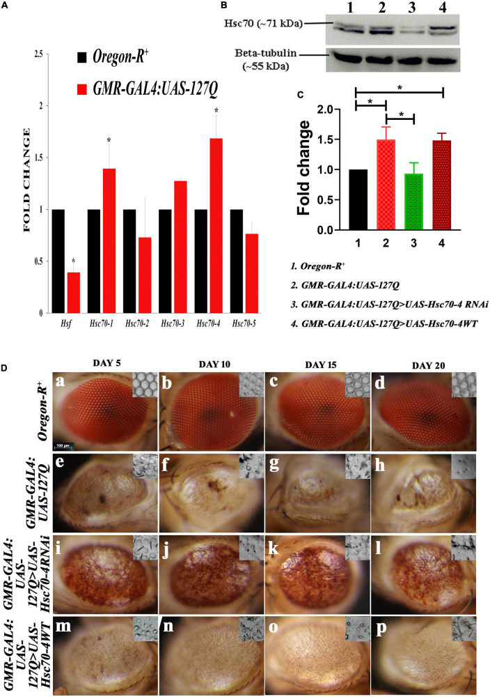
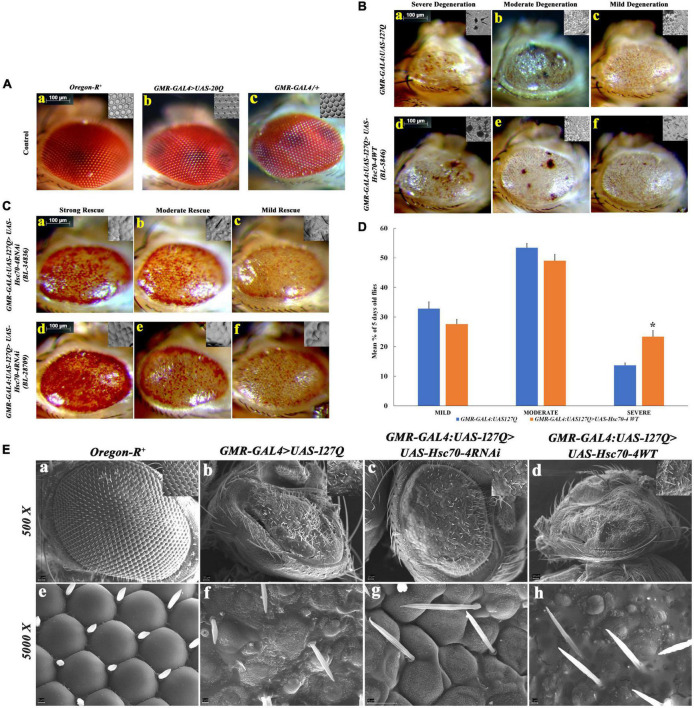
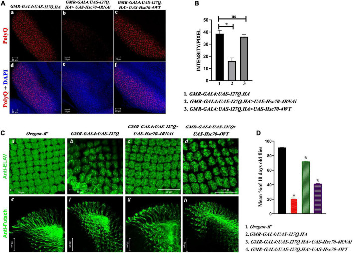
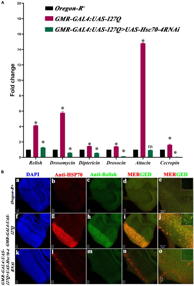
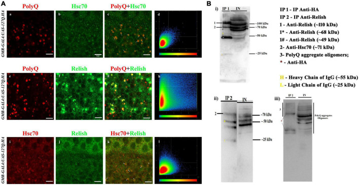
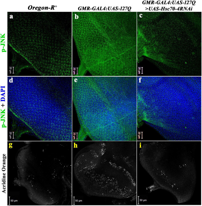
Huntington's disease occurs when the stretch of CAG repeats in exon 1 of the huntingtin (htt) gene crosses the permissible limit, causing the mutated protein (mHtt) to form insoluble aggregates or inclusion bodies. These aggregates are non-typically associated with various essential proteins in the cells, thus disrupting cellular homeostasis. The cells try to bring back normalcy by synthesizing evolutionary conserved cellular chaperones, and Hsp70 is one of the families of heat shock proteins that has a significant part in this, which comprises of heat-inducible and cognate forms. Here, we demonstrate that the heat shock cognate (Hsc70) isoform, Hsc70-4/HSPA8, has a distinct role in polyglutamate (PolyQ)-mediated pathogenicity, and its expression is enhanced in the polyQ conditions in Drosophila. Downregulation of hsc70-4 rescues PolyQ pathogenicity with a notable improvement in the ommatidia arrangement and near-normal restoration of optic neurons leading to improvement in phototaxis response. Reduced hsc70-4 also attenuates the augmented immune response by decreasing the expression of NF-κB and the antimicrobial peptides, along with that JNK overactivation is also restored. These lead to the rescue of the photoreceptor cells, indicating a decrease in the caspase activity, thus reverting the PolyQ pathogenicity. At the molecular level, we show the interaction between Hsc70-4, Polyglutamine aggregates, and NF-κB, which may be responsible for the dysregulation of signaling molecules in polyQ conditions. Thus, the present data provides a functional link between Hsc70-4 and NF-κB under polyQ conditions.

Keywords: Chaperone; HSPA8; Hsc70-4; Immune response; NF-κB; Neurodegeneration; PolyQ; Relish.


Conflict of interest statement

The authors declare that the research was conducted in the absence of any commercial or financial relationships that could be construed as a potential conflict of interest.

Figures

FIGURE 1
FIGURE 1
Alterations in the hsc70 gene isoforms in the polyQ-overexpressing eye phenotype. (A) Transcript expressions of hsc70-1, hsc70-3, and hsc70-4 are upregulated compared to hsc70-2 and hsc70-5, while that of hsf is reduced in GMR-GAL4:UAS-127Q in comparison to Oregon-R+(normalization is done using rp49). (B) Western blot showing the expression of Hsc70 in different genotypes. (C) Graph shows quantitative analysis of Hsc70 expression normalized to beta-tubulin, showing significant upregulation of the protein in lane 2 and 4 compared to lane 1. In contrast, the protein level of Hsc70 in the lane 3 was comparable to lane 1. However, it was significantly downregulated compared to lane 2 (n = 3 and data shown as ±SEM and * indicated significance at the p-value of ≤0.05). (D) Eye degeneration in GMR-GAL4:UAS-127Q background is rescued after the downregulation of hsc70-4. Normal adult eyes in Oregon-R+ (a–d), disrupted in GMR-GAL4:UAS-127Q flies (e–h), as they show distorted ommatidial pattern along with the loss of eye pigmentation due to expanded PolyQ aggregates accumulation. Downregulating hsc70-4 restores the ommatidial arrangement and eye pigmentation in polyQ-driven flies (i–l), while overexpressing hsc70-4 does not correct the ommatidial arrangement and eye pigmentation in polyQ-driven flies (m–p). The nail polish imprint of adult eyes showing the ommatidia pattern is in the inset of each image in this panel (scale bar 100 μm).
FIGURE 2
FIGURE 2
Detailed morphological observations of eye phenotype in hsc70-4 regulated condition in 5-day-old polyQ-expressing flies. (A) Eye phenotype and ommatidial arrangement of three control genotypes: (a) Oregon-R+; (b) GMR-GAL4:UAS-20Q; and (c), GMR-GAL4/+. Oregon-R+ is the negative control, GMR-GAL4/+ is a positive control, and GMR-GAL4:UAS-20Q is also an experimental control. Both of their eye phenotypes are very similar to that of the negative control group. (B) Degeneration of the eye phenotype is categorized into three groups, and based on this categorization, the frequency of eye degeneration is compared between polyQ-expressing flies and hsc70-4 overexpressing polyQ flies. These are (i) mild degeneration in panels (c,f) shows less roughening in the eye (with lesions only at the periphery); (ii) moderate level of degeneration in panels (b,e) where degeneration and necrotic patches are present in either of the eyes; and (iii) severe degeneration in panels (a,d) where the eye size is smaller than the mild eye phenotype along with larger necrotic patches (scale bar 100 μm). (C) Rescue in the eye phenotype after the downregulation of hsc70-4 in GMR-GAL4:UAS-127Q is checked using two different RNAi lines. The rescue phenotype is categorized into three groups: (i) mild rescue phenotype in panels (c,f) shows recovery in the eye pigmentation at the margin and slight improvement in the ommatidial arrangements. (ii) Moderate rescue in panels (b,e) leads to the recovery of the loss of pigmentation throughout the eye and the recovered ommatidia. (iii) High rescue phenotype in panels (a,d) where eye pigmentation is very prominent along with a highly recovered ommatidial pattern (scale bar 100 μm). (D) The graph shows the severity of eye degeneration change upon overexpressing the hsc70-4 in the GMR-GAL4:UAS-127Q background. Slight changes in the percentage of flies with either mild and moderate degenerating eye phenotype have been observed between GMR-GAL4:UAS-127Q and GMR-GAL4:UAS-127Q> UAS-Hsc70-4WT, whereas a significant increase in the percentage of the severe eye phenotype bearing 5-day-old flies which suggests an increase in ocular degeneration after overexpressing hsc70-4. The eye phenotype of each genotype was observed after three independent crosses and n ≥ 100, and data are shown as ±SEM (* indicated significance at the p-value of ≤0.05). (E) SEM image panels show the eye phenotype of different genotypes in detail. (a,e) Oregon-R+, (b,f) GMR-GAL4:UAS-127Q, (c,g) GMR-GAL4:UAS-127Q> UAS-Hsc70-4RNAi, and (d,h) GMR-GAL4:UAS-127Q> UAS-Hsc70-4WT (scale bar 20 μm).
FIGURE 3
FIGURE 3
hsc70-4 downregulation reduces the aggregates accumulation, resulting in the improved arrangement of rhabdomeres and optic neurons. Thus, it enhances the phototaxis response. (A) The panel shows the PolyQ aggregates (in red) accumulation in (a) GMR-GAL4:UAS-127Q; (b) GMR-GAL4:UAS-127Q>UAS-Hsc70-4RNAi; (c) GMR-GAL4:UAS-127Q> UAS-Hsc70-4WT. Panels (d–f) from the panel (A) show the nuclei staining (in blue) along with the aggregates of the respective genotypes (scale bar 20 μm). (B) Quantitative analysis of the intensity/pixel of the immunofluorescence images shows PolyQ aggregates accumulation in the different genotypes from three independent experiments and data shown as ±SEM (ns indicates insignificant change at p-value ≤ 0.05%, and * indicates the significance at a p-value of ≤ 0.05). (C) hsc70-4 downregulation restores the rhabdomere arrangement and the axonal connection disruption in GMR-GAL4:UAS-127Q-driven flies. (a,e) Oregon-R+, (b,f) GMR-GAL4:UAS-127Q, (c,g) GMR-GAL4:UAS-127Q> UAS-Hsc70-4RNAi, (d,h) GMR-GAL4:UAS-127Q> UAS-Hsc70-4WT. Anti-ELAV (in green) to mark rhabdomere and 22C10 staining against Futsch (in green) to visualize the optic neurons (scale bar 20 μm). (D) The graph shows the phototaxis response in the 10-day-old flies of Oregon-R+, GMR-GAL4:UAS-127Q, GMR-GAL4:UAS-127Q> UAS-Hsc70-4RNAi, and GMR-GAL4:UAS-127Q> UAS-Hsc70-4WT (the experiment was performed in triplicate where n ≥100 for each genotype, and data are shown as ±SEM; * indicated significance at the p-value of ≤0.05%).
FIGURE 4
FIGURE 4
Elevated immune responses in the polyQ background get reduced upon hsc70-4 downregulation. (A) The histogram shows qPCR analysis representing the fold change in transcript level of relish and AMPs in GMR-GAL4:UAS-127Q conditions significantly elevated in comparison to the wild-type. But GMR-GAL4:UAS-127Q> UAS-Hsc70-4RNAi shows a significant decline in these respective immune genes (data normalized to rp49, three biological replicated were used, and data are shown as ±SEM; ns means insignificant change at p-value of ≤0.05, and *indicated significance at the p-value of ≤0.05). (B) Reduction in expression of Relish (green) and HSP70 (red) after downregulation of hsc70-4 in GMR-GAL4:UAS-127Q>UAS-Hsc70-4RNAi in comparison to GMR-GAL4:UAS-127Q, in lower magnification (a–d,f–i,k–n) and (e,j,o) shows the higher magnification (scale bar 20 μm). The inset in (e,j,o) was captured at a further 3X zoom to show co-localization (scale bar 10 μm). Genotype description, Oregon-R+ (a–e); GMR-GAL4:UAS-127Q (f–j); GMR-GAL4:UAS-127Q> UAS-Hsc70-4RNAi (k–o).
FIGURE 5
FIGURE 5
PolyQ aggregates colocalized and coprecipitated with Hsc70 and Relish. (A) The panel shows the section image of the eye imaginal disc of GMR-GAL4 UAS 127Q third instar larvae. (a–d) Co-staining of the PolyQ aggregates (in red) and Hsc70 (in green) shows that PolyQ aggregates and Hsc70 colocalize with each other; the arrow demarcates the colocalized regions in yellow (scale bar 5 μm). (e–h) Co-staining of the PolyQ aggregates (in red) and Relish (in green) shows that PolyQ aggregates colocalize with Relish. Arrow demarcates the colocalized regions in yellow (scale bar 5 μm). (i–l) Co-staining of Hsc70 (in red) and Relish (in green) co-stained in the polyQ-expressing eye disc shows the colocalization (in yellow), which is marked by the arrow (scale bar 5 μm). (B) Immunoblot panels illustrated the presence of the interacting proteins in the co-IP elute (denoted as IP) from the crude protein sample (demarcated as IN, means input) of GMR-GAL4:UAS-127Q.HA background. (i) Western blot shows the presence of Relish and Hsc70 in the IP1 sample after precipitation of PolyQ aggregate using anti-HA and the expression pattern in input. Images ii and iii are immunoblot showing the presence of Hsc70 and PolyQ aggregates, respectively, in the IP2 sample after precipitation of Relish using anti-Relish, and the expression pattern was compared with the input sample. IP 1—eluted protein sample after pull-down with anti-HA; IP 2—eluted protein sample after pull-down with anti-Relish; IN—crude protein sample of GMR-GAL4:UAS-127Q.HA genetic background; 1- whole-Relish protein (∼110 kDa), 1* donates a faint band of N-terminal Relish (∼68 kDa), and 1# denotes a sharp band of C-terminal Relish (∼49 kDa); 2—Hsc70 (∼71 kDa); 3—PolyQ aggregates oligomers detected by anti-HA along with *representing the bands of anti-HA; H and L denote heavy chain (∼55 kDa) and light chain (∼25 kDa) of IgG.
FIGURE 6
FIGURE 6
Downregulation of hsc70-4 reduces the elevated p-JNK level in polyQ-overexpressed background, thus rescuing the cell death in the diseased eye disc. (a) The eye disc of Oregon-R+ shows the typical expression pattern of p-JNK (in green), which drastically elevates in panel (b) GMR-GAL4:UAS-127Q, and (c) downregulating hsc70-4 in the polyQ-overexpressed condition reduces the hyperactivated p-JNK. Panels (d–f) show p-JNK staining merged with DAPI in the eye disc of Oregon-R+, GMR-GAL4:UAS-127Q, and GMR-GAL4:UAS-127Q> UAS-Hsc70-4RNAi, respectively (scale bar 20 μm). White puncta in the images (g–i) are acridine orange stained dead cells in the eye disc of third instars of Oregon-R+, GMR-GAL4:UAS-127Q, and GMR-GAL4:UAS-127Q> UAS-Hsc70-4RNAi, respectively, (scale bar 50 μm).

Similar articles

Cited by

References

    1. Ahn S. G., Kim S. A., Yoon J. H., Vacratsis P. (2005). Heat-shock cognate 70 is required for the activation of heat-shock factor 1 in mammalian cells. Biochem. J. 392 145–152. 10.1042/BJ20050412 - DOI - PMC - PubMed
    1. Andrews T. J., Davies-Thompson J., Kingstone A., Young A. W. (2010). Internal and external features of the face are represented holistically in face-selective regions of visual cortex. J. Neurosci. 30, 3544–3552. 10.1523/JNEUROSCI.4863-09.2010 - DOI - PMC - PubMed
    1. Arias E., Cuervo A. M. (2011). Chaperone-mediated autophagy in protein quality control. Curr. Opin. Cell Biol. 23, 184–189. 10.1016/j.ceb.2010.10.009 - DOI - PMC - PubMed
    1. Arya R., Lakhotia S. C. (2006). A simple nail polish imprint technique for examination of external morphology of Drosophila eyes. Curr. Sci. 90, 1179–1180.
    1. Balch W. E., Morimoto R. I., Dillin A., Kelly J. W. (2008). Adapting proteostasis for disease intervention. Science 319, 916–919. 10.1126/science.1141448 - DOI - PubMed

LinkOut - more resources