. 2022 Nov 8:13:1030621.
doi: 10.3389/fphys.2022.1030621. eCollection 2022.

Dopaminergic nuclei in the chick midbrain express serotonin receptor subfamily genes

Affiliations

Dopaminergic nuclei in the chick midbrain express serotonin receptor subfamily genes

Toshiyuki Fujita et al. Front Physiol. .

Abstract

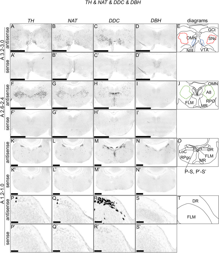
Serotonin (5-hydroxytryptamine, 5-HT) is a phylogenetically conserved modulator of numerous aspects of neural functions. Serotonergic neurons in the dorsal and median raphe nucleus provide ascending innervation to the entire forebrain and midbrain. Another important neural modulatory system exists in the midbrain, the dopaminergic system, which is associated to reward processing and motivation control. Dopaminergic neurons are distributed and clustered in the brain, classically designated as groups A8-A16. Among them, groups A8-A10 associated with reward processing and motivation control are located in the midbrain and projected to the forebrain. Recently, midbrain dopaminergic neurons were shown to be innervated by serotonergic neurons and modulated by 5-HT, with the crosstalk between serotonergic and dopaminergic systems attracting increased attention. In birds, previous studies revealed that midbrain dopaminergic neurons are located in the A8-A10 homologous clusters. However, the detailed distribution of dopaminergic neurons and the crosstalk between serotonergic and dopaminergic systems in the bird are poorly understood. To improve the understanding of the regulation of the dopaminergic by the serotonergic system, we performed in situ hybridization in the chick brainstem. We prepared RNA probes for chick orthologues of dopaminergic neuron-related genes; tyrosine hydroxylase (TH) and dopa decarboxylase (DDC), noradrenaline related genes; noradrenaline transporter (NAT) and dopamine beta-hydroxylase (DBH), and serotonin receptor genes; 5-HTR1A, 5-HTR1B, 5-HTR1D, 5-HTR1E, 5-HTR1F, 5-HTR2A, 5-HTR2B, 5-HTR2C, 5-HTR3A, 5-HTR4, 5-HTR5A, and 5-HTR7. We confirmed that the expression of tyrosine hydroxylase (TH) and NAT was well matched in all chick dopaminergic nuclei examined. This supported that the compensation of the function of dopamine transporter (DAT) by NAT is a general property of avian dopaminergic neurons. Furthermore, we showed that 5-HTR1A and 5-HTR1B were expressed in midbrain dopaminergic nuclei, suggesting the serotonergic regulation of the dopaminergic system via these receptors in chicks. Our findings will help us understand the interactions between the dopaminergic and serotonergic systems in birds at the molecular level.

Keywords: A8; chick (Gallus gallus); dopamine; optic tectum; serotonergic receptor; substantia nigra pars compacta; ventral tegmental area.


Conflict of interest statement

The authors declare that the research was conducted in the absence of any commercial or financial relationships that could be construed as a potential conflict of interest.

Figures

FIGURE 1
FIGURE 1
In situ hybridization of TH, NAT, DDC, and DBH in the P1 chick brainstem. Digoxigenin-labeled RNA antisense [TH, (A,F,K,P), NAT, (B,G,L,Q), DDC, (C,H,M,R), and DBH, (D,I,N,S)] and sense [TH, (A′,F′,K′,P′), NAT, (B′,G′,L′,Q′), DDC, (C′,H′,M′,R′), and DBH, (D′,I′,N′,S′)] probes were used for in situ hybridization in coronal sections of P1 chick brainstems. To evaluate the expression patterns of TH, NAT, DDC, and DBH, sections of 10 P1 chick brainstems were analyzed for TH, 8 for NAT, 7 for DDC, and 7 for DBH. Representative images of the levels of expression in neighboring sections (A3.2–3.0, A2.6–2.4, and A1.2–1.0) from 3 P1 chick brainstems. (P–S and P′–S′) Magnified views of brainstem areas in the box in (O). The levels of expression in these sections were in accordance with those mentioned in the Kuenzel and Masson’s chick atlas (Kuenzel and Masson, 1988). (E,J,O,T) Diagrams of coronal sections shown in panels (A), (F), (K), and (P). Arrowheads indicate signals. A8: A8 cell group; DR, dorsal raphe; FLM, fasciculus longitudinalis medialis; GCt, griseum centrale; LoC, locus coeruleus; MR, median raphe; NIII, nervus oculomotorius; OMN, oculomotor nucleus; P1, post-hatch day 1; RPgc, nucleus reticularis pontis caudalis; RPO, nucleus reticularis pontis oralis; VTA, ventral tegmental area. Scale bars = 2.5 mm (A–I) and (A′–I′), 1 mm (K–N) and (K′–N′), and 100 µm (P–S) and (P′–S′).
FIGURE 2
FIGURE 2
In situ hybridization of 5-HTR1A in the P1 chick brainstem. Digoxigenin-labeled RNA antisense (A–D) and sense (A′–D′) 5-HTR1A probes were used for in situ hybridization in coronal sections of P1 chick brainstems. To evaluate the expression patterns of 5-HTR1A, we analyzed sections from five chick brainstems; representative images of chick brainstem sections are shown. (B) and (B′) and (D) and (D′) Magnified views of brain areas in the box in (A′′) and (C′′). (A′′–D′′) Diagrams of coronal sections shown in the rightmost panels. The levels of expression in sections (A 3.4 and A 2.4) were in accordance with those mentioned in the Kuenzel and Masson’s chick atlas (Kuenzel and Masson, 1988). A8: A8 cell group; BCS, brachium colliculi superiors; FLM, fasciculus longitudinalis medialis; GCt, griseum centrale; ICo, nucleus intercolicularis; MR, median raphe; NIII, nervus oculomotorius; OMN, oculomotor nucleus; P1, post-hatch day 1; PPT, pedunculopontine tegmental nucleus; SGFS, stratum griseum et fibrosum superficiale; SNc, substantia nigra pars compacta; VT, ventriculus tecti mesencephalic; VTA, ventral tegmental area. Scale bars = 2.5 mm (A,C) and (A′,C′) and 1 mm (B,D) and (B′,D′).
FIGURE 3
FIGURE 3
In situ hybridization of 5-HTR1B in the P1 chick brainstem. Digoxigenin-labeled RNA antisense (A–D) and sense (A′–D′) 5-HTR1B probes were used for in situ hybridization in coronal sections of P1 chick brainstems. To evaluate the expression patterns of 5-HTR1B, we analyzed sections from five chick brainstems; representative images of chick brainstem sections are shown. (B) and (B′) and (D) and (D′) Magnified views of brain areas in the box in (A′′) and (C′′). (A′′–D′′) Diagrams of coronal sections shown in the rightmost panels. The levels of expression in sections (A 3.4 and A 2.4) were in accordance with those mentioned in the Kuenzel and Masson’s chick atlas (Kuenzel and Masson, 1988). A8: A8 cell group; BCS, brachium colliculi superiors; FLM, fasciculus longitudinalis medialis; GCt, griseum centrale; ICo, nucleus intercolicularis; MR, median raphe; NIII, nervus oculomotorius; OMN, oculomotor nucleus; P1, post-hatch day 1; PPT, pedunculopontine tegmental nucleus; SGFS, stratum griseum et fibrosum superficiale; SNc, substantia nigra pars compacta; VT, ventriculus tecti mesencephalic; VTA, ventral tegmental area. Scale bars = 2.5 mm (A,C) and (A′,C′) and 1 mm (B,D) and (B′,D′).
FIGURE 4
FIGURE 4
In situ hybridization of 5-HTR1F in the P1 chick brainstem. Digoxigenin-labeled RNA antisense (A,B) and sense (A′,B′) 5-HTR1F probes were used for in situ hybridization in coronal sections of P1 chick brainstems. To evaluate the expression patterns of 5-HTR1F, we analyzed sections from three chick brainstems; representative images of chick brain sections are shown. (A′′) Diagrams of coronal sections shown in the rightmost panels. The levels of expression in sections (A3.4) were in accordance with those mentioned in the Kuenzel and Masson’s chick atlas (Kuenzel and Masson, 1988). (B,B′) Magnified views of the regions of optic tectum. Numbers represent tectal layers according to Ramon y Cajal (Ramon y Cajal, 1911). The leftmost alphabetical system of nomenclature was in accordance with that mentioned in the Kuenzel and Masson’s chick atlas (Kuenzel and Masson, 1988). BCS, brachium colliculi superiors; NIII, nervus oculomotorius; P1, post-hatch day 1; SAC, stratum album centrale; SGC, stratum griseum centrale; SGFS, stratum griseum et fibrosum superficiale; SGP, stratum griseum periventriculare; SO, stratum opticum. Scale bars = 2.5 mm (A,A′) and 250 µm (B,B′).
FIGURE 5
FIGURE 5
In situ hybridization of 5-HTR2A and 5-HTR2C in the P1 chick brainstem. Digoxigenin-labeled RNA antisense [5-HTR2A, (A,C), and 5-HTR2C, (B,D)] and sense [5-HTR2A, (A′,C′), and 5-HTR2C, (B′,D′)] probes were used for in situ hybridization in coronal sections of P1 chick brainstems. To evaluate the expression patterns of 5-HTR2A and 5-HTR2C, we analyzed sections from four chick brainstems for 5-HTR2A and 4 for 5-HTR2C; representative images of chick brainstem sections are shown. (A′′,B′′) Diagrams of coronal sections shown in the rightmost panels. The levels of expression in sections (A2.6) were in accordance with those mentioned in the Kuenzel and Masson’s chick atlas (Kuenzel and Masson, 1988). (C,D) and (C′,D′) Magnified views of the regions of optic tectum. Numbers represent tectal layers according to Ramon y Cajal (Ramon y Cajal, 1911). The leftmost alphabetical system of nomenclature was in accordance with that mentioned in the Kuenzel and Masson’s chick atlas (Kuenzel and Masson, 1988). BCS, brachium colliculi superiors; NIII, nervus oculomotorius; P1, post-hatch day 1; SAC, stratum album centrale; SGC, stratum griseum centrale; SGFS, stratum griseum et fibrosum superficiale; SGP, stratum griseum periventriculare; SO, stratum opticum. Scale bars = 2.5 mm (A,B) and (A′,B′) and 250 µm (C,D) and (C′,D′).
FIGURE 6
FIGURE 6
Double in situ hybridization of 5-HTR1A or 5-HTR1B and TH in the SNc and VTA of the P1 chick brainstem. Digoxigenin-labeled (5-HTR1A and 5-HTR1B) and fluorescein-labeled TH RNA antisense (A,B,D,E) and sense (A′,B′,D′,E′) probes were used for double in situ hybridization in coronal sections of P1 chick brainstems. (A,B) and (A′,B′) Magnified views of dopaminergic nuclei in the box in (C). Black arrowheads indicate 5-HTR1A or 5-HTR1B signals, whereas white arrowheads indicate TH signals. (C) Diagrams of coronal sections shown in the A3.0 sections (Supplementary Figure S4). (D,E) and (D′,E′) Magnified views of dopaminergic nuclei in the box in (F). Black arrowheads indicate 5-HTR1A or 5-HTR1B signals, whereas white arrowheads indicate TH signals. (F) Diagrams of coronal sections shown in the A3.2 sections (Supplementary Figure S4). SNc, substantia nigra pars compacta; VTA, ventral tegmental area. Scale bars = 100 µm.
FIGURE 7
FIGURE 7
Schematic summary of the expression patterns of 5-HTR1A or 5-HTR1B and TH in dopaminergic nuclei in the P1 chick midbrain. Representative expression patterns in sections around A 3.2 are exhibited in colored areas (magenta, 5-HTR1A; orange, 5-HTR1B; grey, TH). The alternating dotted pattern indicates that the cells were sparsely distributed with alternating 5-HTR1A (or 5-HTR1B) and TH signals but no double positive signal. The levels of expression in sections were in accordance with those mentioned in the Kuenzel and Masson’s chick atlas (Kuenzel and Masson, 1988). P1, post-hatch day 1; SNc, substantia nigra pars compacta; VTA, ventral tegmental area.

Similar articles

Cited by

References

    1. Anderegg A., Poulin J. F., Awatramani R. (2015). Molecular heterogeneity of midbrain dopaminergic neurons - moving toward single cell resolution. FEBS Lett. 589, 3714–3726. 10.1016/j.febslet.2015.10.022 - DOI - PMC - PubMed
    1. Atoji Y., Sarkar S., Wild J. M. (2018). Differential projections of the densocellular and intermediate parts of the hyperpallium in the pigeon (Columba livia). J. Comp. Neurol. 526, 146–165. 10.1002/cne.24328 - DOI - PubMed
    1. Atoji Y., Wild J. M. (2019). Projections of the densocellular part of the hyperpallium in the rostral Wulst of pigeons (Columba livia). Brain Res. 1711, 130–139. 10.1016/j.brainres.2019.01.001 - DOI - PubMed
    1. Bacque-Cazenave J., Bharatiya R., Barriere G., Delbecque J. P., Bouguiyoud N., Di Giovanni G., et al. (2020). Serotonin in animal cognition and behavior. Int. J. Mol. Sci. 21, E1649. 10.3390/ijms21051649 - DOI - PMC - PubMed
    1. Bagnoli P., Burkhalter A. (1983). Organization of the afferent projections to the Wulst in the pigeon. J. Comp. Neurol. 214, 103–113. 10.1002/cne.902140111 - DOI - PubMed