Case Reports
. 2022 Nov 17:12:e2021405.
doi: 10.4322/acr.2021.405. eCollection 2022.

Solitary fibrous tumor of the tongue

Affiliations
Case Reports

Solitary fibrous tumor of the tongue

Juliana Mota Siqueira et al. Autops Case Rep. .

Abstract

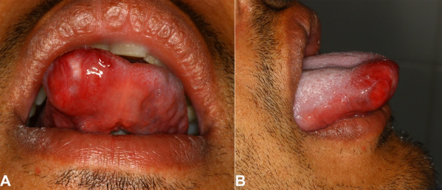
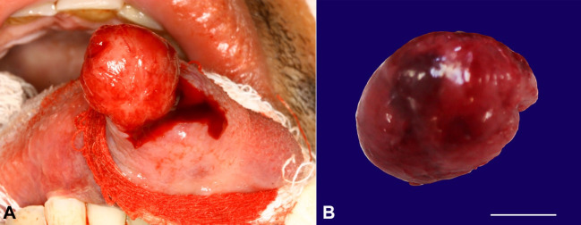
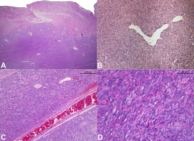
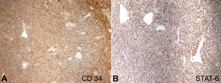
Solitary fibrous tumor (SFT) is a benign mesenchymal neoplasm originally described in pleura with a rare presentation in the oral cavity. Herein, we report a case of a 28-year-old male patient who presented an asymptomatic slow-growing mass in the anterior part of the tongue. Intraoral examination revealed a well-circumscribed mass covered by normal mucosa with a fibrous consistency. Due to non-specific clinical findings, the initial diagnostic hypotheses include benign submucosal neoplasms such as leiomyoma, neurofibroma, SFT, and others. An excisional biopsy was performed. Microscopically, the tumor was surrounded by a thick fibrous capsule; hypo and hypercellular areas were arranged in a storiform pattern with a stroma formed by collagen and abundant vascularization. Tumor cells showed immunopositivity for CD34 and STAT-6 and no expression of CD99, AML, S-100, and Ki-67. According to these findings, the diagnosis of SFT was established. After 24 months, the patient is asymptomatic and has no evidence of recurrence. Although oral involvement is rare, SFT should be included in the differential diagnosis of oral submucosal lesions.

Keywords: Oral Pathology; Oral diagnosis; Solitary Fibrous Tumors.


Conflict of interest statement

Conflict of interest: None

Figures

Figure 1
Figure 1. Intraoral examination shows: A - a well-circumscribed mass covered by an erythematous mucosa in the anterior portion of the tongue (Frontal view); B - right lateral view.
Figure 2
Figure 2. A - Trans-operative aspect of encapsulated lesion on the submucosa; B - size specimen (scale bar = 1 cm).
Figure 3
Figure 3. Photomicrographs of the biopsy specimen show: A - a dense proliferation of neoplastic cells partially surrounded by a thick fibrous capsule (H&E, 2.5X); B - Staghorn-like vessels (H&E, 10X); C - branching with evident perivascular hyalinization (H&E, 10X); D - Sheet of the spindle to ovoid cells with indistinct cytoplasmic borders (H&E, 40X).
Figure 4
Figure 4. Immunohistochemical panel showing the neoplastic cells with: A - diffuse expression of CD34 (10X); B - STAT-6 (10X).

References

    1. Klemperer P, Coleman BR. Primary neoplasms of the pleura: a report of five cases. Arch Pathol (Chic) 1931;11:385–412. - PubMed
    1. Ronchi A, Cozzolino I, Zito Marino F, et al. Extrapleural solitary fibrous tumor: A distinct entity from pleural solitary fibrous tumor. An update on clinical, molecular and diagnostic features. Ann Diagn Pathol. 2018;34:142–150. doi: 10.1016/j.anndiagpath.2018.01.004. - DOI - PubMed
    1. de Morais EF, Martins HDD, Rodrigues KS, de França GM, da Silveira ÉJD, Freitas RDA. Clinicopathologic analysis of oral and maxillofacial solitary fibrous tumor. Am J Clin Pathol. 2020;154(1):15–22. doi: 10.1093/ajcp/aqaa027. - DOI - PubMed
    1. Stanisce L, Ahmad N, Levin K, et al. Solitary fibrous tumors in the head and neck: comprehensive review and analysis. Head Neck Pathol. 2020;14(2):516–524. doi: 10.1007/s12105-019-01058-6. - DOI - PMC - PubMed
    1. Nunes FB, Sant’Ana MSP, Silva AMB, et al. Solitary fibrous tumour of the oral cavity: an update. J Oral Pathol Med. 2020;49(1):14–20. doi: 10.1111/jop.12953. - DOI - PubMed

Publication types